. 2023 Sep;299(9):105135.
doi: 10.1016/j.jbc.2023.105135. Epub 2023 Aug 6.

Characterization of a novel inhibitor for the New Delhi metallo-β-lactamase-4: Implications for drug design and combating bacterial drug resistance

Affiliations

Characterization of a novel inhibitor for the New Delhi metallo-β-lactamase-4: Implications for drug design and combating bacterial drug resistance

James B Thoden et al. J Biol Chem. 2023 Sep.

Abstract

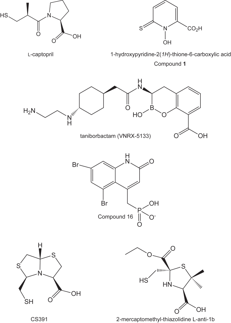
The bacterial metallo-β-lactamases (MBLs) catalyze the inactivation of β-lactam antibiotics. Identifying novel pharmacophores remains crucial for the clinical development of additional MBL inhibitors. Previously, 1-hydroxypyridine-2(1H)-thione-6-carboxylic acid, hereafter referred to as 1,2-HPT-6-COOH, was reported as a low cytotoxic nanomolar β-lactamase inhibitor of Verona-integron-encoded metallo-β-lactamase 2, capable of rescuing β-lactam antibiotic activity. In this study, we explore its exact mechanism of inhibition and the extent of its activity through structural characterization of its binding to New Delhi metallo-β-lactamase 4 (NDM-4) and its inhibitory activity against both NDM-1 and NDM-4. Of all the structure-validated MBL inhibitors available, 1,2-HPT-6-COOH is the first discovered compound capable of forming an octahedral coordination sphere with Zn2 of the binuclear metal center. This unexpected mechanism of action provides important insight for the further optimization of 1,2-HPT-6-COOH and the identification of additional pharmacophores for MBL inhibition.

Keywords: antibiotic resistance; antibiotics; computational biology; crystallography; enzyme kinetics; enzyme structure.


Conflict of interest statement

Conflict of interest The authors declare that they have no conflicts of interest with the contents of this article.

Figures

Figure 1
Figure 1
Inhibitors of New Delhi metallo-β-lactamases.
Figure 2
Figure 2
Structure of NDM-4 (apoenzyme). A ribbon representation of the monomer is shown in (A) with the β-strands and α-helices display in pink and light blue, respectively. Loops L1, L2, and L3 are highlighted in purple. A stereo view of the electron density corresponding to the region defined by Ala 55 to Val 58 is presented in (B). The map was calculated with (2Fo-Fc) coefficients and contoured at 1σ. As can be seen, this region adopts two distinct reverse turn conformations. A close-up stereo view of the binuclear metal center is provided in (C). Metal:ligand bonds are indicated by the dashed lines. Ordered solvent molecules are depicted as red spheres. This figure and Figures 3 and 4 and Figure 7, Figure 8, Figure 9 were prepared with PyMOL (60) (https://pymol.org/2/). NDM, New Delhi metallo-β-lactamase.
Figure 3
Figure 3
Structure of the NDM-4/l-captopril complex. The region defined by Leu 65 to Met 67 adopts two distinct conformations in the NDM-4/l-captopril complex as shown in stereo in (A). The two conformations are labeled A and B. The observed electron density corresponding to l-captopril is presented in stereo in (B). The map shown was calculated with (Fo-Fc) coefficients and contoured at 3σ. The inhibitor, l-captopril, was not included in the X-ray coordinate file used to calculate the omit map, and thus there is no model bias. A close-up stereo view of the binuclear metal center with bound l-captopril is provided in (C). Metal:ligand bonds are indicated by the dashed line. The bound l-captopril is colored in purple bonds. NDM, New Delhi metallo-β-lactamase.
Figure 4
Figure 4
Structure of the NDM-4/Compound 1 complex. The observed electron density for the inhibitor is shown in stereo in (A) and contoured as described in Figure legend 3. A close-up view of the binuclear metal binding region is presented in stereo in (B) with the inhibitor highlighted in purple bonds. A superposition of the binding modes for l-captopril and Compound 1, displayed in greenand purple bonds respectively, is provided in (C). NDM, New Delhi metallo-β-lactamase.
Figure 5
Figure 5
Molecular dynamics simulation analysis. (A) CαRMSD and (B) CαRMSF plots of NDM-4 in the apo unbound state (gray dotted line) and in the bound states with Compound 1 (black solid line) and l-captopril (gray solid line) over the course of the simulation with the largest fluctuation observed in the L1, L2, and L3 regions. The protein–ligand interaction histograms between NDM-4 with (C) Compound 1 and (D) l-captopril are shown to highlight the specific molecular interaction (ionic, hydrogen bond, hydrophobic, and water bridges) required for binding. The interaction fraction is the average number of atomic contacts between the ligand and the protein residue over the course of the simulation. NDM, New Delhi metallo-β-lactamase; RMSF, root-mean-square fluctuation.
Figure 6
Figure 6
Principal component and dynamics cross-correlation analyses. The top three principal components of motion for the (A) apo unbound, (B) Compound 1 bound and (C) l-captopril bound states of NDM-4 over the course of the simulations are shown as PC1, PC2, and PC3 with the regions showing the smallest (red) and largest (blue) movement. The dynamics cross correlation showing positively correlated (red), negatively correlated (blue), and uncorrelated (white) regions of NDM-4. Both correlated and anticorrelated regions are highlighted black boxes. The uncorrelated regions are highlighted in gray boxes. NDM, New Delhi metallo-β-lactamase; PC, principal component.
Figure 7
Figure 7
Comparison of the NDM-4 model presented here to that deposited under PDB accession code5WIG. As expected, the two NDM-4 models are nearly identical with the exception of the loop defined by Leu 65 to Phe 70. The three loop conformations shown here in stereo correspond to our model of the apoenzyme (purple), the coordinates deposited in the Protein Data Bank (aquamarine), and the structure of NDM-4/l-captopril complex (wheat). The l-captopril ligand is displayed in wheat bonds. NDM, New Delhi metallo-β-lactamase.
Figure 8
Figure 8
Comparison of the binding mode of Compound 1 to NDM-4 versus transition state analog mimics. Shown in (A) in stereo is a superposition of the NDM-4/Compound 1 and the NDM-1/taniborbactam complex models. The NDM-4 model is highlighted in wheat bonds and Compound 1 is colored in purple bonds. The NDM-1/taniborbactam complex is presented in blue bonds. The bridging water of the binuclear center is depicted as a red sphere. A stereo superposition of the NDM-4/Compound 1 and the NDM-1/Compound 16 complex models is shown in (B) with the same color coding as described in (A). In both (A) and (B), the coordination geometry about Zn1 and Zn2 in the NDM-4 model is indicated by the dashed lines. NDM, New Delhi metallo-β-lactamase.
Figure 9
Figure 9
Comparison of the binding mode of Compound 1 to NDM-4 versus those observed with thiolate-based inhibitors to NDM-1. A stereo superposition of the NDM-4/Compound 1 and the NDM-1/CS391 complexes is presented in (A). Likewise, a stereo superposition of the NDM-4/Compound 1 and NDM-1/2-mercaptomethyl-thiazolidine L-anti-1b, 4u41 displayed in (B). The color coding is as described in Figure legend 5. NDM, New Delhi metallo-β-lactamase.

References

    1. Fleming A. On the antibacterial action of cultures of a Penicillium, with special reference to their use in the isolation of B. Influenzae. Br. J. Exp. Pathol. 1929;10:226–236.
    1. Lobanovska M., Pilla G. Penicillin's discovery and antibiotic resistance: lessons for the future? Yale J. Biol. Med. 2017;90:135–145. - PMC - PubMed
    1. Abraham E.P., Chain E. An enzyme from bacteria able to destroy penicillin. Nature. 1940;146:837. - PubMed
    1. Murray C.J.L., Ikuta K.S., Sharara F., Swetschinski L., Robles Aguilar G., Gray A., et al. Global burden of bacterial antimicrobial resistance in 2019: a system analysis. Lancet. 2022;399:629–655. - PMC - PubMed
    1. Reygaert W.C. An overview of the antimicrobial resistance mechanisms of bacteria. AIMS Microbiol. 2018;4:482–501. - PMC - PubMed

LinkOut - more resources