. 2023 Aug 7;13(1):12786.
doi: 10.1038/s41598-023-39283-3.

Validation of a non-invasive prenatal test for fetal RhD, C, c, E, K and Fya antigens

Affiliations

Validation of a non-invasive prenatal test for fetal RhD, C, c, E, K and Fya antigens

Brian Alford et al. Sci Rep. .

Abstract

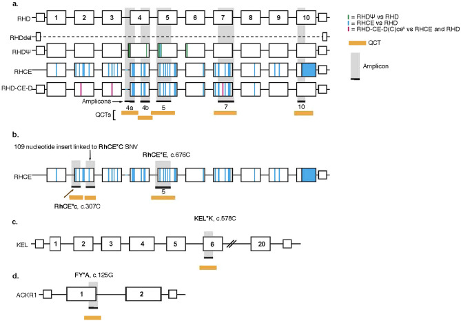
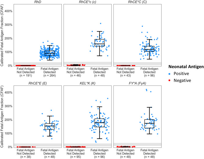
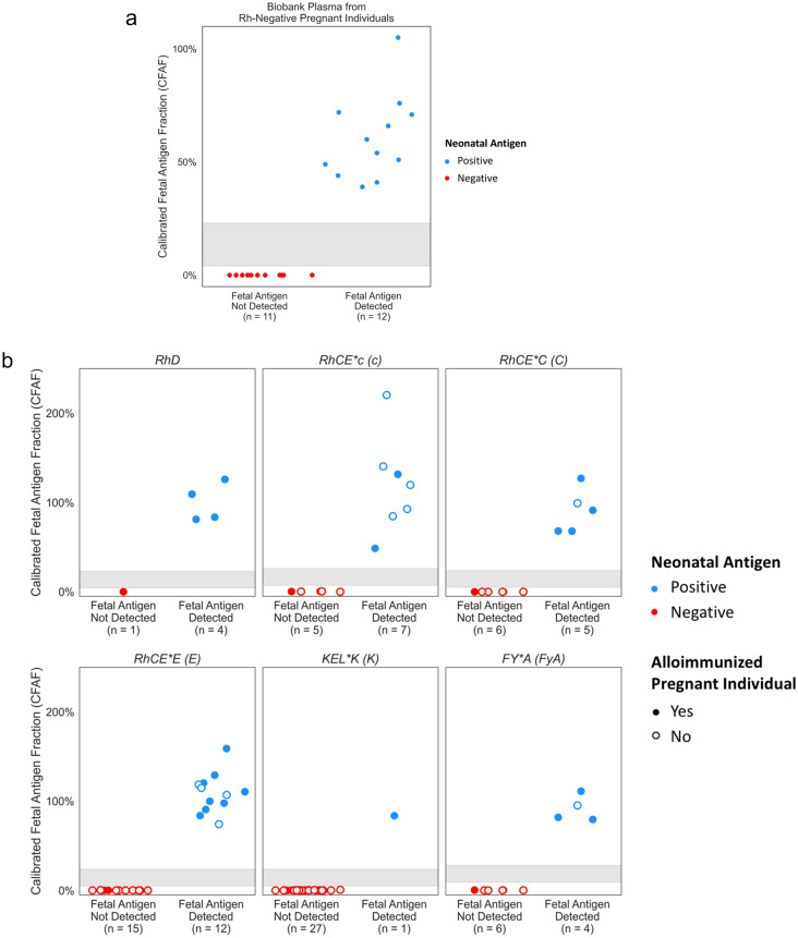
We developed and validated a next generation sequencing-(NGS) based NIPT assay using quantitative counting template (QCT) technology to detect RhD, C, c, E, K (Kell), and Fya (Duffy) fetal antigen genotypes from maternal blood samples in the ethnically diverse U.S. population. Quantitative counting template (QCT) technology is utilized to enable quantification and detection of paternally derived fetal antigen alleles in cell-free DNA with high sensitivity and specificity. In an analytical validation, fetal antigen status was determined for 1061 preclinical samples with a sensitivity of 100% (95% CI 99-100%) and specificity of 100% (95% CI 99-100%). Independent analysis of two duplicate plasma samples was conducted for 1683 clinical samples, demonstrating precision of 99.9%. Importantly, in clinical practice the no-results rate was 0% for 711 RhD-negative non-alloimmunized pregnant people and 0.1% for 769 alloimmunized pregnancies. In a clinical validation, NIPT results were 100% concordant with corresponding neonatal antigen genotype/serology for 23 RhD-negative pregnant individuals and 93 antigen evaluations in 30 alloimmunized pregnancies. Overall, this NGS-based fetal antigen NIPT assay had high performance that was comparable to invasive diagnostic assays in a validation study of a diverse U.S. population as early as 10 weeks of gestation, without the need for a sample from the biological partner. These results suggest that NGS-based fetal antigen NIPT may identify more fetuses at risk for hemolytic disease than current clinical practice, which relies on paternal genotyping and invasive diagnostics and therefore is limited by adherence rates and incorrect results due to non-paternity. Clinical adoption of NIPT for the detection of fetal antigens for both alloimmunized and RhD-negative non-alloimmunized pregnant individuals may streamline care and reduce unnecessary treatment, monitoring, and patient anxiety.


Conflict of interest statement

B.A., B.P.L, S.H., A.N., X.B., A.B., D.C., J.W., and J.H. are employees of BillionToOne and/or hold stock or options to hold stock in the company. B.H. and J.C. are consultants of BillionToOne. K.J.G. reports consulting fees from BillionToOne, Roche, and Aetion. K.J.G. also reports funding from BillionToOne for the transfer of clinical plasma specimens used in this study. D.E.C and T.F.M report no conflict of interests.

Figures

Figure 1
Figure 1
Schematic of the genes associated with the antigens, amplicons, and quantitative counting templates (QCTs). QCTs quantify the molecular count for each genomic loci. (a) The wild type RHD gene is shown along with its common variants, RHD gene deletion and RhDΨ variant, and the homologous RHCE gene and RHD-CE-D hybrid gene. Differences between these variants and homolog are highlighted as indicated in the legend. (b) Schematic of the RHCE gene, single nucleotide variants (SNVs) associated with RhCE*C (c.307C), RhCE*c (c.307T) and RhCE*E (c.676C) genotypes. The amplicon for the RHCE*C quantifies a 109 nucleotide insert highly linked to RhCE*C. The RhCE*E (c.676C) is located in the in amplicon 5 of the RhD assay (c) Schematic of the KEL gene and KEL*K (c.578C) SNV. (d) Schematic of the ACKR1 gene and FY*A (c.125G) SNV. The primer details for the amplicons are in Table S1.
Figure 2
Figure 2
Calibrated fetal antigen fraction (CFAF) and identified fetal antigen status for preclinical samples. Samples were made from sheared genomic DNA samples from replicates of parent–child pairs with an antigen negative parent/ antigen positive fetus (blue) and antigen positive parent/antigen negative child (red) pairs for each antigen type (Table S1). Parent–child samples were mixed to mimic fetal fractions ranging from 1.5 to 12%. Row 1: RhD, RhCE*c and RhCE*C, Row 2: RhCE*E, KEL*K and FY*A. The grayed area on the graphs represents the antigen-specific CFAF intermediate range (Table S2). CFAF values in this range are reported as No Results and a new sample is requested for re-analysis. There were no discordant calls. There were 16 samples where no result was reported because there were too few molecules in the sample (absolute expected molecular (AEM) count below the antigen specific threshold, n = 11) or the CFAF value was within the intermediate range (n = 5). For all 1061 NIPT assays the identified fetal antigen status was concordant with the known fetal antigen status, resulting in an overall sensitivity of 100% (95% CI 99–100%) and specificity of 100% (95% CI 99–100%). The tabulations of these data are in Table 1.
Figure 3
Figure 3
(a, b) Calibrated fetal antigen fraction (CFAF)s and identified fetal antigen status for clinical samples. Pregnant person antigen status was determined by NGS of cfDNA and fetal antigen status was reported for all samples where the pregnant person was identified to be negative for the antigen to mimic the clinical use of this assay. Samples are not unique across plots; the same sample was used for multiple assays if the pregnant person was antigen negative for multiple antigens. (a) Row 1: RhD, RhCE*C and RhCE*c, Row 2: RhCE*E, KEL*K and FY*A. There were 1191 (2.9%) NIPT analyses where fetal antigen was not determined due to low molecular count (absolute expected molecular (AEM) count below the antigen specific threshold, n = 941), CFAF in the intermediate zone (n = 180), or number of fetal antigen molecules detected above the expected range (n = 70). The sensitivity was modeled using a truncated normal distribution for the fetal antigen detected samples and the estimated sensitivity was greater than 99.6%. The tabulations of these data are in Table S5, the individual data points are plotted in Fig. S1a, data are plotted by fetal fraction in Fig. S1c, and the sensitivity of the antigen positive samples is modeled in Fig. S2. (b) The fetal RhD prediction for the 1615 clinical samples where the RhDΨ variant was not detected (panel 1) and the 53 clinical samples where RhDΨ variant was detected (panel 2). In qualitative RhD NIPT assays, samples with RhDΨ variant result in a no-call or inconclusive result. The individual data points are plotted in Fig. S1b.
Figure 4
Figure 4
(a) CFAF is plotted for the 23 samples from the LIFECODES biobank with known fetal RhD serology. All NIPT results were concordant with neonatal serology. Neonate RhD-positive by postnatal serology is indicated in blue and neonate RhD-negative by postnatal serology is indicated in red. The sensitivity and specificity for the detection of fetal RhD based on these 23 biobank samples was 100% (95% CI 73.5–100.0%) and 100% (95% CI 71.5–100.0%). respectively. (b) CFAF plotted for the 30 samples from alloimmunized pregnant individuals for the 93 antigens the pregnant individual was alloimmunized to and/or had a negative genotype. All NIPT results were concordant with neonatal genotype. Neonate antigen positive by postnatal genotyping is indicated by blue and neonate antigen negative in postnatal genotyping is indicated in red. An unfilled dot indicates the pregnant person was alloimmunized for the antigen. For these 93 antigens, the NIPT for detection of fetal antigen had a sensitivity of 100% (95% CI 89.4–100.00%) and a specificity of 100% (95% CI 94.0–100.0%).

References

    1. Silver RM. Practice bulletin no. 181: Prevention of Rh D alloimmunization. Obstet. Gynecol. 2017;130:E57–E70. doi: 10.1097/AOG.0000000000002232. - DOI - PubMed
    1. Strengers PFW. Challenges for plasma-derived medicinal products. Transfus Med. Hemother. 2023;50:116. doi: 10.1159/000528959. - DOI - PMC - PubMed
    1. American College of Obstetricians and Gynecologists’ Committee on Practice Bulletins—Obstetrics. Number 192: Management of Alloimmunization During Pregnancy. (ACOG, 2018).
    1. Minon JM, Gerard C, Senterre JM, Schaaps JP, Foidart JM. Routine fetal RHD genotyping with maternal plasma: A four-year experience in Belgium. Transfusion. 2008;48:373–381. doi: 10.1111/J.1537-2995.2007.01533.X. - DOI - PubMed
    1. Liu S, Ajne G, Wikman A, Lindqvist C, Reilly M, Tiblad E. Management and clinical consequences of red blood cell antibodies in pregnancy: A population-based cohort study. Acta Obstet. Gynecol. Scand. 2021;100:2216–2225. doi: 10.1111/aogs.14261. - DOI - PubMed

Publication types

Substances