. 2023 Sep;8(9):1732-1747.
doi: 10.1038/s41564-023-01433-8. Epub 2023 Aug 7.

Spatially resolved protein map of intact human cytomegalovirus virions

Affiliations

Spatially resolved protein map of intact human cytomegalovirus virions

Boris Bogdanow et al. Nat Microbiol. 2023 Sep.

Abstract

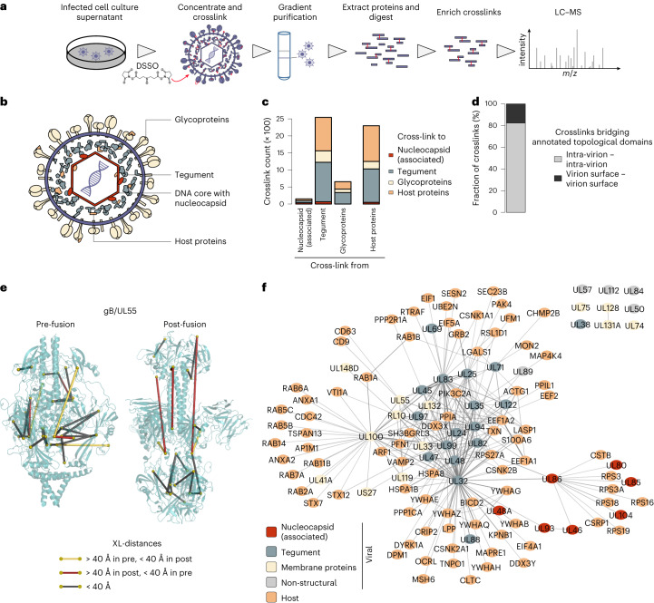
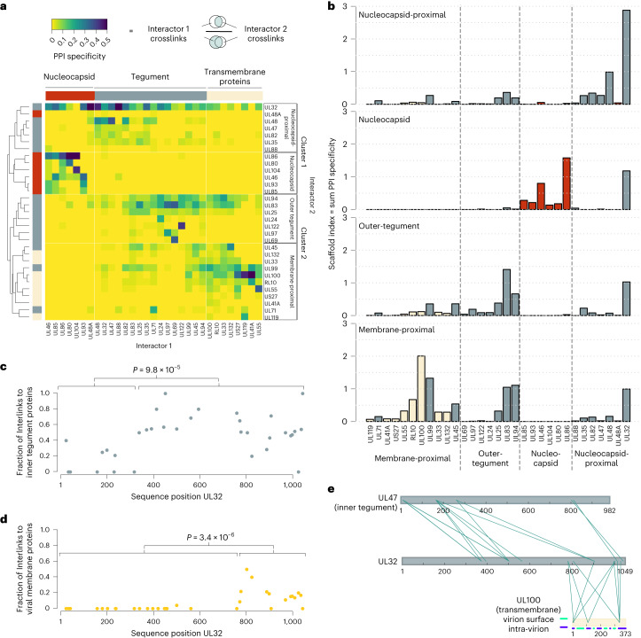
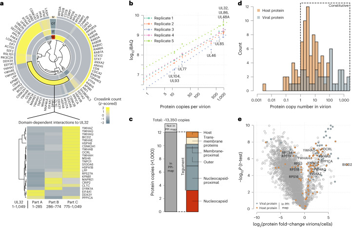
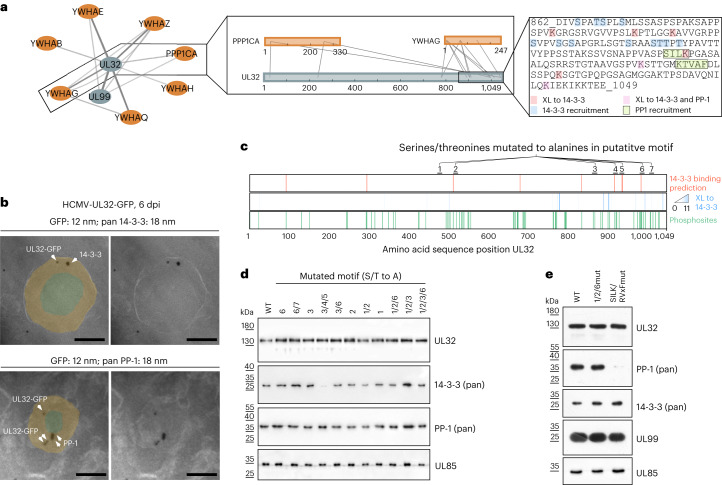
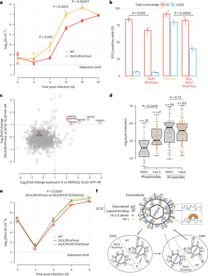
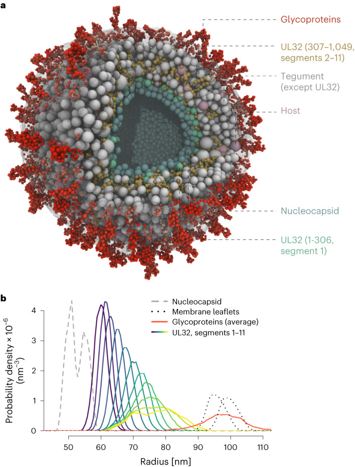
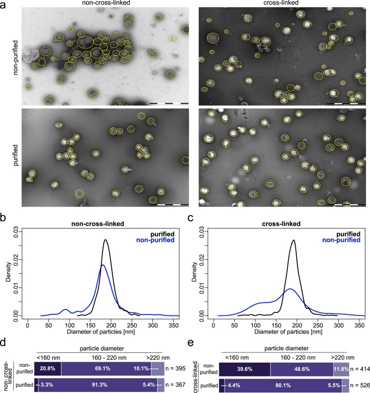
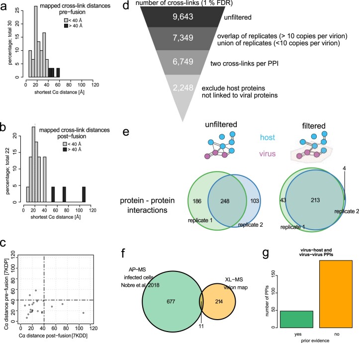
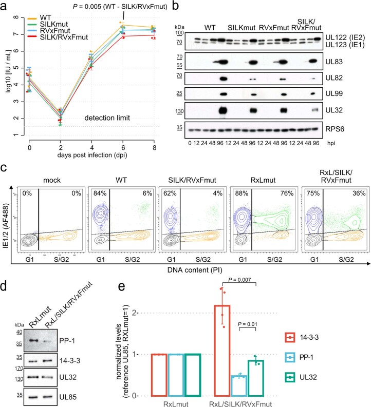
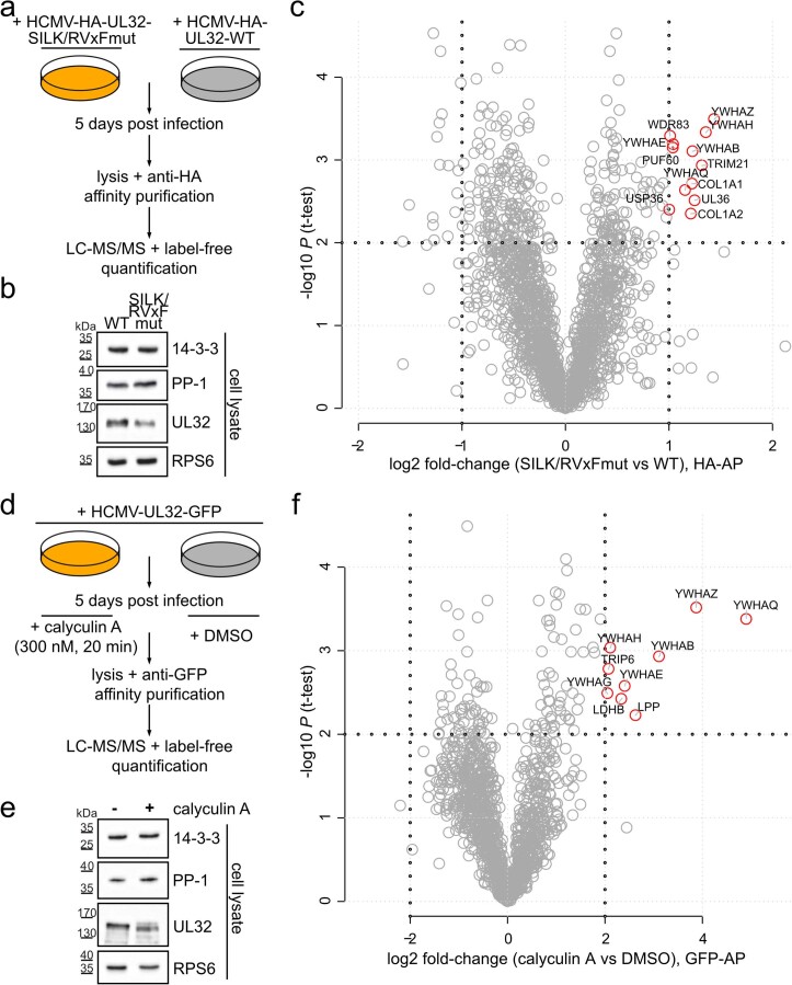
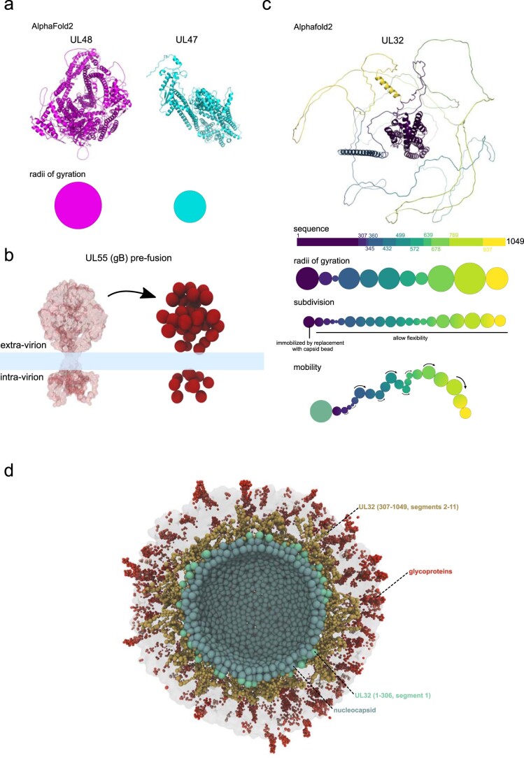
Herpesviruses assemble large enveloped particles that are difficult to characterize structurally due to their size, fragility and complex multilayered proteome with partially amorphous nature. Here we used crosslinking mass spectrometry and quantitative proteomics to derive a spatially resolved interactome map of intact human cytomegalovirus virions. This enabled the de novo allocation of 32 viral proteins into four spatially resolved virion layers, each organized by a dominant viral scaffold protein. The viral protein UL32 engages with all layers in an N-to-C-terminal radial orientation, bridging nucleocapsid to viral envelope. We observed the layer-specific incorporation of 82 host proteins, of which 39 are selectively recruited. We uncovered how UL32, by recruitment of PP-1 phosphatase, antagonizes binding to 14-3-3 proteins. This mechanism assures effective viral biogenesis, suggesting a perturbing role of UL32-14-3-3 interaction. Finally, we integrated these data into a coarse-grained model to provide global insights into the native configuration of virus and host protein interactions inside herpesvirions.


Conflict of interest statement

F.L. is a scientific advisory board member of Absea Biotechnology and VantAI. The remaining authors declare no competing interests.

Figures

Fig. 1
Fig. 1. The spatial proteome of intact cytomegalovirus virions.
a, Workflow for the XL–MS analysis of intact HCMV particles. The experiment was performed in n = 2 biological replicates. b, Schematic depiction of the HCMV virion layers. c, Number of crosslinks between viral proteins with known virion layer localization and between viral and host proteins. d, Proportion of crosslinks within annotated extra-virion and intra-virion domains of viral glycoproteins or intra-virion resident proteins. No crosslinks were observed linking extra-virion domains of viral glycoproteins to their intra-virion domains or to intra-virion resident proteins. e, Crosslink mapping onto structural models of UL55 in pre- and post-fusion conformations. See also Extended Data Fig. 2a–c. f, Network of PPIs inside HCMV particles. The line width (edges) scales with the number of identified crosslinks between interaction partners. Source data
Fig. 2
Fig. 2. Spatial arrangement of viral proteins.
a, Heat map of PPI specificity values of all viral proteins with at least 9 identified crosslinks. PPI specificity values were calculated for each PPI by dividing the number of crosslinks supporting a PPI by the total number of crosslinks of the interaction partner (‘interactor 1’). Interactor 2 proteins (rows) were clustered using 1-Pearson’s correlation distance and complete linkage. Interactor 1 proteins (columns) were manually annotated on the basis of previous knowledge. b, By summing up the PPI specificity values of interactor 2 proteins across all PPIs of the respective subcluster, a scaffold index was calculated and plotted using the same order of proteins as in the columns of a. Bar colours indicate the membership of proteins to nucleocapsid, tegument or viral envelope (transmembrane proteins), identical to a. c,d, For each individual lysine in UL32, the fraction of crosslinks in UL32 linking to other viral inner-tegument proteins (c) or viral membrane proteins (d) is plotted. P values are based on two-sided Wilcoxon rank-sum tests comparing the indicated lysines within the UL32 primary sequence. e, Crosslink network between UL32 and selected proteins of the inner tegument (UL47) and viral envelope (UL100). The membrane topology of UL100 is indicated by blue and green lines. Source data
Fig. 3
Fig. 3. Abundant and layer-specific incorporation of host proteins.
a, Top: heat map depiction of host protein localization across virion layers. Crosslink counts of host proteins to viral proteins of the respective layers (NC, nucleocapsid; IT, inner tegument (nucleocapsid-proximal tegument); OT, outer tegument; M, membrane, including viral transmembrane proteins and membrane-proximal tegument) were z-scored and hierarchically clustered using Euclidean distance. Bottom: z-scored crosslink counts of inner-tegument-localized host proteins to different parts of UL32. b, iBAQ of protein copy numbers using the copy numbers of nucleocapsid-associated proteins as standard (n = 2 biological replicates, with technical duplicates or triplicates). The offset between the replicates is caused by differences in the amount of input material, but the similar slopes demonstrate reproducibility of the quantitative data. c, Protein copies represented in the PPI map and their subvirion localization. d, Histogram of copy numbers from host and viral proteins. Proteins with more than one copy number on average are classified as likely constitutive. e, Relative quantification of proteins comparing virion lysates to cell lysates. Proteins included in the PPI map are highlighted. See Extended Data Fig. 4a for experimental design. Means of fold-change differences and P values of a two-sided t-test without multiple hypothesis correction are based on n = 4 biological replicates. Source data
Fig. 4
Fig. 4. Nearby short linear motifs in UL32 recruit 14-3-3 and PP-1 proteins into virus particles.
a, PPI network including interactors of PP-1 (PPP1CA) and 14-3-3 (YWHAx) proteins, with insets showing crosslinks involving UL32, 14-3-3 protein gamma (YWHAG), PP-1 and the UL32 sequence containing 14-3-3 and PP-1 recruitment sites. The line width scales with the number of identified crosslinks. Host and viral proteins are highlighted orange and grey, respectively. b, EM images of intracellular virions from HCMV-UL32-GFP-infected fibroblasts (MOI = 5). Ultrathin sections were stained with immunogold against GFP (12 nm gold) and either pan 14-3-3 or PP-1 (18 nm gold), as indicated. Approximate areas of tegument and nucleocapsid layers were manually highlighted in light orange and light green. Scale bars, 100 nm. See Extended Data Fig. 5a,b for uncropped micrographs. c, Mutational approach for identifying recruitment sites for 14-3-3 on UL32. Motif predictions above score 0.6 (top bar), crosslink positions to 14-3-3 (middle bar) and identified phosphorylation sites (localization probability >0.75 in n = 2 biological replicates) in virions (bottom bar). d,e, Purified virions from recombinant mutant viruses harbouring alanine substitutions in the designated motifs were assessed for protein levels by immunoblotting. Representative experiments of n = 2 biological replicates are shown. Source data
Fig. 5
Fig. 5. PP-1 recruitment controls early and late events during HCMV biogenesis.
a, Growth curve of mutant and wild-type virus on HELFs (MOI = 0.05). Means ± s.d. of n = 3 biological replicates are depicted. Unpaired two-sided t-tests without multiple hypothesis correction were performed at the indicated timepoints. b, Flow cytometry analysis of IE protein levels as a function of the cell cycle stage. HELFs were infected with the indicated recombinant viruses (MOI = 5, 6 h post infection). The percentage of IE1/2-positive cells in G1 or S/G2 compartments is given, with mean ± s.e. of n = 3 biological replicates. Unpaired two-sided t-tests were performed comparing the fraction of IE1/2-positive cells between the indicated viruses and cell cycle compartments. Representative contour plots of one replicate are shown in Extended Data Fig. 7c. c, Comparison of differential interaction partners to UL32 upon phosphatase inhibition (x axis) or genetic disruption of the SILK/RVxF motifs (y axis). See Extended Data Fig. 9 for volcano plots, experimental design and controls. d, Abundance levels of quantified phosphosites or quantified peptides of pp150/UL32 are depicted with their sum intensity across n = 3 biological replicates in UL32-GFP precipitates (centre line, median; box limits, upper and lower quartiles; whiskers, 1.5× interquartile range). P values from two-sided Wilcoxon rank-sum test as indicated. e, Same as a but with MOI = 5. f, Location of 14-3-3 and PP-1 inside virions and their role during early and late stages of HCMV infection. WT viruses, able to recruit PP-1, dephosphorylate (-P) UL32 efficiently (bold arrows), start viral gene expression (IE) and produce infectious progeny. PP-1-binding-deficient RVxF/SILKmut viruses are impaired (light arrows) at the start of IE gene expression and production of novel progeny. No adjustments for multiple comparisons were performed for a, b, d and e. Source data
Fig. 6
Fig. 6. A coarse-grained model of the HCMV virion.
a, Cross-sectional view through the equilibrium state with transparent membrane. Bead species identity as indicated. UL32 C-terminal segments make occasional contacts with intra-virion domains of viral glycoproteins. b, Probability distributions of UL32 segments (see Extended Data Fig. 10) in the tegument. Nucleocapsid and membrane layers are given as reference. Source data
Extended Data Fig. 1
Extended Data Fig. 1. Estimation of sample purity from cross-linked and non-cross-linked HCMV particles.
(a) Exemplary micrographs of negatively stained HCMV particles. Purification using glycerol tartrate gradients and cross-linking treatment as indicated. Scale bar represents 1000 nm. Manually selected particles are indicated by yellow circles. Representative micrographs of at least 10 per condition. (b, c) Distribution of diameters of manually picked particles of the non-cross-linked (b) or cross-linked (c) samples. Purification led to a more homogenous population of particle diameters. As previously noted25, we also find that the purification leads to membrane blebs, thus representing non-native particle morphologies. (d, e) Two cut-offs were defined to categorize unusually small (<160 nm) and large particles (>220 nm). These correspond to the median of diameters from the purified sample ± two times the standard deviation. The fraction of particles with diameters that fall below 160 nm, exceed 220 nm or fall in between are indicated for the non-cross-linked (d) or cross-linked (e) sample types. Purification results in more homogeneous particle diameters, enriching virions in the expected size range. Thus, our protocol captures the native configuration of the particles at relatively high sample purity. At least 367 particles were counted per condition. Source data
Extended Data Fig. 2
Extended Data Fig. 2. Data filtering and reproducibility.
(a, b) Histogram of distances between Cα atoms obtained from mapping cross-links onto the cryoEM structural models of homotrimeric UL55 in pre- (a) or post-fusion (b) conformations (pdb: 7kdp, 7kdd). See Fig. 1e for visualization of the cross-links on the structural models. (c) Direct comparison of distances between pre- and post-fusion conformations. (d) The number of cross-links is shown that survive the indicated filtering steps. (e) Venn diagrams comparing the number of PPIs between both replicates in unfiltered (Supplementary Table 1) and filtered (Supplementary Table 2) dataset. Homomeric interactions (based on intra-links) were removed for this analysis. (f) Comparison to the AP-MS based interactome from infected cells26, based on viral baits included in the XL-MS PPI-map. (g) Number of PPIs with or without existing evidence in the literature. See also Supplementary Table 3.
Extended Data Fig. 3
Extended Data Fig. 3. Host protein recruitment to HCMV particles.
(a, b) Host proteins included in the PPI map (see Fig. 1f) were compared to host proteins from individual (a) or (b) combined published proteomics dataset15–18 of purified HCMV particles. (c) Cross-link network between ribosomal 40S proteins and UL86 protein. (d) Cross-linked lysines were mapped onto a nucleocapsid hexon (UL86 hexamer), depicted from top (left side) (that is inner tegument side) or bottom (right side) (that is DNA-accommodating side). Electrostatic surface rendering with charges depicted ranging from negatively charged (red) to positively charged (blue) (pdb 5vku). (e) Depiction of cross-links between glycoprotein UL55 and tetraspanins CD9 and CD63. Domain annotations were obtained from uniprot.org.
Extended Data Fig. 4
Extended Data Fig. 4. Quantitative proteomics identifies selective recruitment of host proteins.
(a) Experimental setup to identify selectively recruited proteins (n=4 biological replicates). (b) Spearman’s correlation coefficients comparing log2 transformed LFQ-values of the experiment outlined in a. (c) Cross-comparison of protein copy numbers and enrichment levels of host and viral proteins. Selected proteins are depicted with their gene symbols. Enrichment levels are based on the mean of n=4 biological replicates and copy numbers based on the mean of n=2 biological replicates (with technical duplicates or triplicates). Enrichment level values below log2 -4 are not depicted. (d, e) Cross-link networks between selected host-virus PPIs.
Extended Data Fig. 5
Extended Data Fig. 5. Characterization of the 14-3-3 binding-deficient viral mutants.
(a, b) EM images of the intracellular environment from HCMV-UL32-GFP infected HELFs (MOI=5) prepared using Tokoyasu method. Ultrathin sections were stained with immunogold against GFP (12 nm gold) and pan 14-3-3 or PP-1 (18 nm gold), as indicated. Scale bars: 100 nM. Magnified views of the boxed regions in white are depicted in Fig. 4b. (c) Experimental workflow for SILAC-based comparison of virion protein content between mutant and WT viruses. The experiments were performed in label-swap duplicates. (d) Purified virions from WT or mutant viruses harbouring alanine substitutions in the ¾/5 region (see also Fig. 4c) were assessed for UL32, PP-1, 14-3-3 and UL85 levels by immunoblotting. Control experiment to panel (e). (e) SILAC-based proteomic comparison of log2 protein fold-changes comparing purified particles of WT and 14-3-3 binding mutants for both replicates, based on n=2 biological replicates. (f, g) Western blot analysis of abundance levels of selected proteins of purified virions for different 14-3-3 binding site mutant viruses. Exemplary blots in (f) and quantification based on n=2 biological replicates (with n=2 technical replicates) in (g). The height of the bars corresponds to the mean and the error bars to the standard deviation. P-values based on two-sided t-test without multiple hypothesis correction. Sites 4/5 are most important for 14-3-3 incorporation. Source data
Extended Data Fig. 6
Extended Data Fig. 6. Characterization of the PP-1 binding-deficient viral mutant.
(a) Crystal structure of PP-1 (pdb: 1fjm) with highlighted SILK and RVxF binding grooves as well as lysines cross-linked to UL32. (b) SILAC-based proteomic comparison of protein abundance comparing purified particles of wild-type and PP-1-binding mutant viruses. (c) Cross-links between DYRK1A kinase and UL32. Cross-link reactive lysines are highlighted blue in the sequence of both UL32 and DYRK1A. (d) Average log2 phosphosite fold-changes comparing wild-type and PP-1-binding mutant purified particles (centre line, median; box limits, upper and lower quartiles; whiskers, 1.5× interquartile range, overlaid with all data points). Phosphosites belonging to host proteins, viral proteins (excluding UL32) or to UL32 were compared using a two-sided Wilcoxon rank sum test. The average phosphosite ratio corrected by the protein ratio of n=2 biological replicates (including label-swap) are depicted. Experimental design as in Extended Data Fig. 5c with additional phosphopeptide enrichment.
Extended Data Fig. 7
Extended Data Fig. 7. PP-1-UL32 binding promotes viral growth and the onset of viral gene expression.
(a) Growth curve of mutant and wildtype viruses on HELFs (MOI=5). Means (centre of the error bars) and standard deviations of the mean of n = 3 biological replicates are depicted. Unpaired two-sided t-tests without multiple hypothesis correction were performed comparing the indicated viruses and time points. (b) Kinetics of viral protein levels of wildtype and mutant viruses across the replication cycle (MOI = 5). The experiment was replicated at least three times with similar results. (c) Flow cytometry analysis of Immediate Early (IE) protein levels as a function of the cell cycle stage. HELFs were infected with the indicated recombinant viruses (MOI = 5, 6 h post infection). The percentage of IE1/2-positive cells in G1 or S/G2 compartments is given. (d, e) Western blot analysis of abundance levels of selected proteins of purified virions for RxLmut and RxL/SILK/RVxFmut viruses. Exemplary blots in (d) and quantification based on n=4 (14-3-3) or n=3 (PP-1, UL32) technical replicates in (e). The height of the bars corresponds to the mean and the error bars to the standard deviation. P-values based on two-sided t-tests without multiple hypothesis correction. Control experiments to Fig. 5b. Source data
Extended Data Fig. 8
Extended Data Fig. 8. Hierarchical gating strategy for analysis of cell cycle-dependent viral gene expression by flow cytometry.
First, a contour plot was created displaying on a linear scale the forward light scatter (FSC) and sideward light scatter (SSC) of measured particles. A population P1 was gated that excludes damaged cells and cell aggregates from further analysis. Based on the area (A) and width (W) of the propidium iodide (PI) fluorescence signal (PerCP channel), a cell population P2 was defined that excludes cell doublets and cells with a greater than 2n DNA content from further analysis (upper right contour plot). Based on cellular DNA content and on the Alexa Fluor 488 fluorescence (FITC channel) from IE1/IE2 immunostaining, four subpopulations of P2 were defined (lower right contour plot): P3 consists of IE-positive G1 cells; P4 of IE-positive G2/M cells, P5 of IE-negative G1 cells, P6 of IE-negative G2/M cells. For better visibility, these four cell populations were depicted in a ‘zoom-in’ representation mode (lower left contour plot). The described gating strategy was used for all flow cytometry data presented in Fig. 5b and Extended Data Fig. 7c. Contour levels were set to 15% probability. Outliers falling outside the lowest contour level are displayed as dots.
Extended Data Fig. 9
Extended Data Fig. 9. Recruitment of PP-1 functionally antagonizes 14-3-3 binding to UL32.
(a, d) Experimental strategy to quantify changes in the UL32 interactome that were induced by loss of PP-1 binding sites (a) or pharmacologic inhibition of PP-1 phosphatase activity (d). Both experiments were performed in n=3 biological replicates. See also Fig. 5c,d. (b, e) HCMV infected cells were lysed at 5 days post infection. Before the cell lysates were used as input for HA or GFP affinity purification, they were controlled for UL32, PP-1 and 14-3-3 expression by immunoblot analysis. The ribosomal protein RPS6 indicates equal loading. Experiments were replicated with similar results at least three times. (c, f) Volcano plot analysis of LC-MS/MS data showing the average log2 fold-change of identified proteins and the corresponding p-values of a two-sided t-test without multiple hypothesis correction. Cut-offs were set manually at P = 0.01 and log2 fold-change = 1 (c) or log2 fold-change = 2 (f). Proteins passing these cut-offs were labelled with gene symbols. Source data
Extended Data Fig. 10
Extended Data Fig. 10. Parametrization of proteins for coarse-grained modelling.
(a) Bead sizes for tegument and host proteins other than UL32 were approximated by the gyroscopic radii from their corresponding AF2 predictions, which is exemplarily shown for UL47 and UL48 proteins. (b) Representation of glycoproteins in the coarse-grained model via clustering of ɑ-carbon from cryoEM or AF2 models into beads, exemplarily shown for gB pre-fusion structure. The transmembrane domains are substituted by beads from the membrane model (light blue). (c) The UL32 AF2 prediction was segmented into 11 parts based on a molecular dynamics simulation. Segment dimensions were calculated from the segment-specific radii of gyration. As the radius of gyration overestimates the volume for a disordered protein, the chain of beads was further subdivided to yield a chain of more but smaller beads. The most n-terminal bead was immobilized by replacing it with a bead from the nucleocapsid shell. The chain of beads was allowed to have segment-specific flexibility at hinges (indicated arrows). (d) Cross-sectional view of the HCMV virion model with tegument proteins other than UL32 removed.

References

    1. Liu, F. & Zhou, Z. H. in Human Herpesviruses: Biology, Therapy, and Immunoprophylaxis (eds Arvin, A. et al.) Ch. 3 (Cambridge Univ. Press, 2007). - PubMed
    1. Connolly SA, Jardetzky TS, Longnecker R. The structural basis of herpesvirus entry. Nat. Rev. Microbiol. 2021;19:110–121. - PMC - PubMed
    1. Yu X, et al. Biochemical and structural characterization of the capsid-bound tegument proteins of human cytomegalovirus. J. Struct. Biol. 2011;174:451–460. - PMC - PubMed
    1. Laine RF, et al. Structural analysis of herpes simplex virus by optical super-resolution imaging. Nat. Commun. 2015;6:5980. - PMC - PubMed
    1. Bohannon KP, Jun Y, Gross SP, Smith GA. Differential protein partitioning within the herpesvirus tegument and envelope underlies a complex and variable virion architecture. Proc. Natl Acad. Sci. USA. 2013;110:E1613–E1620. - PMC - PubMed

Publication types