Review
. 2023 Aug 7;10(1):36.
doi: 10.1186/s40580-023-00385-3.

Lipid nanoparticle-based mRNA delivery systems for cancer immunotherapy

Affiliations
Review

Lipid nanoparticle-based mRNA delivery systems for cancer immunotherapy

Jieun Han et al. Nano Converg. .

Abstract

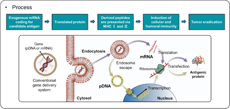
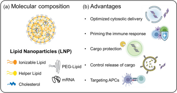
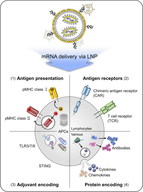
Cancer immunotherapy, which harnesses the power of the immune system, has shown immense promise in the fight against malignancies. Messenger RNA (mRNA) stands as a versatile instrument in this context, with its capacity to encode tumor-associated antigens (TAAs), immune cell receptors, cytokines, and antibodies. Nevertheless, the inherent structural instability of mRNA requires the development of effective delivery systems. Lipid nanoparticles (LNPs) have emerged as significant candidates for mRNA delivery in cancer immunotherapy, providing both protection to the mRNA and enhanced intracellular delivery efficiency. In this review, we offer a comprehensive summary of the recent advancements in LNP-based mRNA delivery systems, with a focus on strategies for optimizing the design and delivery of mRNA-encoded therapeutics in cancer treatment. Furthermore, we delve into the challenges encountered in this field and contemplate future perspectives, aiming to improve the safety and efficacy of LNP-based mRNA cancer immunotherapies.

Keywords: Cancer immunotherapy; Lipid nanoparticles (LNPs); Messenger RNA (mRNA); Tumor-associated antigens (TAAs).


Conflict of interest statement

The authors declare that they have no competing interests.

Figures

Fig. 1
Fig. 1
Schematic of the gene delivery process, highlighting pDNA and mRNA pathways. The diagram illustrates the utilization of exogenous mRNA encoding a target antigen, which results in protein translation and peptide presentation through MHC Class I and II molecules. This process activates both cellular and humoral immune responses, ultimately contributing to tumor elimination
Fig. 2
Fig. 2
LNPs composition and advantages as gene carriers. a Depiction of the molecular composition of LNPs, showcasing a variety of components such as ionizable lipids, helper lipids, cholesterol, polyethylene glycol (PEG)-lipids, and the cargo gene (mRNA). b Advantages: LNPs provide several benefits as gene carriers, including enhanced cytosolic delivery, immune response priming, cargo protection, controlled cargo release, and targeting of antigen-presenting cells (APCs)
Fig. 3
Fig. 3
Schematic of strategies for LNP-based mRNA delivery. This diagram displays various approaches to LNP-based mRNA delivery, including: (1) Antigen presentation—delivery of mRNA encoding TAAs to be presented by antigen-presenting cells (APCs); (2) Antigen receptor—delivery of mRNA encoding CARs or TCRs for T cell activation; (3) Adjuvant—the mRNA can encode factors that activate TLR3/7/8 or STING, amplifying immune responses; (4) Protein—delivery of mRNA encoding therapeutic proteins, such as cytokines or antibodies, for direct anti-cancer effects
Fig. 4
Fig. 4
Representative mRNA-LNPs for cancer vaccines. a Schematic illustration of the mRNA-loaded LNPs and the experimental method employed. b Prophylactic antitumor activity of A11-LNPs in E.G7-OVA tumor-bearing mice. c Therapeutic antitumor activity of A-11-LNPs, MC3-LNP, RNA-LPX, and B-8-LNPs in E.G7-OVA tumor-bearing mice, intravenously (i.v.) injected with OVA mRNA-loaded formulations at two doses of 0.03 mg mRNA/kg on days 8 and 11 (n = 5). df Expression of activation markers CD40 (d), CD80 (e), and CD86 (f) in splenic dendritic cells (DCs) 24 h after an i.v. injection of OVA mRNA-loaded formulations at a dose of 0.03 mg mRNA/kg (n = 3). (* p < 0.05, ** p < 0.01). af: Reproduced from a previous report [70] with Elsevier.) (g) Experimental timeline for vaccination and blood withdrawal. h OVA-specific antibody titers in mice treated with 113-O12B/mOVA and ALC-0315/mOVA on day 12. (i) Representative flow cytometry diagrams of IFN-γ-positive cells within CD3 + CD8 + T cells 7 days after the second vaccination. j Tumor volumes in the B16F10-OVA tumor model. (k) Lungs collected 18 days after the i.v. injection of B16F10-OVA cells. gk: Reproduced from a previous report [72] with permission from PNAS)
Fig. 5
Fig. 5
CAR-encoded mRNA-LNP delivery system. a Schematic representation of T-cell targeting using CAR mRNA-loaded LNPs. b Fabrication of LNPs with various components using microfluidic technology. c Expression rate of CAR in primary T cells analyzed by flow cytometry, with both purified LNP and electroporation (EP) groups showing an increase in CAR expression on T cells. d Cell viability assessment, highlighting that the EP group exhibited the lowest cell viability among primary T cells. (Reproduced from a previous report [6] with permission from American Chemical Society)
Fig. 6
Fig. 6
Adjuvant-encoded mRNA-LNP delivery system. a Cytokine levels in C57BL/6 mouse serum following treatment with STING-encoded mRNA-LNPs. b Flow cytometry analysis of IFN-γ, TNF-α, and IL-2 in the spleen after treatment with STING-encoded mRNA-LNPs. c Decreased lung metastasis observed in the E6/7 + NTFIX + DMXAA (STING-encoded mRNA-LNP) group after tumor challenge. d Survival rate in mice treated with STING-encoded mRNA-LNPs. (Reproduced from a previous report [101] with permission from Elsevier)
Fig. 7
Fig. 7
Cytokine-encoded mRNA-LNP Delivery System. a Schematic representation of tumor challenge with cytokine-encoded mRNA-LNP systems. b Tumor volume and survival rate in the B16F10 tumor model treated with IL-12-encoded mRNA-LNPs. The IL-12-alb-lum mRNA-LNP formulation demonstrated effective tumor suppression efficacy. ab: Reproduced from a previous report [106] with permission from Springer Nature). c Tumor volume measured for IL-23, IL-36γ, and OX40L-encoded mRNA-LNPs, showing anticancer effects in the MC38-R tumor model. d Tumor volume suppression due to the abscopal effect of IL-23, IL-36γ, and OX40L-encoded mRNA-LNPs. cd: Reproduced from a previous report [111] with permission from American Association for the Advancement of Science). e Schematic of IL-12, IL-27, and GM-CSF-encoded mRNA-LNP systems. f Cytokine concentrations following treatment with IL-27, IL-12, or GM-CSF mRNA-loaded MC3-LNPs or DAL4-LNPs. g Tumor size measured in the B16F10 murine model treated with DAL4-LNP-encapsulated mRNA. h Survival rate in the B16F10 murine model treated with mRNA-loaded DAL4-LNPs. eh: Reproduced from a previous report [7] with permission from Elsevier)
Fig. 8
Fig. 8
Antibody-encoded mRNA-LNP Delivery System. a Schematic representation of mRNAs encoding the heavy and light chains of trastuzumab. b Trastuzumab concentrations in C57BL/6 mouse serum 24 h after injection of cKK-E12 LNPs with trastuzumab mRNA via the tail vein at different doses. c Pharmacokinetics of trastuzumab in C57BL/6 mouse serum after a single i.v. dose of 8 mg/kg Herceptin (Genentech) or 2 mg/kg cKK-E12 LNPs with trastuzumab mRNA. d Growth of HER2-negative (MDA-MB-231) and HER2-positive (MDA-MB-231-HER2) tumors in mice treated with trastuzumab mRNA. Arrows indicate the days of mRNA-LNP injections. ad: Reproduced from a previous report [8] with permission from Elsevier). e Structures of the IVT bi-(scFv)2 and Fab-(scFv)2 RiboMABs. f Ex vivo cytotoxicity (left) and concentration (Cp) (right) of endogenously translated CD3 × CLDN6 RiboMAB in the plasma of NSG mice after i.v. administration of polymer/lipid-formulated mRNA. g Mice were treated with CD3 × CLDN6 or luciferase mRNA (n = 6/group; three doses of 3 µg/mouse i.v. weekly) or with purified CD3 × CLDN6 protein (200 µg/kg) or vehicle (n = 7/group; three doses intraperitoneally (i.p.) weekly, total of ten doses). Tumor growth for individual mice (left, mRNA; right, recombinant protein) are shown. h Mice were treated with two doses of CD3 × CLDN6 mRNA (n = 4) or luciferase mRNA as a negative control (n = 4) (both 3 µg/mouse i.v. weekly). Tumor-infiltrating lymphocytes (human CD3 + cells; left) and CLDN6-expressing tumor cells (right) were quantified by immunohistochemistry in three consecutive tumor sections. eh: Reproduced from a previous report [121] with permission from Springer Nature). i Binding of mRNA-encoded rituximab expressed in BHK cells to Raji cells. Depicted is the median of phycoerythrin (PE) fluorescence of all living cells. jm mRNA-encoded mAb protects mice from lethal tumor challenge. Each group comprised 12 mice. j Tumor development assessed by whole-body luminescence imaging at indicated times after tumor challenge. k Survival of mice receiving i.v. injections of either 10 or 50 µg of mRNA-LNP encoding rituximab. l Representative luminescence images of mice treated with two different doses of mRNA-LNP encoding rituximab or untreated mice at day 13 after tumor challenge. m Tumor development of mice receiving i.v. injections of 50 µg of mRNA-LNP encoding rituximab or control antibody or 200 µg of recombinant rituximab. The experiment was assessed by whole-body luminescence imaging at indicated times after tumor challenge. (Reproduced from a previous report [122] with permission from EMBO Press)

References

    1. Zhang Z, Zhou L, Xie N, Nice EC, Zhang T, Cui Y, Huang C. Signal Transduct Target Ther. 2020;5:113. doi: 10.1038/s41392-020-00213-8. - DOI - PMC - PubMed
    1. Zhang S, Wang J, Kong Z, Sun X, He Z, Sun B, Luo C, Sun J. Biomaterials. 2022;282:121433. doi: 10.1016/j.biomaterials.2022.121433. - DOI - PubMed
    1. Shin S, Lee J, Han J, Li F, Ling D, Park W. Tissue Eng. Regen. Med. 2022;19:205. doi: 10.1007/s13770-021-00403-7. - DOI - PMC - PubMed
    1. Phan NM, Nguyen TL, Kim J. Tissue Eng. Regen. Med. 2022;19:237. doi: 10.1007/s13770-022-00430-y. - DOI - PMC - PubMed
    1. Sun X, Zeng L, Huang Y. J. Gene Med. 2019;21:e3089. doi: 10.1002/jgm.3089. - DOI - PubMed

LinkOut - more resources