. 2023 Aug 8;42(1):198.
doi: 10.1186/s13046-023-02745-7.

GATA2 co-opts TGFβ1/SMAD4 oncogenic signaling and inherited variants at 6q22 to modulate prostate cancer progression

Affiliations

GATA2 co-opts TGFβ1/SMAD4 oncogenic signaling and inherited variants at 6q22 to modulate prostate cancer progression

Xiayun Yang et al. J Exp Clin Cancer Res. .

Abstract

Background: Aberrant somatic genomic alteration including copy number amplification is a hallmark of cancer genomes. We previously profiled genomic landscapes of prostate cancer (PCa), yet the underlying causal genes with prognostic potential has not been defined. It remains unclear how a somatic genomic event cooperates with inherited germline variants contribute to cancer predisposition and progression.

Methods: We applied integrated genomic and clinical data, experimental models and bioinformatic analysis to identify GATA2 as a highly prevalent metastasis-associated genomic amplification in PCa. Biological roles of GATA2 in PCa metastasis was determined in vitro and in vivo. Global chromatin co-occupancy and co-regulation of GATA2 and SMAD4 was investigated by coimmunoprecipitation, ChIP-seq and RNA-seq assays. Tumor cellular assays, qRT-PCR, western blot, ChIP, luciferase assays and CRISPR-Cas9 editing methods were performed to mechanistically understand the cooperation of GATA2 with SMAD4 in promoting TGFβ1 and AR signaling and mediating inherited PCa risk and progression.

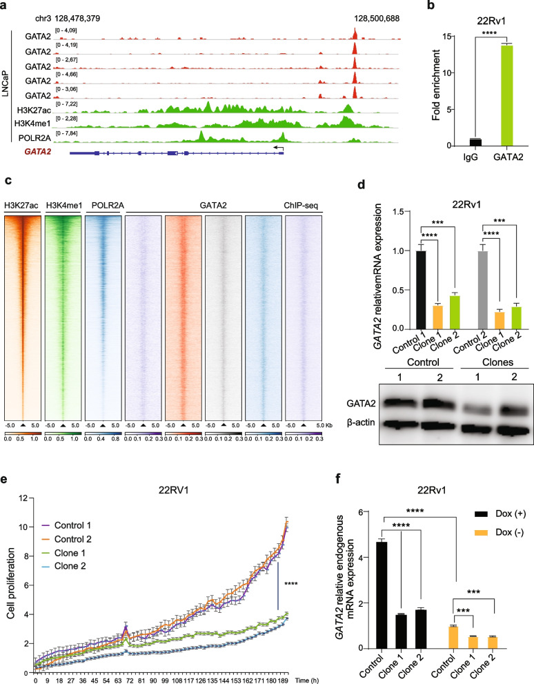
Results: In this study, by integrated genomics and experimental analysis, we identified GATA2 as a prevalent metastasis-associated genomic amplification to transcriptionally augment its own expression in PCa. Functional experiments demonstrated that GATA2 physically interacted and cooperated with SMAD4 for genome-wide chromatin co-occupancy and co-regulation of PCa genes and metastasis pathways like TGFβ signaling. Mechanistically, GATA2 was cooperative with SMAD4 to enhance TGFβ and AR signaling pathways, and activated the expression of TGFβ1 via directly binding to a distal enhancer of TGFβ1. Strinkingly, GATA2 and SMAD4 globally mediated inherited PCa risk and formed a transcriptional complex with HOXB13 at the PCa risk-associated rs339331/6q22 enhancer, leading to increased expression of the PCa susceptibility gene RFX6.

Conclusions: Our study prioritizes causal genomic amplification genes with prognostic values in PCa and reveals the pivotal roles of GATA2 in transcriptionally activating the expression of its own and TGFβ1, thereby co-opting to TGFβ1/SMAD4 signaling and RFX6 at 6q22 to modulate PCa predisposition and progression.

Keywords: GATA2; Prostate cancer; SMAD4; TGFβ1; rs339331.


Conflict of interest statement

The authors have declared that no competing interests exists.

Figures

Fig. 1
Fig. 1
GATA2 genomic amplification and upregulation are correlated with tumor progression and poor prognosis in PCa patients. a Genome-wide CRISPR loss-of-function identification of the essential genes for cell survival in PCa cell LNCaP. Lower ATARiS scores indicated higher essentiality of the indicated genes for cell growth and survival. Orange dots represented causal amplification genes defined by their significant positive linear correlations between copy number gain and expression levels in the CPGEA or EU PCa cohort. Green dots highlighted ARHOXB13MYC and BRD4 that are known to be crucial for PCa cell proliferation and survival whereas TP53 is not favorable for PCa cell growth and survival. b Proportion of GATA2 genomic amplificaiton across 17 cohorts of PCa tumors in different populations. c-d GATA2 expression in prostate tumor tissues with GATA2 diploid or copy number gain or amplification (left panel), and Pearson correlation between GATA2 mRNA expression and copy number changes (right panel). Gain: with presence of one copy; AMP, amplification: with the presence of two copies. P values determined by the Mann–Whitney U test or Pearson correlaiton. AMP, amplification. e Box plots showing GATA2 upregulation in human primary and metastasis PCa. P values determined by the Kruskal–Wallis H test or the Mann–Whitney U test. f Elevated GATA2 expression correlated with higher Gleason score. P values determined by the Kruskal–Wallis H test. g-h Kaplan–Meier plots indicated increased biochemical recurrence and metastasis risks of PCa patients with tumors expressing higher GATA2 levels in two independent cohorts. Patient groups stratified by the median value of GATA2 expression levels. P values assessed by a log-rank test. i-j Higher expression levels of GATA2 exhibited predictive values for biochemical relapse and metastasis in PCa patient group with an intermediate risk (Gleason Score 7). P values assessed by a log-rank test. k Fraction of PCa tumors harboring GATA2 copy number gain is elevated in metastasis than in primary PCa in multiple independent cohorts of PCa patients. P values were examined by the Fisher's exact test. l-m Top gene set depleted in PCa tumors with GATA2 copy number gain/amplification vs diploid (l) or with GATA2 high (higher than the 50th percentile) versus low (lower than the 50th percentile) mRNA abundance (m) in the TCGA PCa cohort. NES, normalized enrichment score. FDR values calculated by the GSEA analysis. n Gleason scores were higher in PCa patient group with GATA2 copy gain in multiple independent PCa datasets. P values examined by the Fisher’s exact test. o-q PCa patients with GATA2 copy gain were associated with elevated risks for biochemical relapse (o), metastasis (p), and decreased disease-specific survival (q). P values assessed by a log-rank test
Fig. 2
Fig. 2
GATA2 drives its own expression via a positive feedback regulatory circuit. a Genome browser representation of ChIP-seq signals of active enhancer marks (H3K27ac and H3K4me1), RNA polymerase II (POLR2A), and transcription factor GATA2 at 3q21.3 GATA2 locus in the LNCaP cells. b ChIP-qPCR analysis of GATA2 chromatin binding at the GATA2 enhancer in 22Rv1 cells. c Heatmaps of H3K27ac, H3K4me1, RNA polymerase II and GATA2 ChIP-seq signals around H3K27ac binding sites in LNCaP cells. H3K27ac binding sites were rank-ordered based on H3K27ac ChIP-seq intensities. d CRISPR/Cas9-mediated deletion of the GATA2-occupied GATA2 enhancer. GATA2 expression was analyzed by qRT-PCR and western blot in the two positive clones and control cells, β-actin used as loading control. e Cell growth potential was measured in real time by the Incucyte detecting system. The cell proliferation rate was detected every three hours. f Ectopic expression of GATA2 in the enhancer knockout clones showed a lower mRNA expression of endogenous GATA2 comparing to control cells
Fig. 3
Fig. 3
GATA2 potentiates PCa proliferation and metastasis. a-c Knockdown of GATA2 inhibited cell proliferation. Cells were transfected with control or GATA2 siRNAs or shRNAs, and cell proliferation was measured by XTT assay (absorbance at 450 nm) at the indicated time points. d-e Dox-induced GATA2 overexpression in RWPE2 (d) and PC3 cells (e) potentiated cell migration determined by wound healing assay. f-g GATA2 promoted cell invasion in RWPE2 (f) and PC3 (g) cells. h-i GATA2 promoted PCa metastasis in vivo. SCID male mice were tail-vain injected with RWPE2-GATA2 cells (h) or left ventricle injected with PC3-GATA2 cells (i), then administrated water with or without Dox (2 μg/mL). After 4 weeks, the lung (h) or bone (i) metastatic nodules and fluorescence intensity were measured by IVIS. j-l GATA2 promoted prostate cancer cell proliferation in vivo. SCID male mice were subcatenously inoculated with 22Rv1 scramble cells or GATA2 downregulated cells. Tumor size (j), tumor volume (k) and tumor weight (l) were measured. m-q GATA2 knockdown repressed cell cycle progression driver MYC (m) and promoted the expression of cell proliferation inhibitor P21 (n) as well as stimulated the expression of metastasis suppressor PTEN (o) and inhibited metastasis drivers VEGF (p) and TWIST1 (q) examined by qRT-PCR. Error bars are mean s.e.m, n = 3 independent experiments. *P < 0.05, **P < 0.01, ***P < 0.001, ****P < 0.0001 determined by unpaired Student’s t-test
Fig. 4
Fig. 4
GATA2 physically interacts with and co-opts to SMAD4 for genome-wide chromatin co-occupancy and co-regulation of PCa genes and cancer metastasis pathways. a Proteins interacting with GATA2 in four indenpdent PPI databases. 10 proteins were condordently identified to interact with GATA2 in the four databases. b Interaction between endogenous GATA2 and SMAD4 was examined by immunoprecipitation (IP) using LNCaP cell lysates. c Directly interaction between GATA2 and SMAD4 was confirmed by GST-pull down assay. d Heatmap representation of GATA2 and SMAD4 chromatin binding intensities within 3 kb around the center of binding peaks in 1F5 cells. ChIP-seq signals were displayed in a descending order for clustered categories of GATA2 unique, GATA2 and SMAD4 common, and SMAD4 unique binding regions. e Venn diagram exhibiting common differentially expressed genes upon knockdown of GATA2 or SMAD4 followed by RNA-seq in 1F5 cells. FDR < 0.1. f Common regulated pathways of GATA2 and SMAD4 from MSigDB gene sets Chemical and Genetic Perturbations, Hallmark and Reactome. FDR < 0.05. g Several functional categories including cell cycle progression, metastasis and TGFβ signaling commonly enriched with downregulated genes upon siRNA-mediated knockdown of GATA2 or SMAD4 in 1F5 cells. h-i GSEA plots displaying pathways related to cell cycle progression, metastasis and TGFβ signaling enriched in GATA2 (h) or SMAD4 (i) upregulated genes. j Validation of common enriched pathways, which include metastasis/EMT-related gene and TGFβ pathway gene signatures, from GATA2 and SMAD4 target gene sets in additional resources. k-l Expression levels of GATA2 or SMAD4 were significantly correlated with the EMT score (k) or TGFβ signaling score (l) in PCa tumors
Fig. 5
Fig. 5
GATA2 cooperates with SMAD4 to promote TGFβ1 signaling. a-c TGFβ signaling inhibitor LY2157299 compromised GATA2-induced cell migration. 1F5-GATA2 (a), 22Rv1-GATA2 (b) and V16A-GATA2 (c) cells were treated with or without Dox (1 μg/ml) and LY2157299 (10 μM). Cell migration was determined by wound healing assay. d Knockdown of GATA2 decreased the expression of TGFβ1. 1F5, V16A and 22Rv1 cells were transfected with control siRNA or siRNAs specifically targeted on GATA2. Seventy-two hours later, the mRNA expression level of TGFβ1 was analyzed by qRT-PCR. e–f Downregulation of GATA2 suppressed the activity of TGFβ1/SMAD signaling. 1F5 and V16A cells were transfected with control siRNA or siRNAs against GATA2. Seventy-two hours later, the protein expression levels of GATA2 and p-SMAD3 were analyzed by western blot, β-actin used as loading control. g-l Upregulation of GATA2 activated TGFβ1/SMAD signaling. Cells were incubated with or without Dox (1 μg/ml) for 48 h or 72 h. Then the cell pellets were collected and the expression of TGFβ1, p-SMAD2 and p-SMAD3 were detected by qRT-PCR and western blot, respectively. m V16A cells were co-transfected with SBE4-luc, GATA2 or SMAD4 with or without 5 ng/mL TGFβ1 recombinant protein. Forty-eight hours later, cell luciferase activity was measured. n-q Significant positive linear expression correlation between GATA2 & SMAD4 and TGFβ signaling scores was observed in multiple independent PCa cohorts. P values assessed by the Pearson's product-moment correlation test. n = 3 independent experiments. **P < 0.01, ***P < 0.001, ****P < 0.0001 determined by unpaired Student’s t-test
Fig. 6
Fig. 6
GATA2 binds to a distal enhancer of TGFβ1 and regulates TGFβ1 expression in PCa cells. a Luciferase reporter assays showing increased promoter activity of TGFβ1 when co-transfected with GATA2 expression vector in V16A cells. b Genome browser representations of GATA2 ChIP-seq enriched profiles at far upstream of TGFβ1 in PCa LNCaP and 1F5 cells. Chromosome coordinates presented the human genome build hg38. c ChIP-qPCR verification of GATA2 chromatin binding at the TGFβ1 upstream enhancer region in LNCaP, 22Rv1 and VCaP cells. d Hi-C analysis of chromatin interactions between the potential GATA2-occupied TGFβ1 enhancer and TGFβ1 promoter locus (chr19: 41,355,000–41,531,050). e 3C-qPCR analysis of chromatin interactions between the enhancer locus and TGFβ1 promoter region (chr19: 41,355,000–41,531,050). f-g CRISPR/Cas9-mediated deletion of the GATA2-occupied TGFβ1 enhancer. 22Rv1 cells were transfected with control or TGFβ1 enhancer-targeting sgRNAs. Three clones were picked up and confirmed by Sanger sequencing. TGFβ1 expression was analyzed by qRT-PCR (f) and western blot (g) in the three clones and control cells, β-actin used as loading control. h-i Kaplan Meier plots indicated increased biochemical recurrence and metastasis risks of PCa patients with higher TGFβ1 expression levels in TCGA cohort. P values assessed by log-rank test. All the error bars are mean s.e.m, n = 3 independent experiments. ****P < 0.0001, determined by unpaired Student’s t-test
Fig. 7
Fig. 7
GATA2 co-opts with SMAD4 to regulate AR signaling. a-c AR signaling antagonists Enzalutamide compromised GATA2-driven cell migration in 1F5 (a), V16A (b) and 22Rv1 (c) cells. d GATA2 and SMAD4 expression levels positively correlated with AR signaling scores in human PCa tumors in multiple independent cohorts. P values examined by the Pearson's product-moment correlation test. e Upper panel: Integrated heatmaps of RNA-seq and ChIP-seq representations for the joint direct targeting genes of GATA2 and SMAD4 identified in 1F5 cells. GATA2 and SMAD4 ChIP-seq signals were illustrated for the genes shown, deeper color indicating higher enrichment. Lower panel: chromatin-binding of GATA2 and SMAD4 on the proxy region of representative PCa gene KLK3 with indicated genomic interval. f The Z-score sum of expression levels of GATA2 & SMAD4 direct target gene signature showed positive linear correlation with AR signaling score in human PCa tumors in multiple independent cohorts. P values examined by the Pearson's product-moment correlation test. g-h Knockdown of GATA2 (g) or SMAD4 (h) inhibited the mRNA expression levels of AR targeting genes KLK2 and KLK3 in 1F5 and V16A cells. All the error bars represent s.e.m, n = 3 technical replicates. *P < 0.05, **P < 0.01, ****P < 0.0001, determined by unpaired Student’s t-test
Fig. 8
Fig. 8
GATA2 shows a global impact on inherited PCa risk and forms a transcriptional complex with SMAD4 and HOXB13 at rs339331/6q22 enhancer to drive the expression of PCa risk gene RFX6. a The enrichment analysis indicated a substantial increase for PCa GWAS risk SNPs enriched in the common chromatin binding sites of GATA2 and SMAD4. b Circos overview of PCa risk loci enriched in the GATA2 and SMAD4 common chromatin binding regions in 1F5 cells. The outer ring represented a circular ideograph of the human genome annotated with chromosome numbers. Tag SNPs were positioned in each locus followed by corresponding proxy SNPs with a cutoff LD, R2 ≥ 0.5. The eQTL genes were indicated adjacent to proxy SNPs. c Genome browser represented of rs339331 residing within GATA2, SMAD4 and HOXB13 ChIP-seq chromatin binding regions in PCa cell lines. Lower panel: rs339331 is located within the GATA2 DNA-binding motif. Chromosome coordinates indicate as the human genome build hg38. d ChIP-qPCR for GATA2 and SMAD4 chromatin binding at the rs339331-containing region in 22Rv1 cells after ETH or DHT (100 nM) treatment for 24 h. HOXB13 was shown as as a positive control. e-f GATA2 and SMAD4 favor binding to the T risk allele than C at rs339331 as determined by ChIP followed by AS-qPCR (e) and ChIP followed by PCR amplification and Sanger sequencing (f). g-j PCa tumors carrying rs339331 risk allele TT were associated with shorter biochemical recurrence-free (g) and metastasis-free (i) survival in patient group expressing higher RFX6 levels. rs339331 could not stratifiy PCa patient with lower RFX6 expression (h, j). P values assessed by the log-rank test. k-l Knockdown of GATA2 or SMAD4 decreased the expression of RFX6 in LNCaP 1F5 and V16A cells. mn Scatter plot illustration of linear correlation between GATA2 and RFX6 expression levels in human prostates. o-p Kaplan–Meier plots demonstrated increased risks of biochemical recurrence and metastasis for PCa patients with elevated GATA2 and RFX6 expression levels in TCGA and Taylor cohorts. q-r Synergistic expression effect of RFX6 and GATA2 exhibited predictive values for biochemical relapse and metastasis in PCa patient group with an intermediate risk of Gleason Score 7. Samples with RFX6 deep loss were ruled out in the analysis. Patients were stratified by the median value of RFX6 and GATA2 expression levels. P values assessed by the log-rank test. s The effect of combination TGFβ signaling inhibitor and GATA2 inhibitor on the expression of RFX6 and KLK3 in 1F5 cells. LNCaP cells were treated with or without 10 μM K7174 together with 10 μM LY2157299 for 48 h. qRT-PCR was following performed to analyze the mRNA expression of the correlated genes. All the error bars represent s.e.m, n = 3 technical replicates. *P < 0.01 **P < 0.01, ***P < 0.001, ****P < 0.0001, determined by two-tailed Student’s t-test. N.S: Non-significant
Fig. 9
Fig. 9
An extensive mechanistic cooperation between GATA2 and TGFβ1/SMAD4 signaling contributes to PCa predisposition and progression. Schematic showing that prevalent genomic copy gain of GATA2 in PCa and a previously unappreciated autoregulation mechanism direct GATA2 overexpression, thereby promoting PCa cell proliferation and metastatic progression. Mechanistically, GATA2 cooperates with SMAD4 physically and on chromatin, and drives the expression of TGFβ1 via a distal enhancer, hence activating TGFβ1/SMAD4 signaling and orchestrating decreased expression of cell cycle inhibitor P21 as well as enhanced transcription of metastasis-associated genes, such as TGFβ1 and TWIST1. Moreover, GATA2 is cooperative with SMAD4 and the prostate-lineage-specific transcription factor HOXB13 to mediate inherited PCa risk, indicating chromatin-binding preference to the 6q22 PCa risk-asociated T allele of the SNP rs339331, resulting increased expression of the eGene RFX6 contributing to PCa severity. Collectively, GATA2 upregulation contributes to PCa predisposition and tumor progression through controlling oncogenic signaling and this extensive somatic-germline interplay mechanism in PCa

References

    1. Sung H, Ferlay J, Siegel RL, Laversanne M, Soerjomataram I, Jemal A, et al. Global Cancer Statistics 2020: GLOBOCAN Estimates of Incidence and Mortality Worldwide for 36 Cancers in 185 Countries. CA Cancer J Clin. 2021;71(3):209–249. doi: 10.3322/caac.21660. - DOI - PubMed
    1. DeMarzo AM, Nelson WG, Isaacs WB, Epstein JI. Pathological and molecular aspects of prostate cancer. Lancet. 2003;361(9361):955–964. doi: 10.1016/S0140-6736(03)12779-1. - DOI - PubMed
    1. Helsen C, Van den Broeck T, Voet A, Prekovic S, Van Poppel H, Joniau S, et al. Androgen receptor antagonists for prostate cancer therapy. Endocr Relat Cancer. 2014;21(4):T105–118. doi: 10.1530/ERC-13-0545. - DOI - PubMed
    1. Shen MM, Abate-Shen C. Molecular genetics of prostate cancer: new prospects for old challenges. Genes Dev. 2010;24(18):1967–2000. doi: 10.1101/gad.1965810. - DOI - PMC - PubMed
    1. Taylor BS, Schultz N, Hieronymus H, Gopalan A, Xiao Y, Carver BS, et al. Integrative genomic profiling of human prostate cancer. Cancer Cell. 2010;18(1):11–22. doi: 10.1016/j.ccr.2010.05.026. - DOI - PMC - PubMed