Anteroposterior Combined Surgery of a Rare Massive Epithelioid Hemangioendothelioma at the Cervicothoracic Junction
- PMID: 37551289
- PMCID: PMC10404447
- DOI: 10.7759/cureus.43032
Anteroposterior Combined Surgery of a Rare Massive Epithelioid Hemangioendothelioma at the Cervicothoracic Junction
Abstract
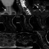
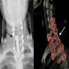
Epithelioid hemangioendothelioma is a rare mesenchymal tumor of vascular endothelial origin. Non-soft tissue epithelioid hemangioendothelioma can also be seen in different organs. Although chemotherapy has been used in some patients, complete surgical removal of the tumor tissue has proven to be the most durable solution. A 15-year-old female patient was admitted to our institution with right arm and neck pain. The patient complained of numbness and weakness in the right hand. Computerized tomography indicated an expansile lesion exhibiting osteolytic features located predominantly on the right side of the corpus, pedicle, lamina, and lateral processes of the C7-T1 vertebra. The patient underwent a surgical procedure involving the application of a bilateral C4-5-6 lateral mass screw, left C7-T1 pedicle screw, and bilateral T2-3 pedicle screw and fusion. The complete residual neoplasm was surgically removed during the procedure. Due to the rarity of epithelioid hemangioendothelioma, the existing literature on this topic is confined to case reports, supplemented by a small number of retrospective descriptive case series that aimed to improve our understanding of the clinical, pathological, and molecular features of the condition, as well as to guide potential treatment strategies.
Keywords: combined surgery; craniocervical junction; epitheloid hemangioendothelioma; pediatric neurosurgery; spinal tumor.
Copyright © 2023, Cine et al.
Conflict of interest statement
The authors have declared that no competing interests exist.
Figures


References
-
- Thoracic epithelioid hemangioendothelioma: clinical demonstration and therapeutic procedures. Graça LL, Almeida Cunha S, Lopes RS, Carvalho L, Prieto D. Port J Card Thorac Vasc Surg. 2022;29:39–44. - PubMed
-
- Epithelioid hemangioendothelioma in children: the European Pediatric Soft Tissue Sarcoma Study Group experience. Orbach D, Van Noesel MM, Brennan B, et al. Pediatr Blood Cancer. 2022;69:0. - PubMed
-
- New molecular insights, and the role of systemic therapies and collaboration for treatment of epithelioid hemangioendothelioma (EHE) Stacchiotti S, Tap W, Leonard H, Zaffaroni N, Baldi GG. Curr Treat Options Oncol. 2023;24:667–679. - PubMed
Publication types
LinkOut - more resources
Full Text Sources
Miscellaneous
