

Wound healing of the corneal epithelium: a review
- PMID: 37551323
- PMCID: PMC10388779
- DOI: 10.2478/abm-2021-0026
Wound healing of the corneal epithelium: a review
Abstract
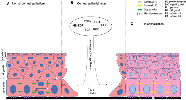
The corneal epithelium (CE) forms the outermost layer of the cornea. Despite its thickness of only 50 μm, the CE plays a key role as an initial barrier against any insults to the eye and contributes to the light refraction onto the retina required for clear vision. In the event of an injury, the cornea is equipped with many strategies contributing to competent wound healing, including angiogenic and immune privileges, and mechanotransduction. Various factors, including growth factors, keratin, cytokines, integrins, crystallins, basement membrane, and gap junction proteins are involved in CE wound healing and serve as markers in the healing process. Studies of CE wound healing are advancing rapidly in tandem with the rise of corneal bioengineering, which employs limbal epithelial stem cells as the primary source of cells utilizing various types of biomaterials as substrates.
Keywords: cornea; corneal; epithelium; markers; regeneration; wound healing.
© 2021 Norzana Abd Ghafar et al., published by Sciendo.
Figures
References
-
- Ashby BD, Garrett Q, Willcox MDP. Corneal injuries and wound healing – review of processes and therapies. Austin J Clin Ophthalmol. 2014;1:1–25. id1017.
-
- Leong Y-Y, Tong L. Barrier function in the ocular surface: from conventional paradigms to new opportunities. Ocul Surf. 2015;13:103–9. - PubMed
-
- Liu C-Y, Kao WW-Y. Corneal epithelial wound healing. Prog Mol Biol Transl Sci. 2015;134:61–71. - PubMed
-
- Thoft RA, Friend J. The X, Y, Z hypothesis of corneal epithelial maintenance. Invest Opthalmol Vis Sci. 1983;24:1442–3. - PubMed
Publication types
LinkOut - more resources
Full Text Sources
Miscellaneous