. 2023 Oct;10(29):e2303779.
doi: 10.1002/advs.202303779. Epub 2023 Aug 8.

Biosynthetic Plastics as Tunable Elastic and Visible Stent with Shape-Memory to Treat Biliary Stricture

Affiliations

Biosynthetic Plastics as Tunable Elastic and Visible Stent with Shape-Memory to Treat Biliary Stricture

Wei Wang et al. Adv Sci (Weinh). 2023 Oct.

Abstract

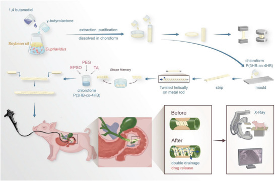
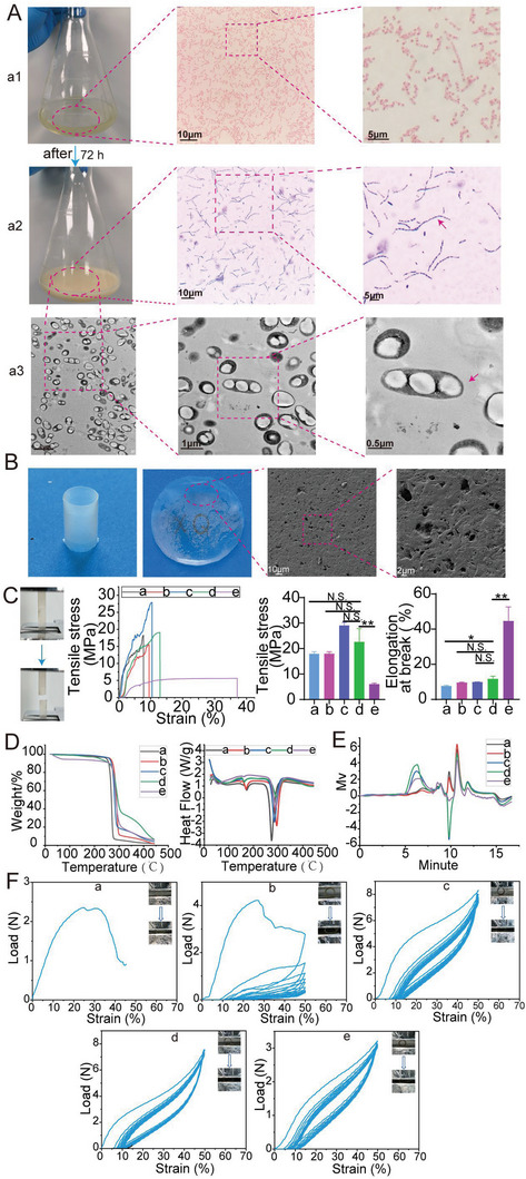
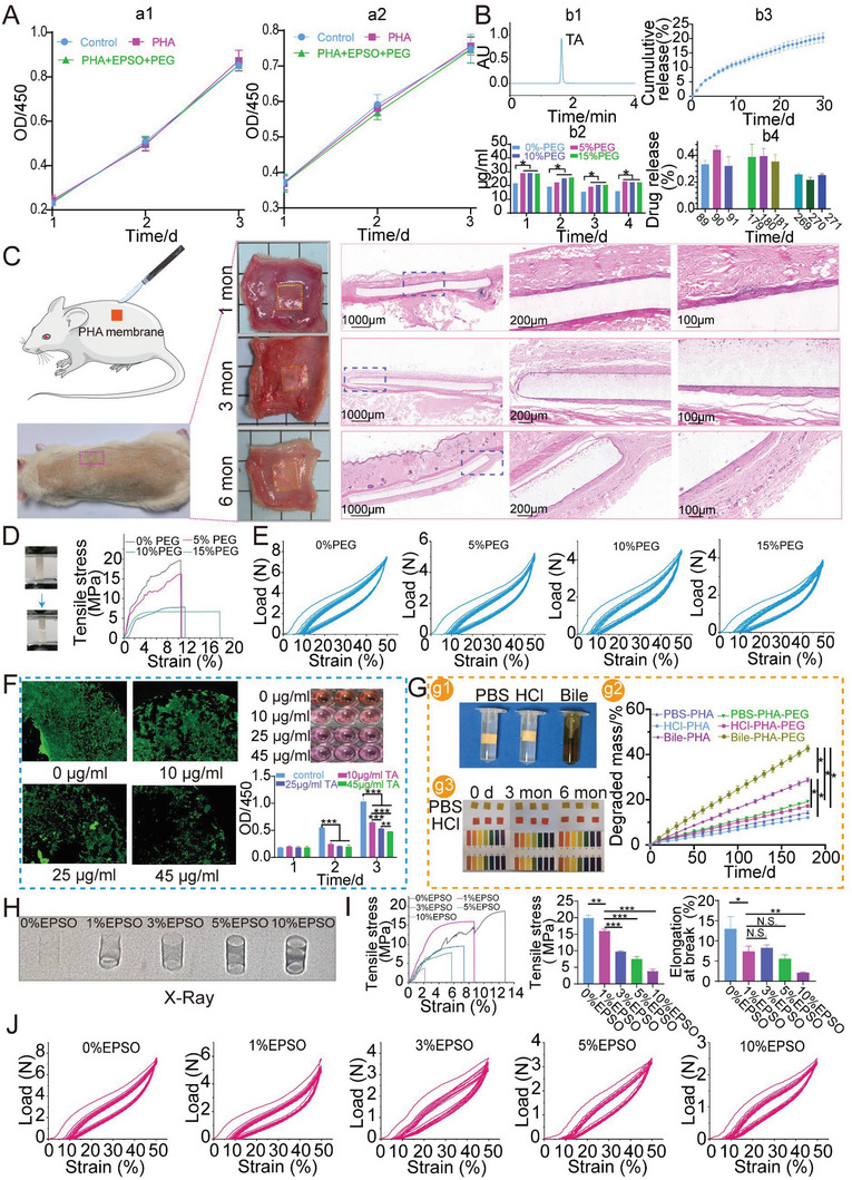
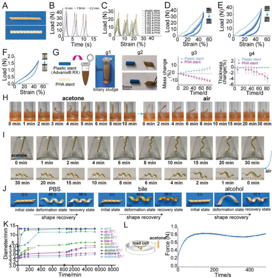
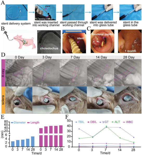
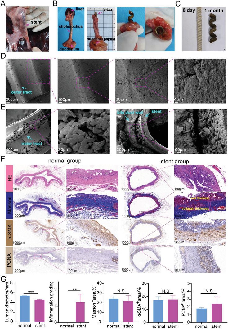
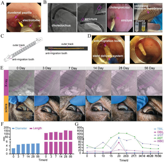
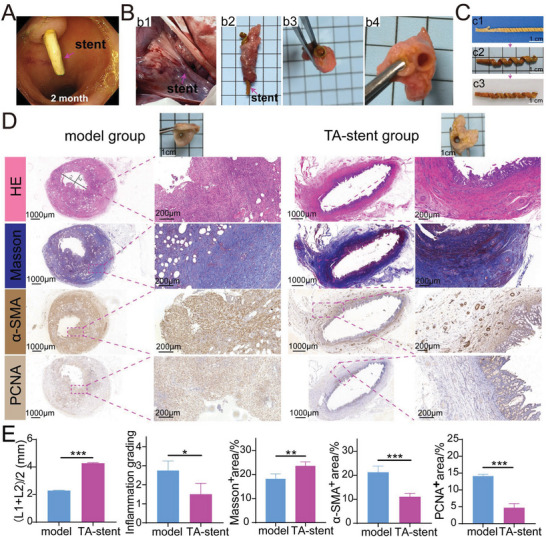
Common biliary tract is ≈4 mm in diameter to deliver bile from liver to small intestine to help digestion. The abnormal narrowing leads to severe symptoms such as pain and nausea. Stents are an effective treatment. Compared with non-degradable stents which require repeated removal, biodegradable stents have the advantage of reducing secondary injury related to endoscopic operation and patient burden. However, current biodegradable materials may cause tissue hyperplasia and the treatment method does not target etiology of stricture. So recurrence rates after biodegradable stent implantation are still high. Here, a biodegradable helical stent fabricated from biosynthetic P(3HB-co-4HB) is reported. Tunable properties can be acquired through altering culture substrates. Stent shows shape memory in various solvents. The stent has an optimized design with helical structure and outer track. The self-expanding of helical structure and double drainage realized by outer track greatly improve drainage of bile. Importantly, stent-loading triamcinolone acetonide can inhibit proliferation of fibroblasts and reduce incidence of restricture. Therapeutic effect is also demonstrated in minipigs with biliary stricture. The results of minipig experiments show that biliary duct in treatment group is unobstructed and tissue hyperplasia is effectively inhibited.

Keywords: biliary stricture; biodegradable stent; endoscopy; polyhydroxyalkanoate; shape memory; triamcinolone acetonide.


Conflict of interest statement

The authors declare no conflict of interest.

Figures

Scheme 1
Scheme 1
The fabrication process of stent and insertion into bile duct endoscopically for the treatment of biliary stricture. TA: Triamcinolone acetonide. PEG: Polyethylene glycol. EPSO: Ethiodized poppy seed oil.
Figure 1
Figure 1
The production and physicochemical properties of P(3HB‐co‐4HB). A) The production process of P(3HB‐co‐4HB). a1) Gram staining of Cupriavidus necator strain; a2) Staining of intracellular P(3HB‐co‐4HB) with Sudan Black; a3) Observation of intracellular accumulation of P(3HB‐co‐4HB) with TEM; The red arrow indicating the intracellular P(3HB‐co‐4HB). B) Images of film and tubular stent constructed with extracted P(3HB‐co‐4HB) and SEM images of P(3HB‐co‐4HB) film. C–F) Physicochemical properties of P(3HB‐co‐4HB) with different monomer ratios. Five materials (a–e) were characterized by tensile tests (C), thermal gravity analyzing (D), gel permeation chromatography (E), and compression tests (F).
Figure 2
Figure 2
The biocompatibility of P(3HB‐co‐4HB), the incorporation of PEG and EPSO to increase TA release and stent visibility respectively. A) Toxicity of P(3HB‐co‐4HB) on 3T3 fibroblast cells (a1) and mouse extrahepatic bile duct epithelial cells (a2). PHA stands for P(3HB‐co‐4HB) in this figure. B) Releasing characteristics of P(3HB‐co‐4HB) film loading TA. b1) The HPLC curve of TA standard. b2) TA releasing curves of P(3HB‐co‐4HB)‐TA films with different contents of PEG. b3) TA releasing curves in the first month. b4) TA releasing curves at 3, 6, and 9 months. C) Biocompatibility of P(3HB‐co‐4HB) in rats. D) Tensile properties of P(3HB‐co‐4HB) films with different contents of PEG. E) Compressive properties of P(3HB‐co‐4HB) stents with different contents of PEG. F) Inhibition against 3T3 cells of different concentrations of TA in vitro. G) Degradation profiles of P(3HB‐co‐4HB) films in PBS, HCl and bile in vitro. g1) Images of three groups. g2) pH changes during degradation of P(3HB‐co‐4HB). g3) Degradation curve of P(3HB‐co‐4HB) films. H) Visibility of P(3HB‐co‐4HB) stents with different contents of EPSO under X‐ray. I) Tensile properties of P(3HB‐co‐4HB) with different contents of EPSO. J) Compression properties of P(3HB‐co‐4HB) stents with different contents of EPSO.
Figure 3
Figure 3
Characterization and shape memory behaviors of P(3HB‐co‐4HB) helical stent. A) The photograph of P(3HB‐co‐4HB) helical stent with outer track. B) The cyclic compression performance of P(3HB‐co‐4HB) helical stent with different pitches (strain 25%). C) The cyclic compression performance of plastic stent and P(3HB‐co‐4HB) helical stent with different thicknesses and diameters (strain 25%). PHA stands for P(3HB‐co‐4HB) in this figure. D) The cyclic compression performance of plastic stent (strain 50%). E) The cyclic compression performance of P(3HB‐co‐4HB) stent (2.8 mm in diameter, 0.4 mm in thickness, strain 50%). F) The longitudinal cyclic tensile performance of P(3HB‐co‐4HB) helical stent (2.8 mm in diameter, 0.4 mm in thickness). G) Adhesive properties of P(3HB‐co‐4HB) helical stents and polyethylene plastic stents against biliary sludge in vitro. g1) Aged bile with biliary sludge. g2) Images of plastic stent and P(3HB‐co‐4HB) stent immersed in bile for 3 months. g3) Mass changes of two stents at different time points. g4) Thickness changes of two stents at different times. H) Shape change of P(3HB‐co‐4HB) helical stent before and after immersion in acetone at room temperature. I) Shape change of P(3HB‐co‐4HB) helical stent when hanging in acetone bottle and being taken out. J) The shape recovery of P(3HB‐co‐4HB) stent in 37 °C PBS, bile and alcohol at different time points. K) Self‐expansion of P(3HB‐co‐4HB) helical stent in air, PBS, bile, alcohol, and acetone at room temperature (rt) and 37 °C. L) Expansion force of P(3HB‐co‐4HB) helical stent in acetone at different times.
Figure 4
Figure 4
Stent without tooth insertion in vitro and in minipigs with normal bile duct. A) Endoscopic release of P(3HB‐co‐4HB) helical stent in vitro. B) Observation of normal bile duct in minipig through ERCP and the implantation process of stent without tooth. C) Endoscopy images of stent at day 0 and 28. D) The images of P(3HB‐co‐4HB) stent under X‐ray and sclera color at different times. E) Changes in diameter and length of P(3HB‐co‐4HB) helical stent in vivo. F) Changes of blood indexes in minipig at different times.
Figure 5
Figure 5
Gross and microscopic view of stents after dissection and histologic staining of the bile duct in a normal minipig. A) Position of common bile duct with a stent (without tooth) in the minipig. B) The dissected bile duct in stent group. C) The images of the initial stent and stent removed from the minipig. D) SEM images of the initial stent. E) SEM images of stents and bile duct from the minipig. F) Histologic staining of biliary tissue in normal group and stent (without tooth) group. G) Quantification analysis of lumen diameter, inflammation grading, Masson+ area, α‐SMA+ area, and PCNA+ area.
Figure 6
Figure 6
Biliary stricture model, stent insertion and therapeutic evaluation. A) Modeling of biliary stricture by cauterizing the bile duct through electrotome. B) Success of biliary stricture model was evaluated by ERCP and cholangioscopy. After successful modeling, the minipig appeared jaundice and serum color became significantly yellow. C) A P(3HB‐co‐4HB) stent with anti‐migration tooth. D) Stent was placed endoscopically. E) Changes of the stent and scleral colors under X‐ray at different times. The dotted red line marks the position of stent. F) Changes of stent diameter and length at different times. G) Changes of blood indexes of minipig at different times. Days after stent placement were shown in parentheses.
Figure 7
Figure 7
Gross and microscopic view of stents after dissection and histologic staining of the bile duct in modeled minipigs. A) Observation of stent under endoscope after insertion of 2 months. B) The position of the common bile duct and the stent. It could be seen the bile duct and stent was unobstructed. C) Shape memory characteristics of the stent at different times. c1) The initial stent (IS). c2) Fresh stent just removed from biliary duct (FS). c3) The dried stent (DS) removed from biliary duct. D) Histologic staining of bile duct in two group. L1: The longest distance in lumen, L2: The shortest distance in lumen, (L1+L2)/2 was used to estimate the lumen diameter because the lumen is irregular. E) Quantification analysis of lumen diameter, inflammation grading, Masson+ area, α‐SMA+ area and PCNA+ area.

Similar articles

Cited by

References

    1. a) Almeida G. G., Donato P., Eur. J. Radiol. 2020, 125, 108899; - PubMed
    2. b) Hu B., Sun B., Cai Q., Lau J. Y. W., Ma S., Itoi T., Moon J. H., Yasuda I., Zhang X., Wang H.‐P., Ryozawa S., Rerknimitr R., Li W., Kutsumi H., Lakhtakia S., Shiomi H., Ji M., Li X., Qian D., Yang Z., Zheng X., Gastrointest. Endosc. 2017; - PubMed
    3. c) Vitale G. C., Tran T. C., Davis B. R., Vitale M., Vitale D., Larson G., J. Am. Coll. Surg. 2008, 206, 918. - PubMed
    1. Ramchandani M., Lakhtakia S., Costamagna G., Tringali A., Püspöek A., Tribl B., Dolak W., Devière J., Arvanitakis M., van der Merwe S., Laleman W., Ponchon T., Lepilliez V., Gabbrielli A., Bernardoni L., Bruno M. J., Poley J. W., Arnelo U., Lau J., Roy A., Bourke M., Kaffes A., Neuhaus H., Peetermans J., Rousseau M., Reddy D. N., Gastroenterology 2021, 161, 185. - PubMed
    1. Kaffes A., Endoscopy 2019, 51, 1115. - PubMed
    1. Lawrence C., Romagnuolo J., Payne K. M., Hawes R. H., Cotton P. B., Gastrointest. Endosc. 2010, 72, 558. - PubMed
    1. Dumonceau J. M., Tringali A., Papanikolaou I. S., Blero D., Mangiavillano B., Schmidt A., Vanbiervliet G., Costamagna G., Devière J., García‐Cano J., Gyökeres T., Hassan C., Prat F., Siersema P. D., van Hooft J. E., Endoscopy 2018, 50, 910. - PubMed

LinkOut - more resources