. 2023 Oct;10(28):e2301852.
doi: 10.1002/advs.202301852. Epub 2023 Aug 8.

Deubiquitinase OTUD5 as a Novel Protector against 4-HNE-Triggered Ferroptosis in Myocardial Ischemia/Reperfusion Injury

Affiliations

Deubiquitinase OTUD5 as a Novel Protector against 4-HNE-Triggered Ferroptosis in Myocardial Ischemia/Reperfusion Injury

Lulu Liu et al. Adv Sci (Weinh). 2023 Oct.

Abstract

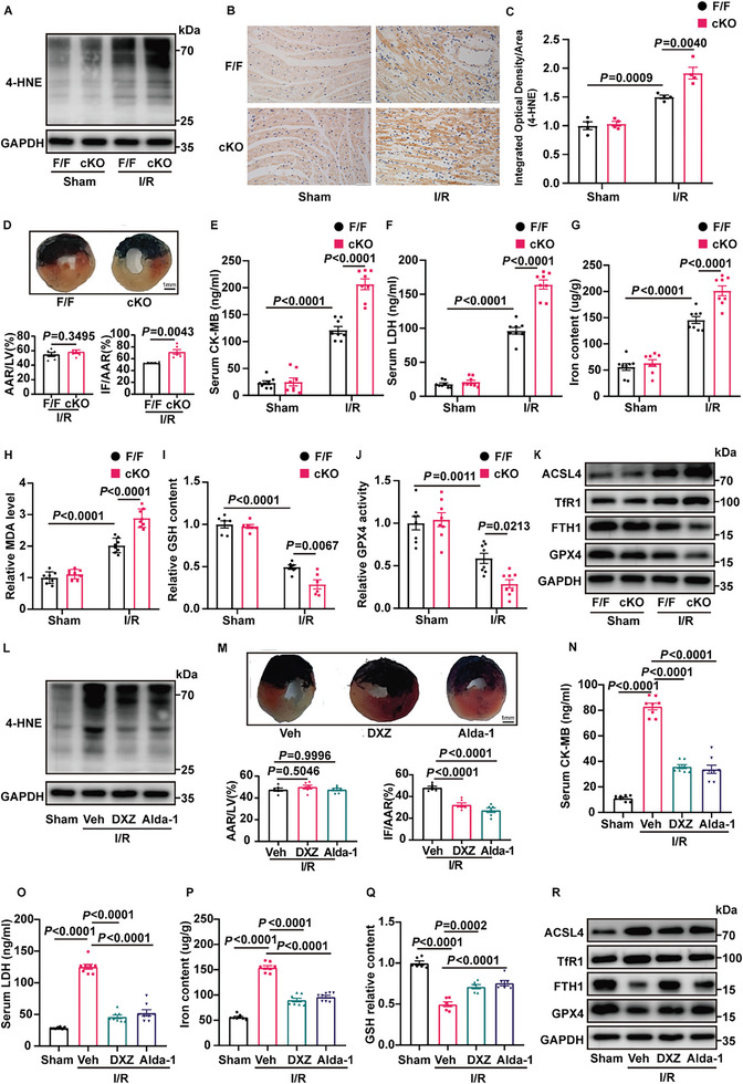
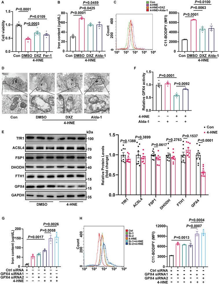
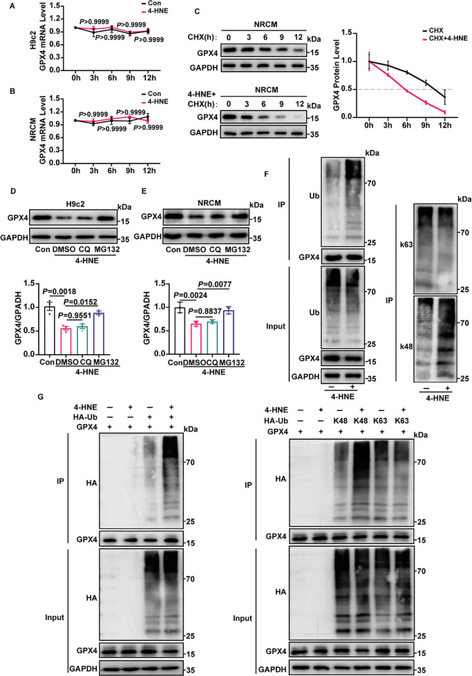
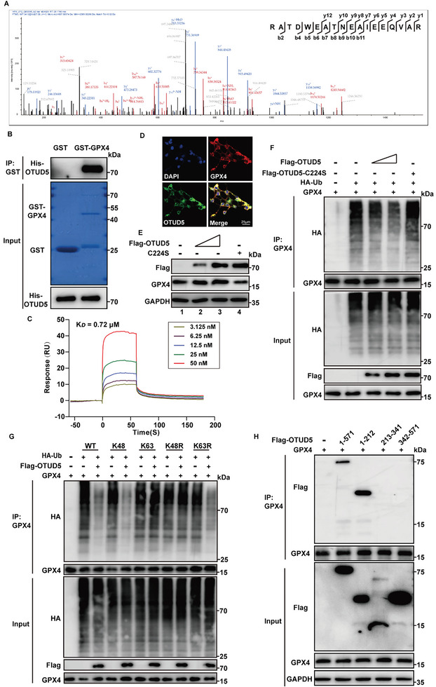
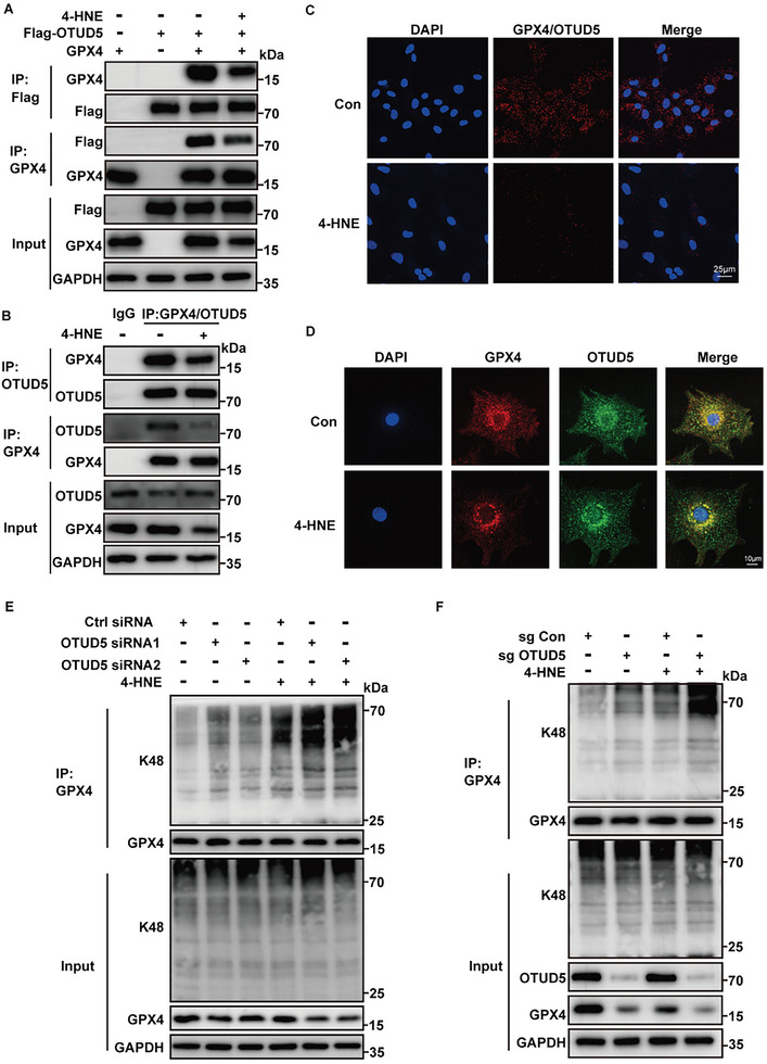
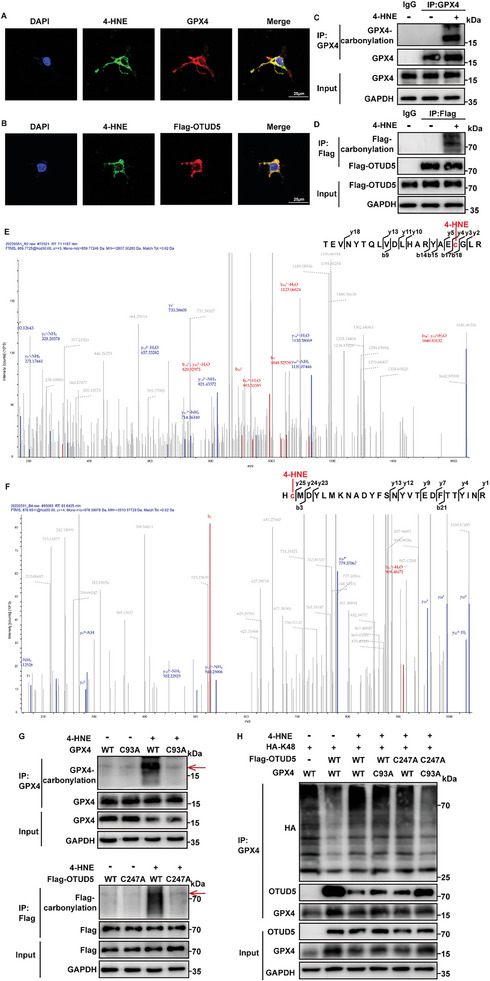
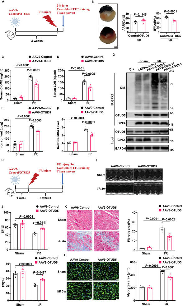
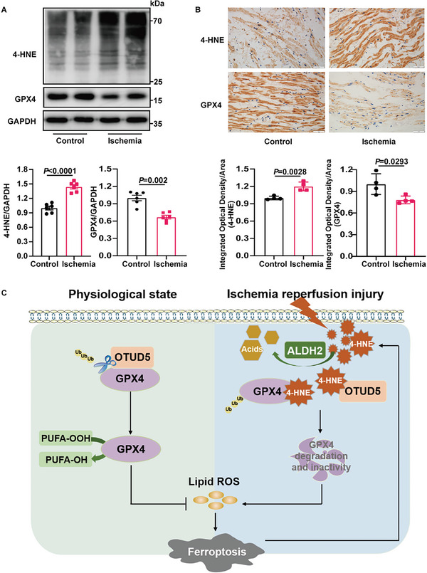
Despite the development of advanced technologies for interventional coronary reperfusion after myocardial infarction, a substantial number of patients experience high mortality due to myocardial ischemia-reperfusion (MI/R) injury. An in-depth understanding of the mechanisms underlying MI/R injury can provide crucial strategies for mitigating myocardial damage and improving patient survival. Here, it is discovered that the 4-hydroxy-2-nonenal (4-HNE) accumulates during MI/R, accompanied by high rates of myocardial ferroptosis. The loss-of-function of aldehyde dehydrogenase 2 (ALDH2), which dissipates 4-HNE, aggravates myocardial ferroptosis, whereas the activation of ALDH2 mitigates ferroptosis. Mechanistically, 4-HNE targets glutathione peroxidase 4 (GPX4) for K48-linked polyubiquitin-related degradation, which 4-HNE-GPX4 axis commits to myocyte ferroptosis and forms a positive feedback circuit. 4-HNE blocks the interaction between GPX4 and ovarian tumor (OTU) deubiquitinase 5 (OTUD5) by directly carbonylating their cysteine residues at C93 of GPX4 and C247 of OTUD5, identifying OTUD5 as the novel deubiquitinase for GPX4. Consequently, the elevation of OTUD5 deubiquitinates and stabilizes GPX4 to reverse 4-HNE-induced ferroptosis and alleviate MI/R injury. The data unravel the mechanism of 4-HNE in GPX4-dependent ferroptosis and identify OTUD5 as a novel therapeutic target for the treatment of MI/R injury.

Keywords: 4-hydroxy-2-nonenal (4-HNE); ferroptosis; glutathione peroxidase 4 (GPX4); myocardial ischemia reperfusion injury; ovarian tumor (OTU) deubiquitinase 5 (OTUD5).


Conflict of interest statement

The authors declare no conflict of interest.

Figures

Figure 1
Figure 1
ALDH2 regulates 4‐HNE accumulation and ferroptosis in MI/R. A) Protein expression of 4‐HNE in ALDH2flox/flox (F/F) and cardiomyocyte‐specific ALDH2‐knockout (cKO) mice hearts subjected to MI/R (30‐min ischemia/24‐h reperfusion, n = 4). B,C) Representative immunohistochemical staining results indicating 4‐HNE expression in F/F and cKO mice heart tissues 24 h after MI/R surgery (n = 4). Scale bar:50 µm. D) Representative photographs and statistical analysis of infarct size (IF) and area at risk (AAR) in mice hearts stained with Evans blue dye (EBD) and triphenyl tetrazolium chloride (TTC) (n = 6). The hearts were collected 24 h after MI/R surgery. Scale bar: 1 mm. E,F) Serum levels of CK‐MB and LDH in F/F and cKO mice with sham or MI/R injury (30‐min ischemia/24‐h reperfusion, n = 8). G‐J) Iron content, MDA, GSH, and GPX4 activity in F/F and cKO mice 24 h after MI/R surgery (n = 6–8). K) Western blotting of the ferroptosis marker protein (ACSL4, TfR1, FTH1, and GPX4) in F/F and cKO mice hearts subjected to MI/R (30‐min ischemia/24‐h reperfusion, n = 4). L) Representative western blotting of 4‐HNE in WT mice subjected to MI/R (30‐min ischemia/24‐h reperfusion). The mice were treated with DXZ (50 mg/kg), Alda‐1 (25 mg kg−1) or vehicle (solvent control) (n = 4). M) EBD/TTC staining of heart sections collected from the DXZ (50 mg kg−1), Alda‐1 (25 mg kg−1), and vehicle groups(n = 6). The hearts were collected 24 hours after MI/R surgery, and the infarct area and at‐risk area were evaluated. Scale bar: 1 mm. N,O) Serum levels of CK‐MB and LDH in mice treated with DXZ (50 mg kg−1), Alda‐1 (25 mg kg−1) and vehicle 24 h after MI/R surgery (n = 8). P,Q) Iron content, and GSH levels in WT mice with DXZ (50 mg/kg) or Alda‐1 (25 mg/kg) subjected to MI/R (30‐min ischemia/24‐h reperfusion, n = 6–8). R) Protein levels of ACSL4, TfR1, FTH1, and GPX4 in WT mice heart tissues from DXZ (50 mg kg−1), Alda‐1 (25 mg kg−1) and vehicle groups. The hearts were collected from 24 hours after MI/R surgery (n = 4). Data are expressed as mean ± SEM. Unpaired two‐tailed Student's t‐test (analysis in AAR/LV) and Mann‐Whitney test (analysis in IF/AAR) were used for the analysis in (D). One‐way analysis of variance (ANOVA) was used for the analysis in E–J,M–Q).
Figure 2
Figure 2
4‐HNE reduces GPX4 expression and induces cardiomyocyte ferroptosis. A) Cell viability was measured in H9c2 cells treated with 4‐HNE (40 µM) for 6 h in the presence of DXZ (10 µM), Fer‐1 (1 µM) or DMSO (n = 6). B,C) Iron content and lipid ROS of H9c2 cells under the indicated treatments (n = 3). D) Representative images of mitochondrial injury in H9c2 cells observed using transmission electron micrographs (n = 3). Scale bars:10 µm (top) and 1 µm (bottom). E) Western blotting analysis of the ferroptosis proteins under 4‐HNE treatment (40 µM,6 h) in NRCMs (n = 9). F, The activity of GPX4 under 4‐HNE (40 µM) treatment in the presence or absence of Alda‐1 (20 µM) for 6 h (n = 4). G,H) Iron content and lipid ROS in NRCMs treated with 4‐HNE (40 µM,6 h) after transfection with control siRNA (NC) or two siRNAs targeting GPX4 (n = 3). Data are expressed as mean ± SEM. Unpaired two‐tailed Student's t‐test was used for the analysis in (E). One‐way ANOVA was used for the analysis in A–C,F–H).
Figure 3
Figure 3
4‐HNE promotes the degradation of GPX4. A,B) The mRNA levels of GPX4 in 4‐HNE (40 µM) treated H9c2 cells and NRCMs at the indicated time points were detected by RT‐qPCR (n = 4). C) The protein levels of GPX4 in cycloheximide (CHX, 10 µM) treated NRCMs at the indicated time points with or without 4‐HNE (40 µM) incubation were detected by western blotting (n = 3). D,E) MG132 (10 µM) or chloroquine (CQ, 10 µM) were preincubated for 30 min, and H9c2 cells and NRCMs were treated with 4‐HNE (40 µM,6 h). Protein levels of GPX4 were analyzed by western blotting (n = 3). F) Representative images showed the total, K48‐, and K63‐linked ubiquitylation by western blotting in immunoprecipitation (IP) assays using anti‐GPX4 antibody (n = 3). G) GPX4, HA‐ubiquitin (HA‐Ub), HA‐K48, and HA‐K63 were transfected into HEK293T cells treated with or without 4‐HNE (40 µM,6 h), and IP assays were performed (n = 3). Data are expressed as mean ± SEM. Two‐way ANOVA was used for the analysis in A,B). One‐way ANOVA was used for the analysis in D,E).
Figure 4
Figure 4
OTUD5 regulates GPX4 stability. A) OTUD5 was identified in the protein mixture enriched by anti‐GPX4 antibody. The graph represented peptide fragment RATDWEATNEAIEEQVAR, which was specifically referred to OTUD5 protein. B) Purified recombinant GST, GST‐GPX4 were incubated with His‐OTUD5 in vitro and the direct interaction between GPX4 and OTUD5 was demonstrated by the GST pull‐down assay (n = 3). C) Surface plasmon resonance (SPR) sensorgrams of the binding of an increasing amount of OTUD5 to GPX4 ligand captured on a CM5 chip. The increase in RUs from baseline was measured and used to calculate the equilibrium dissociation constant (K D ) for OTUD5 binding to immobilized GPX4 ligand. D) Colocalization of GPX4 (red) with OTUD5 (green) was detected in HEK293T cells by immunofluorescence analysis (n = 5). Scale bar:25 µm. E) GPX4 was transfected into HEK293T cells along with increasing amounts of Flag‐OTUD5 or C224S. GPX4 protein levels were detected by western blotting (n = 3). F) GPX4 ubiquitination was analyzed in HEK293T cells transfected with GPX4, HA‐ubiquitin (HA‐Ub), or increasing amounts of Flag‐OTUD5 or C224S (n = 3). G) GPX4, HA‐Ub, or its lysine residue mutants were transfected into HEK293T cells with or without Flag‐OTUD5. Co‐IP assays were performed using anti‐GPX4 antibody (n = 3). H) HEK293T cells transfected with the indicated constructs were subjected to Co‐IP with anti‐GPX4 antibody (n = 3).
Figure 5
Figure 5
OTUD5 mediates the effects of 4‐HNE on the degradation of GPX4. A) Co‐immunoprecipitation (Co‐IP) was performed with lysates from HEK293T cells transfected with GPX4 and Flag‐OTUD5 plasmids, with or without 4‐HNE treatment (40 µM for 6 h, n = 3). B) Co‐IP was performed on lysates of NRCMs incubated with or without 4‐HNE (40 µM,6 h). Lysates were extracted for Co‐IP with GPX4/OTUD5 specific antibody or control IgG, followed by probing with antibodies specific for GPX4/OTUD5 (n = 4). C) Representative images of proximity ligation assay (red fluorescent dots) between GPX4 and OTUD5 in NRCMs with or without 4‐HNE treatment (40 µM for 6 h, n = 5). Scale bar: 25 µm. D) Colocalization of GPX4 (red) with OTUD5 (green) in NRCMs in the presence or absence of 4‐HNE treatment (40 µM for 6 h, n = 5). Scale bar:10 µm. E) Co‐IP analysis of endogenous GPX4 ubiquitination in NRCMs transfected with control siRNA (NC) or with two siRNAs targeting OTUD5 after 4‐HNE stimulation (40 µM for 6 h, n = 3). F) OTUD5 WT (sg Con) and KO (sg OTUD5) H9c2 cells were treated with 4‐HNE (40 µM,6 h), then GPX4 protein level and the ubiquitination of GPX4 were detected by Immunoblot assays (n = 3).
Figure 6
Figure 6
4‐HNE‐induced carbonylation inhibits the interaction between OTUD5 and GPX4. A,B) Staining of 4‐HNE (green), GPX4 or Flag‐OTUD5(red) in HEK293T cells (n = 5). Scale bar: 25 µm. C,D) Immunoblot assays of carbonylation of GPX4 and OTUD5 in HEK293T cells transfected with GPX4 and Flag‐OTUD5 detected by selective labeling with m‐APA (n = 3). E,F) LC‐MS spectra of 4‐HNE modification of GPX4(E) and OTUD5(F) at the Cys residue. G) Immunoblot assays of carbonylation in HEK293T cells transfected with GPX4 WT, GPX4 mutants (C93A) or Flag‐OTUD5 WT, Flag‐OTUD5 mutants (C247A) by oxyblot technology (n = 3). The targeted protein bands are indicated by the red arrows. H) HEK293T cells were co‐transfected with indicated plasmids. Co‐IP assays were performed using anti‐GPX4 antibody and the levels of GPX4 ubiquitination were examined by western blotting (n = 3).
Figure 7
Figure 7
OTUD5 overexpression prevents cardiac ischemia/reperfusion injury in ALDH2 cKO mice. A) Schematic diagram showing that AAV9‐Control or AAV9‐OTUD5 were injected via tail vein, and 3 weeks later mice were subjected to cardiac ischemia/reperfusion (I/R) injury for 24 h. B) Representative sections and quantitative data for infarct size (IF) and area at risk (AAR) in mice hearts subjected to I/R injury (30‐min ischemia/24‐h reperfusion, n = 6). Scale bar: 1 mm. C,D) Serum levels of CK‐MB and LDH in mice with sham or MI/R injury (30‐min ischemia/24‐h reperfusion, n = 8). E,F) Iron content and MDA levels in mice subjected to MI/R (30‐min ischemia/24‐h reperfusion, n = 8). G) The ubiquitination of GPX4 was determined by immunoprecipitation with anti‐GPX4 antibody in mice subjected to MI/R injury (30‐min ischemia/24‐h reperfusion, n = 4). H) Schematic diagram showing that AAV9‐Control or AAV9‐OTUD5 were injected via tail vein, and 1 week later mice were subjected to cardiac ischemia/reperfusion (I/R) injury for 3 weeks. I,J) Echocardiography for left ventricular ejection fraction (EF, %) and fractional shortening (FS, %) in mice after I/R injury (30‐min ischemia/3‐week reperfusion, n = 5). K,L) Masson Trichrome staining for cardiac fibrosis (Scale bar: 50 µm) and WGA staining for cardiac hypertrophy (Scale bar: 20 µm) in heart tissues of mice after MI/R (30‐min ischemia/3‐week reperfusion, n = 5). Data are expressed as mean ± SEM. Unpaired two‐tailed Student's t‐test was used for the analysis in B). One‐way ANOVA was used for the analysis in C–F,J–L).
Figure 8
Figure 8
Myocardial ischemia induces 4‐HNE accumulation and GPX4 reduction in human samples. A) Immunoblots of 4‐HNE and GPX4 proteins in the right auricle from control patients and patients with ischemia (n = 6). B) Representative immunohistochemical staining of the control and ischemic patients (n = 4). Scale bar:50 µm. C) Schematic illustration of 4‐HNE‐induced ferroptosis in myocardial ischemia‐reperfusion. At basal conditions, OTUD5 deubiquitinates and stabilizes GPX4. In myocardial ischemia/reperfusion, 4‐HNE binds competitively to OTUD5 and GPX4, blocks the interaction between OTUD5 and GPX4, and promotes GPX4 ubiquitination and degradation, which induces ferroptosis and aggravates myocardial injury. Data are expressed as mean ± SEM. Unpaired two‐tailed Student's t‐test was used for the analysis in A,B).

References

    1. Hausenloy D. J., Yellon D. M., J. Clin. Invest. 2013, 123, 92. - PMC - PubMed
    1. Tsao C. W., Aday A. W., Almarzooq Z. I., Alonso A., Beaton A. Z., Bittencourt M. S., Boehme A. K., Buxton A. E., Carson A. P., Commodore‐Mensah Y., Elkind M. S. V., Evenson K. R., Eze‐Nliam C., Ferguson J. F., Generoso G., Ho J. E., Kalani R., Khan S. S., Kissela B. M., Knutson K. L., Levine D. A., Lewis T. T., Liu J., Loop M. S., Ma J., Mussolino M. E., Navaneethan S. D., Perak A. M., Poudel R., Rezk‐Hanna M., et al., Circulation 2022, 145, e153. - PubMed
    1. Ibáñez B., Heusch G., Ovize M., Van de Werf F., Journal of the American College of Cardiology 2015, 65, 1454. - PubMed
    1. Heusch G., Nat. Rev. Cardiol. 2020, 17, 773. - PubMed
    1. Dixon S. J., Lemberg K. M., Lamprecht M. R., Skouta R., Zaitsev E. M., Gleason C. E., Patel D. N., Bauer A. J., Cantley A. M., Yang W. S., Morrison B. III, Stockwell B. R., Cell 2012, 149, 1060. - PMC - PubMed

Publication types

MeSH terms

Substances

LinkOut - more resources