Clinical Trial
. 2023 Nov 1;13(11):2412-2431.
doi: 10.1158/2159-8290.CD-23-0669.

MRTX1719 Is an MTA-Cooperative PRMT5 Inhibitor That Exhibits Synthetic Lethality in Preclinical Models and Patients with MTAP-Deleted Cancer

Affiliations
Clinical Trial

MRTX1719 Is an MTA-Cooperative PRMT5 Inhibitor That Exhibits Synthetic Lethality in Preclinical Models and Patients with MTAP-Deleted Cancer

Lars D Engstrom et al. Cancer Discov. .

Abstract

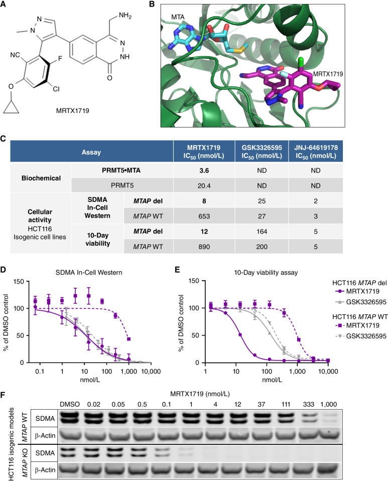
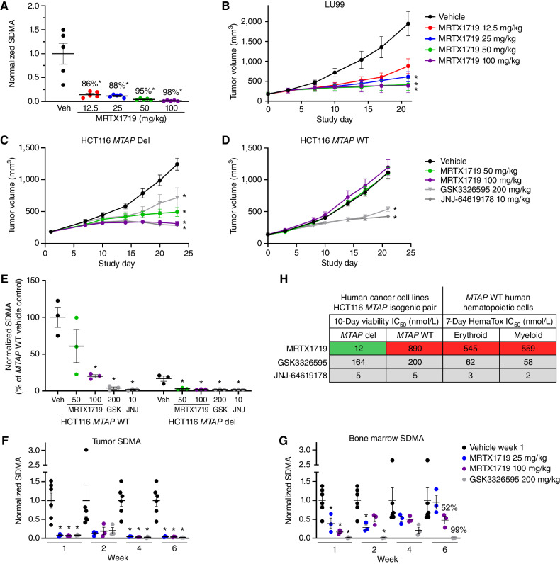
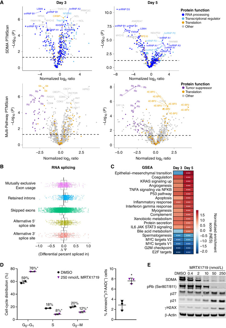
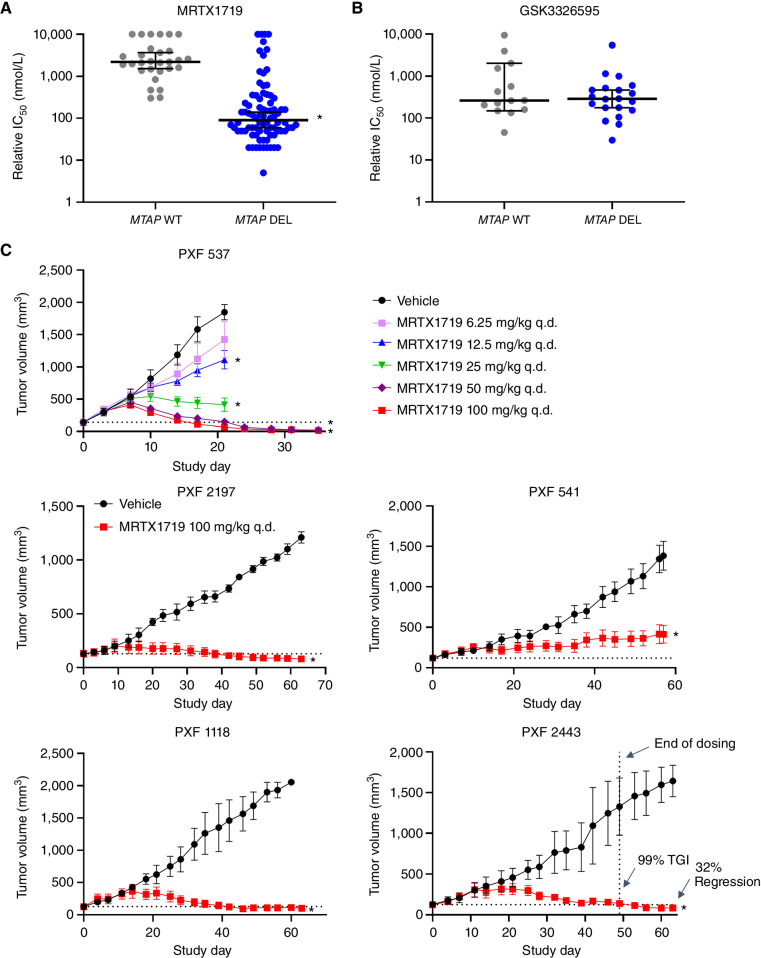
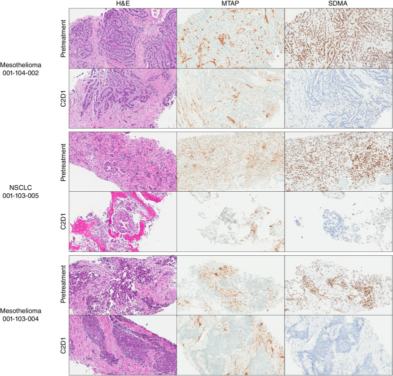
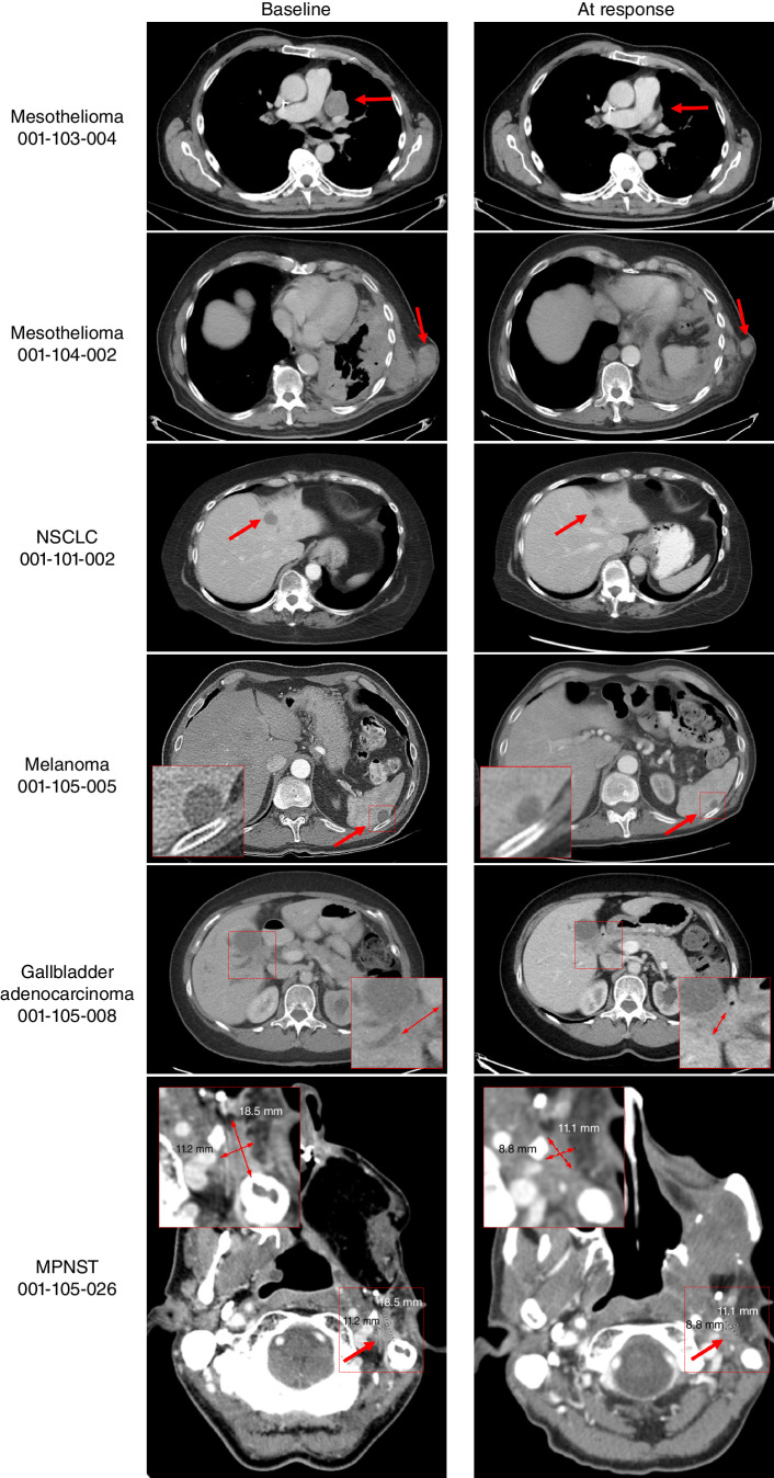
Previous studies implicated protein arginine methyltransferase 5 (PRMT5) as a synthetic lethal target for MTAP-deleted (MTAP del) cancers; however, the pharmacologic characterization of small-molecule inhibitors that recapitulate the synthetic lethal phenotype has not been described. MRTX1719 selectively inhibited PRMT5 in the presence of MTA, which is elevated in MTAP del cancers, and inhibited PRMT5-dependent activity and cell viability with >70-fold selecti-vity in HCT116 MTAP del compared with HCT116 MTAP wild-type (WT) cells. MRTX1719 demonstrated dose-dependent antitumor activity and inhibition of PRMT5-dependent SDMA modification in MTAP del tumors. In contrast, MRTX1719 demonstrated minimal effects on SDMA and viability in MTAP WT tumor xenografts or hematopoietic cells. MRTX1719 demonstrated marked antitumor activity across a panel of xenograft models at well-tolerated doses. Early signs of clinical activity were observed including objective responses in patients with MTAP del melanoma, gallbladder adenocarcinoma, mesothelioma, non-small cell lung cancer, and malignant peripheral nerve sheath tumors from the phase I/II study.

Significance: PRMT5 was identified as a synthetic lethal target for MTAP del cancers; however, previous PRMT5 inhibitors do not selectively target this genotype. The differentiated binding mode of MRTX1719 leverages the elevated MTA in MTAP del cancers and represents a promising therapy for the ∼10% of patients with cancer with this biomarker. See related commentary by Mulvaney, p. 2310. This article is featured in Selected Articles from This Issue, p. 2293.


Figures

Figure 1. MRTX1719 binds PRMT5/MTA and selectively inhibits MTAP del cancer cells with reduced activity in MTAP WT cells. A, Chemical structure of MRTX1719. B, X-ray crystal structure of MRTX1719 cocomplexed with PRMT5/MEP50 and MTA (right; Protein Data Bank code 7S1S; ref. 14). C, MRTX1719 was run in a PRMT5/MEP50 biochemical assay that measures the activity of the complex in the absence and presence of MTA at an approximate IC50 concentration of MTA in the assay (2 μmol/L). MRTX1719, GSK3326595, and JNJ-64619178 were run in SDMA In-Cell Western (SYM11 antibody; C and D) and 10-day viability assays (C and E) in MTAP del and WT HCT116 cell lines. ND, not determined. F, Cropped SDMA Western blot showing protein bands that correlate with the molecular weight of SmD3 in MTAP del and WT HCT116 cell lines following 4 days of treatment with a range of concentrations of MRTX1719, with β-actin run as a loading control. KO, knockout.
Figure 1.
MRTX1719 binds PRMT5/MTA and selectively inhibits MTAP del cancer cells with reduced activity in MTAP WT cells. A, Chemical structure of MRTX1719. B, X-ray crystal structure of MRTX1719 cocomplexed with PRMT5/MEP50 and MTA (right; Protein Data Bank code 7S1S; ref. 14). C, MRTX1719 was run in a PRMT5/MEP50 biochemical assay that measures the activity of the complex in the absence and presence of MTA at an approximate IC50 concentration of MTA in the assay (2 μmol/L). MRTX1719, GSK3326595, and JNJ-64619178 were run in SDMA In-Cell Western (SYM11 antibody; C and D) and 10-day viability assays (C and E) in MTAP del and WT HCT116 cell lines. ND, not determined. F, Cropped SDMA Western blot showing protein bands that correlate with the molecular weight of SmD3 in MTAP del and WT HCT116 cell lines following 4 days of treatment with a range of concentrations of MRTX1719, with β-actin run as a loading control. KO, knockout.
Figure 2. MRTX1719 exhibits selective, dose-dependent inhibition of PRMT5-dependent SDMA modification in MTAP del tumor xenografts in vivo. A, MRTX1719 was administered daily via daily oral gavage for 22 days to immunocompromised mice bearing LU99 tumor xenografts (average initial tumor volume ∼180 mm3) at 12.5, 25, 50, and 100 mg/kg. Tumors were collected 4 hours after dose, and SDMA was analyzed by immunoblot and quantified with densitometry. Average normalized SDMA values were divided by the average value in vehicle-treated tumors to calculate percent inhibition. Data shown represent the average of 2 to 3 tumors per treatment group ± SEM. Statistics were determined using a two-tailed Student t test with significance indicated (*, P < 0.05). Veh, vehicle. B, MRTX1719 was administered at the indicated doses via daily oral gavage to mice bearing established LU99 cell line–derived tumor xenografts as in A. Data, mean tumor volume ± SEM. Statistics were determined using a two-way ANOVA with significance indicated (*, P < 0.05). A and B were previously published in Smith et al. (14). C and D, MRTX1719, GSK3326595, or JNJ-64619178 was administered via daily oral gavage at the doses indicated to mice bearing established HCT116 MTAP del (C) or MTAP WT (D) cell line–derived tumor xenografts. Dosing was initiated when tumors were ∼150 mm3. Data, mean tumor volume ± SEM. Statistics were determined using a two-way ANOVA with significance indicated (*, P < 0.05). E, Tumors from the HCT116 MTAP WT or HCT116 MTAP del cell line–derived xenografts in C and D were collected 4 hours after dose, and SDMA was analyzed by immunoblot. Data shown represent the average of 3 tumors per treatment group ± SEM. Statistics were determined using a two-tailed Student t test with significance indicated (*, P < 0.05). GSK, GSK3326595; JNJ, JNJ-64619178. F and G, MRTX1719 and GSK3326595 were administered via daily oral gavage to immunocompromised mice bearing established LU99 xenograft tumors at the indicated doses for the indicated number of weeks. Tumor (F) and bone marrow (G) were collected 4 hours after last dose, and SDMA was analyzed by Western blot densitometry. Representative SDMA bands were analyzed from 3 to 5 tumor lysates and bone marrow lysates from 3 to 5 mice per treatment group, and data are shown as mean normalized SDMA levels ± SEM. Statistics were determined using a two-tailed Student t test with significance indicated (*, P < 0.05). H, MRTX1719, JNJ-64619178, and GSK3326595 were run in human erythroid and myeloid 7-day HemaTox assays (STEMCELL Technologies) and were compared with IC50 values from 10-day HCT116 MTAP del and MTAP WT cell line viability assays.
Figure 2.
MRTX1719 exhibits selective, dose-dependent inhibition of PRMT5-dependent SDMA modification in MTAP del tumor xenografts in vivo. A, MRTX1719 was administered daily via daily oral gavage for 22 days to immunocompromised mice bearing LU99 tumor xenografts (average initial tumor volume ∼180 mm3) at 12.5, 25, 50, and 100 mg/kg. Tumors were collected 4 hours after dose, and SDMA was analyzed by immunoblot and quantified with densitometry. Average normalized SDMA values were divided by the average value in vehicle-treated tumors to calculate percent inhibition. Data shown represent the average of 2 to 3 tumors per treatment group ± SEM. Statistics were determined using a two-tailed Student t test with significance indicated (*, P < 0.05). Veh, vehicle. B, MRTX1719 was administered at the indicated doses via daily oral gavage to mice bearing established LU99 cell line–derived tumor xenografts as in A. Data, mean tumor volume ± SEM. Statistics were determined using a two-way ANOVA with significance indicated (*, P < 0.05). A and B were previously published in Smith et al. (14). C and D, MRTX1719, GSK3326595, or JNJ-64619178 was administered via daily oral gavage at the doses indicated to mice bearing established HCT116 MTAP del (C) or MTAP WT (D) cell line–derived tumor xenografts. Dosing was initiated when tumors were ∼150 mm3. Data, mean tumor volume ± SEM. Statistics were determined using a two-way ANOVA with significance indicated (*, P < 0.05). E, Tumors from the HCT116 MTAP WT or HCT116 MTAP del cell line–derived xenografts in C and D were collected 4 hours after dose, and SDMA was analyzed by immunoblot. Data shown represent the average of 3 tumors per treatment group ± SEM. Statistics were determined using a two-tailed Student t test with significance indicated (*, P < 0.05). GSK, GSK3326595; JNJ, JNJ-64619178. F and G, MRTX1719 and GSK3326595 were administered via daily oral gavage to immunocompromised mice bearing established LU99 xenograft tumors at the indicated doses for the indicated number of weeks. Tumor (F) and bone marrow (G) were collected 4 hours after last dose, and SDMA was analyzed by Western blot densitometry. Representative SDMA bands were analyzed from 3 to 5 tumor lysates and bone marrow lysates from 3 to 5 mice per treatment group, and data are shown as mean normalized SDMA levels ± SEM. Statistics were determined using a two-tailed Student t test with significance indicated (*, P < 0.05). H, MRTX1719, JNJ-64619178, and GSK3326595 were run in human erythroid and myeloid 7-day HemaTox assays (STEMCELL Technologies) and were compared with IC50 values from 10-day HCT116 MTAP del and MTAP WT cell line viability assays.
Figure 3. MRTX1719 treatment disrupts PRMT5-regulated processes in MTAP del cancer cells. A, Volcano plots of posttranslational modifications of trypsin-digested peptides from LU99 cells treated for 3 or 5 days with 1 μmol/L MRTX1719 vs. DMSO as measured by LC-MS/MS following antibody enrichment using the SDMA PTMScan kit (top) or the Multi-Pathway PTMScan kit (bottom) that includes phosphorylation (Cell Signaling Technology). B, RNA-seq data were generated from LU99 cells treated with MRTX1719 or DMSO for 3 days, and transcripts were analyzed for alternative RNA splicing using rMATS. Transcripts with retained introns were increased in MRTX1719-treated vs. DMSO-treated cells. C, GSEA was performed on RNA-seq data from 3- and 5-day MRTX1719-treated LU99 cells compared with DMSO-treated. ***, FDR <0.25. D, LU99 cells were treated with 250 nmol/L MRTX1719 for 3 days and analyzed on a Guava flow cytometer. Cell-cycle distribution analysis (left) was performed, and apoptotic cells were analyzed for surface Annexin V–positive, 7-AAD–negative staining (right). Summary data from 2 or 3 independent experiments are presented as the mean ± SD. Statistics were determined using a two-tailed Student t test with significance indicated (*, P < 0.05). E, Cell lysates from LU99 cells treated for 3 days with MRTX1719 and analyzed for selected protein markers by Western blot.
Figure 3.
MRTX1719 treatment disrupts PRMT5-regulated processes in MTAP del cancer cells. A, Volcano plots of posttranslational modifications of trypsin-digested peptides from LU99 cells treated for 3 or 5 days with 1 μmol/L MRTX1719 vs. DMSO as measured by LC-MS/MS following antibody enrichment using the SDMA PTMScan kit (top) or the Multi-Pathway PTMScan kit (bottom) that includes phosphorylation (Cell Signaling Technology). B, RNA-seq data were generated from LU99 cells treated with MRTX1719 or DMSO for 3 days, and transcripts were analyzed for alternative RNA splicing using rMATS. Transcripts with retained introns were increased in MRTX1719-treated vs. DMSO-treated cells. C, GSEA was performed on RNA-seq data from 3- and 5-day MRTX1719-treated LU99 cells compared with DMSO-treated. ***, FDR <0.25. D, LU99 cells were treated with 250 nmol/L MRTX1719 for 3 days and analyzed on a Guava flow cytometer. Cell-cycle distribution analysis (left) was performed, and apoptotic cells were analyzed for surface Annexin V–positive, 7-AAD–negative staining (right). Summary data from 2 or 3 independent experiments are presented as the mean ± SD. Statistics were determined using a two-tailed Student t test with significance indicated (*, P < 0.05). E, Cell lysates from LU99 cells treated for 3 days with MRTX1719 and analyzed for selected protein markers by Western blot.
Figure 4. MRTX1719 selectively inhibits MTAP del cancer cell line growth in vitro and in vivo. A and B, MRTX1719 in vitro activity across a panel of MTAP WT and MTAP del cell line models (5-day viability assay, Crown Biosciences). Dot plots showing median with 95% confidence intervals of IC50 values for MRTX1719 (A) and GSK3326595 (GSK-595; B) in MTAP WT and MTAP del cell line models. A smaller cohort of models was tested with GSK-595. Median values: MRTX1719-MTAP del: IC50 = 90 nmol/L; MTAP WT: IC50 = 2.2 mmol/L; GSK-595-MTAP del: IC50 = 262 nmol/L; MTAP WT: IC50 = 286 nmol/L. Statistics were determined using a two-tailed Student t test with significance indicated (*, P < 0.05). C, MRTX1719 was administered orally at the doses indicated to five mesothelioma PDX models. n = 3–5 mice per treatment group. Data, average tumor volume ± SEM. Statistics were determined using a two-way ANOVA with significance indicated (*, P < 0.05). TGI, tumor growth inhibition.
Figure 4.
MRTX1719 selectively inhibits MTAP del cancer cell line growth in vitro and in vivo. A and B, MRTX1719 in vitro activity across a panel of MTAP WT and MTAP del cell line models (5-day viability assay, Crown Biosciences). Dot plots showing median with 95% confidence intervals of IC50 values for MRTX1719 (A) and GSK3326595 (GSK-595; B) in MTAP WT and MTAP del cell line models. A smaller cohort of models was tested with GSK-595. Median values: MRTX1719-MTAP del: IC50 = 90 nmol/L; MTAP WT: IC50 = 2.2 mmol/L; GSK-595-MTAP del: IC50 = 262 nmol/L; MTAP WT: IC50 = 286 nmol/L. Statistics were determined using a two-tailed Student t test with significance indicated (*, P < 0.05). C, MRTX1719 was administered orally at the doses indicated to five mesothelioma PDX models. n = 3–5 mice per treatment group. Data, average tumor volume ± SEM. Statistics were determined using a two-way ANOVA with significance indicated (*, P < 0.05). TGI, tumor growth inhibition.
Figure 5. PRMT5-mediated SDMA modification in MTAP del patient tumor biopsies is extinguished following daily oral administration of 200 mg MRTX1719. Pretreatment and C2D1 tumor biopsies were stained for MTAP and SDMA protein expression by IHC. Adjacent slides were also stained with hematoxylin and eosin (H&E). MTAP and SDMA H-scores were determined by a pathologist and scored in Supplementary Fig. S7A.
Figure 5.
PRMT5-mediated SDMA modification in MTAP del patient tumor biopsies is extinguished following daily oral administration of 200 mg MRTX1719. Pretreatment and C2D1 tumor biopsies were stained for MTAP and SDMA protein expression by IHC. Adjacent slides were also stained with hematoxylin and eosin (H&E). MTAP and SDMA H-scores were determined by a pathologist and scored in Supplementary Fig. S7A.
Figure 6. Activity of MRTX1719 in patients with MTAP del cancers. Mesothelioma patient 001-103-004: Pretreatment and follow-up (FU) C6D20 scans, indicating 56% reduction of target lesions, including the pleural disease shown here. The patient continues on study. Mesothelioma patient 001-104-002: Activity of MRTX1719 in patient epithelioid mesothelioma with MTAP deletion. Pretreatment and FU 3 scans, indicating 30% reduction of target lesions, including soft tissue lesions shown here. The patient continues on study. NSCLC patient 001-101-002: Baseline: dominant mass left hepatic dome measuring 29 × 19 mm, previous 20 × 19 mm. Stable other tiny scattered hypodense hepatic lesions too small to accurately characterize. C5D1: left hepatic dome metastasis, measuring 17 × 17 mm. Stable and mild decreasing size of hepatic metastases. The patient continues on study. Melanoma patient 001-105-005: Baseline and cycle 9 scans of a patient with MTAP del melanoma. Partial response was confirmed at cycle 9, and the patient continues on study. Gallbladder adenocarcinoma patient 001-105-008: Pretreatment and cycle 9 scans of a patient with treatment-refractory MTAP loss gallbladder indicating 43% reduction of target lesions. The patient continues on study. MPNST patient 001-105-026: Patient received MRTX1719 800 mg q.d. Restaging after 4 cycles shows a partial response with a 38.9% decrease of target lesions. The patient continues on study.
Figure 6.
Activity of MRTX1719 in patients with MTAP del cancers. Mesothelioma patient 001-103-004: Pretreatment and follow-up (FU) C6D20 scans, indicating 56% reduction of target lesions, including the pleural disease shown here. The patient continues on study. Mesothelioma patient 001-104-002: Activity of MRTX1719 in patient epithelioid mesothelioma with MTAP deletion. Pretreatment and FU 3 scans, indicating 30% reduction of target lesions, including soft tissue lesions shown here. The patient continues on study. NSCLC patient 001-101-002: Baseline: dominant mass left hepatic dome measuring 29 × 19 mm, previous 20 × 19 mm. Stable other tiny scattered hypodense hepatic lesions too small to accurately characterize. C5D1: left hepatic dome metastasis, measuring 17 × 17 mm. Stable and mild decreasing size of hepatic metastases. The patient continues on study. Melanoma patient 001-105-005: Baseline and cycle 9 scans of a patient with MTAP del melanoma. Partial response was confirmed at cycle 9, and the patient continues on study. Gallbladder adenocarcinoma patient 001-105-008: Pretreatment and cycle 9 scans of a patient with treatment-refractory MTAP loss gallbladder indicating 43% reduction of target lesions. The patient continues on study. MPNST patient 001-105-026: Patient received MRTX1719 800 mg q.d. Restaging after 4 cycles shows a partial response with a 38.9% decrease of target lesions. The patient continues on study.

Comment in

References

    1. Huang A, Garraway LA, Ashworth A, Weber B. Synthetic lethality as an engine for cancer drug target discovery. Nat Rev Drug Discov 2020;19:23–38. - PubMed
    1. Muller FL, Aquilanti EA, DePinho RA. Collateral lethality: a new therapeutic strategy in oncology. Trends Cancer 2015;1:161–73. - PMC - PubMed
    1. Gu F, Pfeiffer RM, Bhattacharjee S, Han SS, Taylor PR, Berndt S, et al. . Common genetic variants in the 9p21 region and their associations with multiple tumours. Br J Cancer 2013;108:1378–86. - PMC - PubMed
    1. Blanc RS, Richard S. Arginine methylation: the coming of age. Mol Cell 2017;65:8–24. - PubMed
    1. Kim H, Ronai ZA. PRMT5 function and targeting in cancer. Cell Stress 2020;4:199–215. - PMC - PubMed

Publication types

Substances