. 2023 Oct 9;58(19):1917-1932.e6.
doi: 10.1016/j.devcel.2023.07.009. Epub 2023 Aug 7.

The mammalian midbody and midbody remnant are assembly sites for RNA and localized translation

Affiliations

The mammalian midbody and midbody remnant are assembly sites for RNA and localized translation

Sungjin Park et al. Dev Cell. .

Abstract

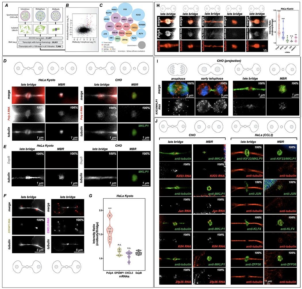
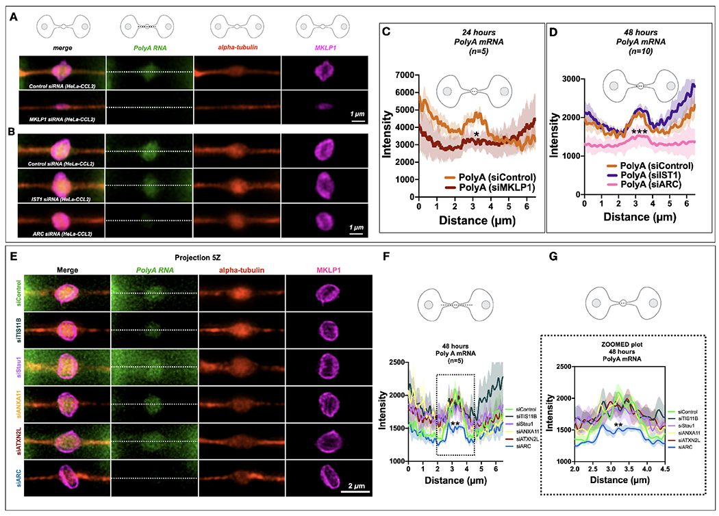
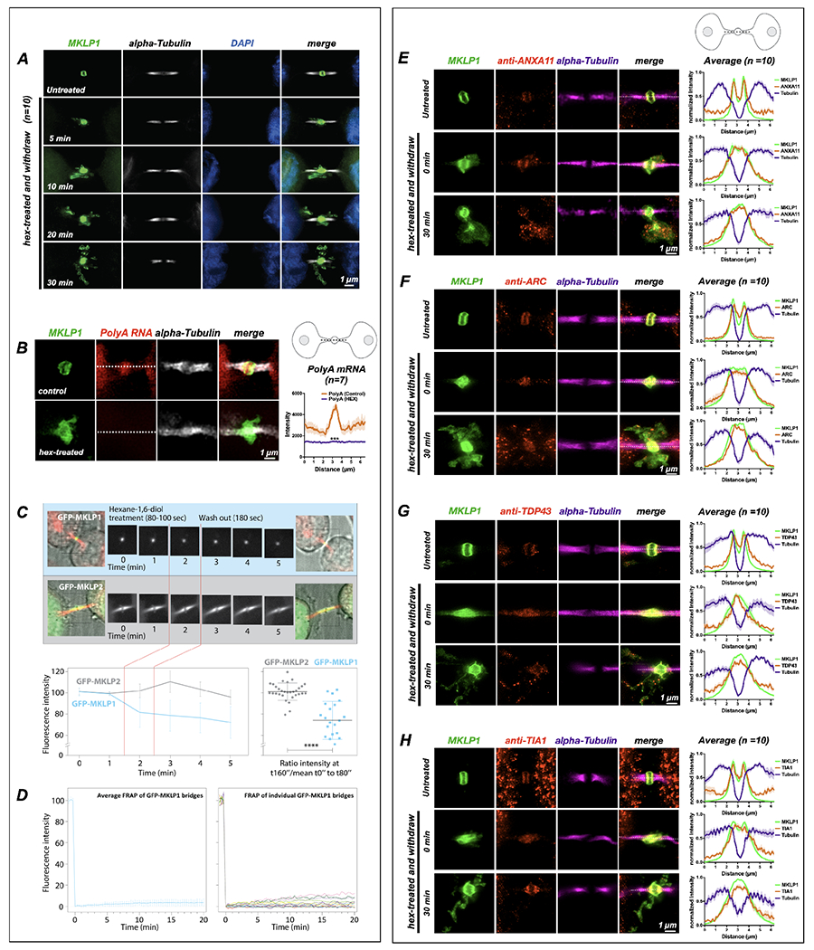
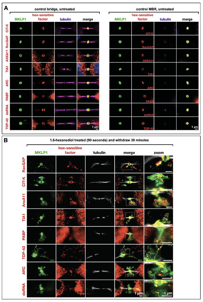
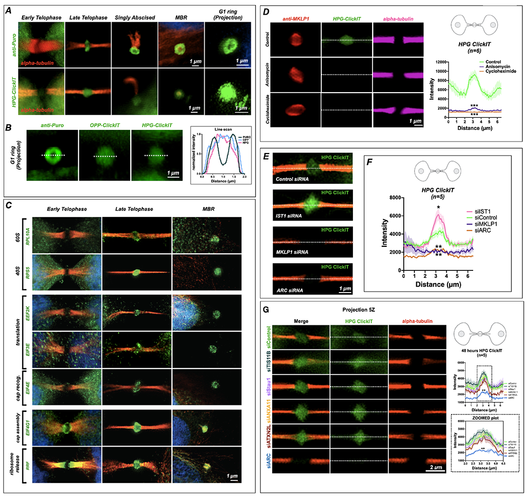
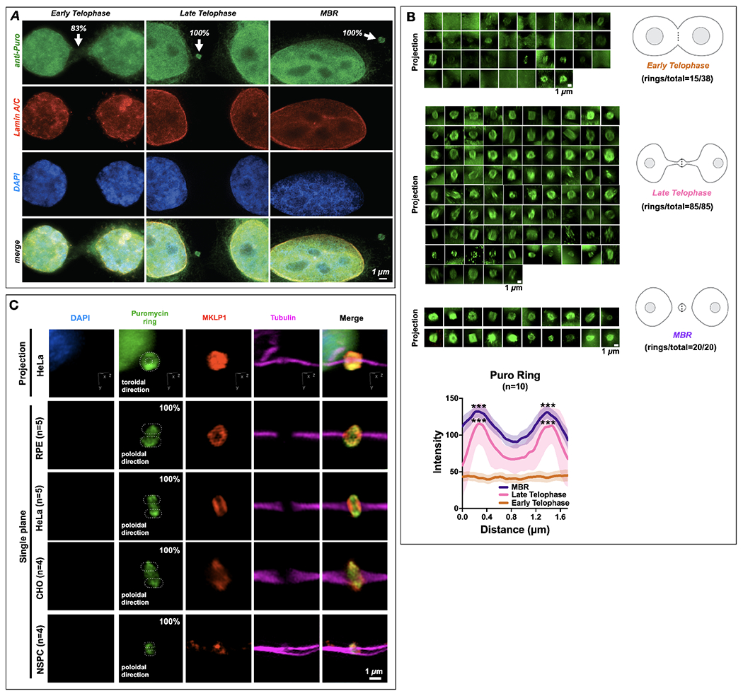
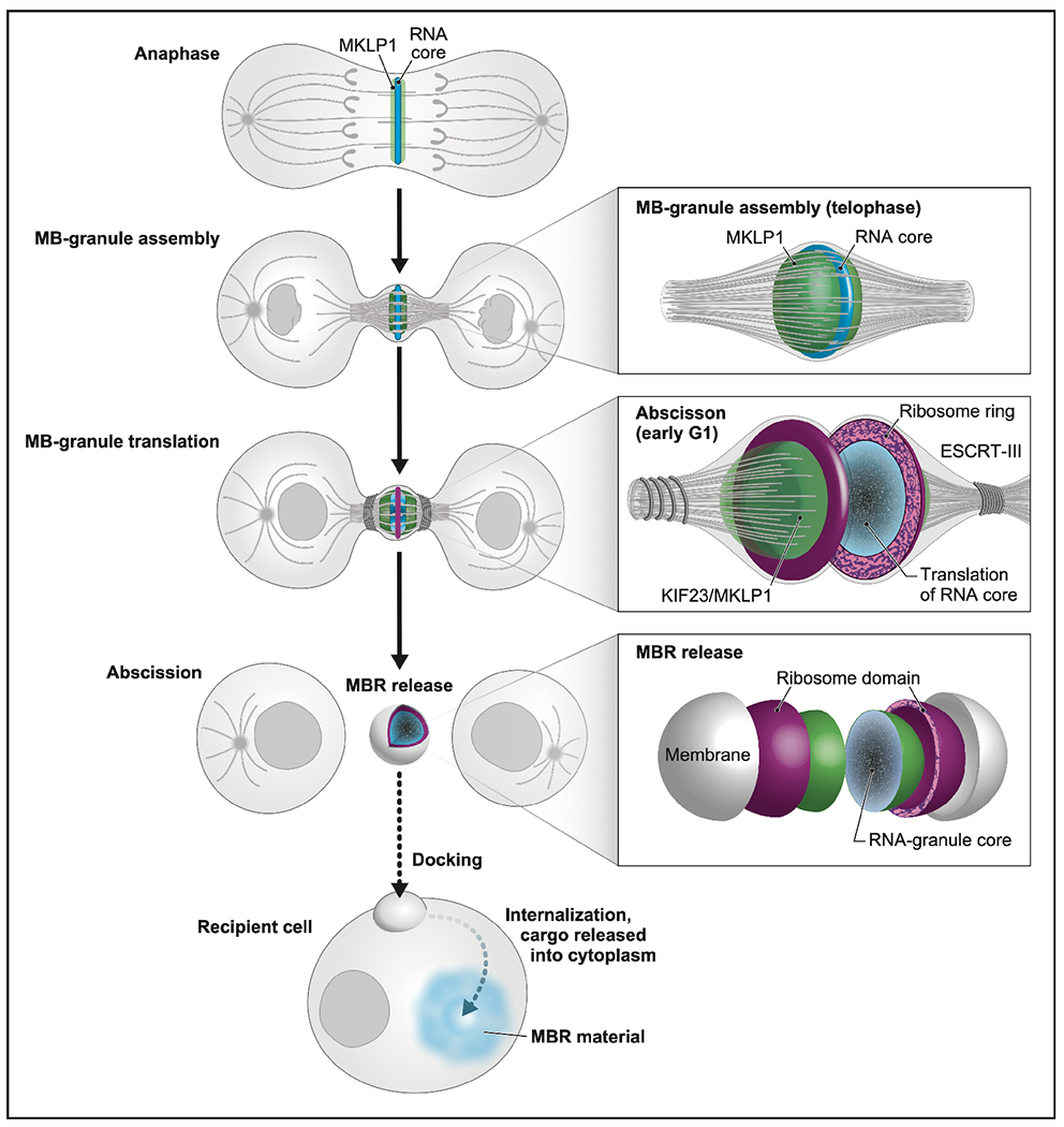
Long ignored as a vestigial remnant of cytokinesis, the mammalian midbody (MB) is released post-abscission inside large extracellular vesicles called MB remnants (MBRs). Recent evidence suggests that MBRs can modulate cell proliferation and cell fate decisions. Here, we demonstrate that the MB matrix is the site of ribonucleoprotein assembly and is enriched in mRNAs that encode proteins involved in cell fate, oncogenesis, and pluripotency, which we are calling the MB granule. Both MBs and post-abscission MBRs are sites of spatiotemporally regulated translation, which is initiated when nascent daughter cells re-enter G1 and continues after extracellular release. MKLP1 and ARC are necessary for the localization and translation of RNA in the MB dark zone, whereas ESCRT-III is necessary to maintain translation levels in the MB. Our work reveals a unique translation event that occurs during abscission and within a large extracellular vesicle.

Keywords: Arc; ESCRT-III; EV; MB; MBR; MBsome; RBP; abscission; cytokinesis; intercellular bridge; large extracellular vesicle; midbody; midbody remnant; mitosis; translation.


Conflict of interest statement

Declaration of interests The authors declare no competing interests.

Figures

Figure 1:
Figure 1:. Midbodies and midbody remnants are sites of RNA storage. MKLP1 and ARC are necessary for mRNA localization and maintenance in the dark zone.
(A-C) RNA-Seq analysis of the MB transcriptome. mRNA was sequenced from three stages of the cell cycle: interphase, metaphase, and late cytokinesis (or “MB stage”). Tubulin structures were purified, and associated RNAs were isolated and analyzed by RNA-Seq. Of 21,607 distinct CHO transcripts identified, 20,821 could be annotated by gene ontology. Of those, 7,986 had ≥100 reads in all cell cycle stages and were further analyzed as plotted in (B). Raw data can be found in Supp.Tables 1–4. (B) Transcripts with ≥100 reads in all three populations were compared and plotted based on their log2 enrichment scores (RPKM/RPKM). Dotted lines at x = 1 and y = 1 indicate minimum values for 2-fold enrichment. The 22 transcripts enriched in the MB relative to both interphase and metaphase are highlighted in red. (C) Enrichment score (relative diameter) and gene ontology groups of the 22 MB-enriched transcripts; colors correspond to gene ontology biological process terms (Fig. 1; see also Supp. Tables 1–4). (D) Single-molecule RNAscope (RNA in situ) hybridization revealed mRNA enrichment in the MB and released MBRs. PolyA-positive mRNAs (red) localized to mitotic MBs and post-mitotic MBRs in both CHO and HeLa Kyoto cells, in contrast with the bacterial DapB negative control (E). (E) The bacterial DapE was not found at the midbody in HeLa Kyoto or CHO cells midbodies or MBRs. (F-G) Two mRNAs. EPEMP1 and CNCL5, identified from interphase enriched RNAseq data (see Table 4) were not enriched in the midbody. *Significance was determined and denoted by * or n.s. n.s denotes not significant. (H) Localization of PolyA, MKLP1, KLF4, JUN and ANXA11 to the midbody in HeLa Kyoto cells. Here we observed an enrichment of PolyA and MKLP1 RNAs but less so the other transcription factors, KLF4 and JUN, and ANXA11 (plot). *Significance was determined by comparing data to DapB 1E. n.s. denotes not significant. See S2C for CHO cell RNAscope quantification for similar probes. (I) mRNA encoding Kif23, an MB-resident kinesin required for abscission, localized to the spindle overlap from anaphase through abscission; however, in early telophase, Kif23/MKLP1 was also found in the cytoplasm in distinct puncta as well as at the MB dark zone. In late telophase (or G1), puncta were found in the dark zone but were also highly enriched in cell bodies; the released MBR contained Kif23/MKLP1 NA molecules; tubulin is shown in green. (J) mRNAs identified as MB-enriched by RNA-Seq co-localized to the MB and MBR in CHO cells. In HeLa cells, their complementary proteins were localized to the dark zone and the MBR. RNAscope experiments demonstrated that four mRNAs (Kif23, Jun, Klf4, and Zfp36) localized within the MB matrix, or alpha-tubulin-free zone, of the mitotic MB during G1, and post-mitotically in the MBRs. See S2C for CHO cell RNAscope quantification for similar probes. *Significance was determined by comparing data to DapB 1E. Proteins encoded by these transcripts similarly localized to mitotic MBs and post-mitotic MBRs in HeLa cells. All data were done in triplicate and quantifications are noted on each figure at a minimum of n=10 for each stage.
Fig. 2.
Fig. 2.. MKLP1 and Arc are important for PolyA localization or translation at the MB
(A-B) PolyA signals (green) localized to the MB matrix surrounded by MKLP1 signal (magenta) in HeLa cells. RNAscope fixation techniques led to loss of the MB dark zone as seen by the tubulin bulge along the intercellular canal (red). Scale bars are 1 μm unless noted. (C) Quantification of the line scans revealed that loss of MKLP1 by siRNA knockdown led to a decrease in polyA mRNA in MKLP1 siRNA-treated cells. *denotes significance (D) Loss of ESCRT-III/IST1 did not affect RNA levels, but loss of ARC led to decreased levels of polyA mRNA in the MB. *denotes significance (E-G) The RBP Arc leads to a decrease of PolyA RNA localization or maintenance at the MB, whereas loss of TIS11B, Stau1, ANXA11 or ATXN2L do not lead to a decrease in PolyA RNA signal. Note there is a slight insignificant decrease in siTIS11B treated cells. (F-G) Line scans across the bridge are shown (F) and a zoomed portion (G)(dotted line) shows the area of the dark zone. *denotes significance
Figure 3:
Figure 3:. Midbody proteins and RNAs behave as ribonucleoprotein granules.
(A) Synchronized HeLa cells (n=10) were treated at the MB stage for 90 seconds with 1,6-hexanediol and then were allowed to recover in normal medium for specified times (T = minutes post-hexanediol). The MB kinesin MKLP1 protein dispersed upon hexanediol addition, reforming spatially disseminated aggregates over time that surrounded the bridge in projected Z-series images. The MB structural component alpha-tubulin was unaffected by hexanediol treatment. (B) Treatment with 1,6-hexanediol (hex) also affected polyA localization at the dark zone (n=7). We observed a loss of polyA and dissolution of the MKLP1 signal in the intercellular bridge. (C) Live imaging of hexanediol-treated HeLa cells expressing a GFP-MKLP1 fusion protein and incubated with fluorescent SiR-tubulin (red) revealed a rapid and sustained partial loss (30% decrease) of MKLP1 levels at the native MB location; in contrast, the closely related mitotic kinesin MKLP2 fused to GFP exhibited no change in intensity after hexanediol treatment. The 30% loss of MKLP1-GFP after hexanediol treatments reveals that this kinesin is specifically sensitive to 1,6-hexanediol. (D) FRAP analysis of GFP-MKLP1 MBs showed no recovery after photobleaching, suggesting little mobility of GFP-MKLP1 within the MB granule in native MBs. (E-H) A functional range of MB matrix proteins (ANXA11, ARC, TDP-43, and TIA1) dispersed and reaggregated in apposition to MKLP1 upon hexanediol treatment (T=0 seconds) and after a long recovery time (T=30 seconds)(n=10 for E-H). Interestingly, all hexanediol-sensitive components tested reaggregated in domains complementary, but tightly apposed, to MKLP1. Of note, we often observed that only a portion of MB factors moves farther away from their original location in the intercellular bridge after hexanediol treatment. For example, the bulk of TIA1 remained diffuse in the dark zone immediately after treatment, but TIA1 quickly assembled back to its normal localization pattern after 30 minutes. MB expression in untreated controls was similar to MKLP1 for all hexanediol-sensitive MB factors (Fig. 4A, B). See also Fig. 4 for a timed series of hexanediol-mediated dissolution and reaggregation of RacGAP, TIA1, ANXA11, and ARC. Scale bars are 1 μm.
Fig. 4.
Fig. 4.. Hexanediol-sensitive proteins and double-stranded RNAs localize to the midbody matrix and are sensitive to hexanediol.
(A) A range of MB-localized proteins and double-stranded RNAs exhibited sensitivity to 1-6’ hexanediol (hex) treatment, causing their dispersal and progressive reaggregation over time. These factors localized to the MB matrix (red) in mitotic MBs. MKLP1 (green) was used as a marker of the MB matrix, and alpha-tubulin staining (magenta) was used to visualize the dark zone interruption. All assays are done in triplicate and a minimum of n=5 for each. (B) The factors in (A) all remained co-localized with MKLP1 following abscission and release of the MB as an MBR. All assays are done in triplicate and a minimum of n=5 for each. Scale bars: 1 μm.
Figure 5:
Figure 5:. The midbody is a translation which is regulated by IST1, MKLP1 and ARC.
(A) SUnSET labeling (α-Puro) revealed that the MB is a translation platform during abscission. Translation was undetected in early MBs (early telophase) but observed at high levels in late MBs (late telophase/G1), in abscising midbodies, and in released extracellular MBRs. Projection revealed that translation occurred in a toroid shape encircling the MB matrix or dark zone. (See Fig. 6B for quantification of α-Puro rings per stage) HPG-ClickIT analysis revealed a similar pattern, which suggests that active translation occurred in the dark zone. The HPG-ClickIT pattern appeared as a hazy disk surrounded by a faint ring or cloud. (B) The images show the translation patterns from α-Puro (ring) and the OPP-ClickIT and HPG-ClickIT reagents (hazy disk), which indicate a site of recent translation. The graph shows the normalized intensity of the ring (puro) and disk (OPP-ClickIT or HPG-ClickIT) patterns. (C) Coincident with the puromycin rings, rings were observed for all translation factors previously identified by midbody proteomics (Skop, 2004). Here, 40S and 60S ribosomal subunits (RPL10A and RPS5), translation elongation factors (EEF2K and EIF3E), a cap recognition factor (EIF4E), and a cap assembly regulator (EIF4G1) were first robustly detected in late-stage MBs (abscission/G1) and remained detectable in MBRs. The translational regulators EIF4G1 (cap assembly) and RRF (ribosome release) were present in lateral MB domains in early telophase but re-localized to the translation/ribosome ring at the abscission/G1 transition. (D) The robust control HPG-ClickIT signal in the MB dark zone was significantly reduced after treatment with the translation inhibitors anisomycin and cycloheximide. Asterix denote significance. (E) Several candidate MB markers were tested, and ESCRT-III/IST1 was found to regulate the levels of active translation in the MB. Here, ESCRT-III/IST1 loss leads to significantly increased levels of HPG-ClickIT (green). MKLP1 and ARC both lead to a loss of HPG-ClickIT (green) signal. Con: control. (F) Quantification of the HPG-ClickIT signals in control, IST1, MKLP1, and ARC siRNA knockdown cells (n=5). Con: control. Asterix denote significance. (G) Arc siRNA treatment leads to a decrease of translation activity in the midbody as visualized by HPG-ClickIT in HeLa (CCL2). Knockdown of TIS11B, Stau1, ANXA11, ATXN2L did not lead to a loss of HPG-ClickIT signal. Line scans across the bridge are shown (top graph) and a zoomed portion (dotted line in top corresponds to zoomed part in bottom graph) shows the area of the dark zone (n=5 for each assay). Asterix denote significance for ARC. Scale bars are 1 μm unless noted.
Figure 6.
Figure 6.. The midbody is a site of spatiotemporally regulated translation which also occurs in different cell types.
(A) Translational onset (α-Puro; arrowheads at MB) occurred precisely as cells formally exited mitosis at the G1 transition, coincident with the mature reformation of the nuclear envelope (detected by lamin A/C) and the de-condensation of chromatin (DNA detected by DAPI staining). DAPI: 4’,6-diamidino-2-phenylindole. Quantification is noted at each stage in figure; 83% Early Telophase (n =10/12), 100% Late Telophase (n =3/3) and 100% MBR (n =3/3). (B) Quantification of the number of distinct puromycin rings observed at different points during the late stages of mitosis, namely early telophase (ET), late telophase (LT), and MBR. The α-Puro label was primarily found in late telophase/G1 and continued in the MBR stage after MBR release. Quantification is noted next to each stage. Line scans denoted by the dotted line in each schematic was quantified for data sets and plotted (n=10). Here, the puromycin ring is seen prominently during Late Telophase and MBR stages. Asterix denotes significance. (C) Retinal pigment epithelium cells (RPE)(n=5), HeLa CCL2 cells(n=5), CHO cells (n=4), and neural stem/progenitor cells (NSPCs)(n=4) all had puromycin rings labeled with MKLP1 within the bridge (tubulin). DAPI: 4’,6-diamidino-2-phenylindole. Quantification is noted next to each cell type, which are 100% for each cell type. Scale bars are 1 μm unless noted.
Figure 7.
Figure 7.. Model of the unique life cycle of the midbody granule and biogenesis of the midbody remnant, a unique actively translating extracellular vesicle with RNA cargo.
We present a model in which the MB not only plays its traditionally considered role in abscission, but also mediates a form of intercellular communication reported previously by Crowell et al, Peterman et al, and Chaigne et al., via a RNA cargo. In anaphase, MB-targeted RNAs and associated RNA-binding proteins, such as MKLP1/KIF23 and ARC (both green), begin to form small phase-separated RNP condensates (blue) at the spindle microtubule overlap. Actomyosin ring constriction drives intercellular bridge formation and accretion of a single large MB granule in telophase. At the abscission/G1 transition, ribosomes (magenta) and translation factors surround the RNA core (blue). Translation is active throughout the entire MB granule (blue) and is followed by assembly of the abscission machinery and scission. The MBR is released, which harbors an MB granule core surrounded by a shell of active translation. We propose that MBRs dock to and are internalized by recipient cells (dotted arrow), and this process is followed by the transfer of MB granule cargo, including RNA, across endo-lysosomal membranes into the cytoplasm (dotted arrow in cell) as suggested previously by Crowell et al, Peterman et al,, and Chaigne et al,. We hypothesize that the instructional information resides in the MB granule RNA and serves as templates for either direct translation or epigenetic modulation.

References

    1. Skop AR, Liu H, Yates J, Meyer BJ, and Heald R (2004). Dissection of the Mammalian Midbody Proteome Reveals Conserved Cytokinesis Mechanisms. Science 305, 61–66. 10.1126/science.1097931. - DOI - PMC - PubMed
    1. Mierzwa BE, Chiaruttini N, Redondo-Morata L, Filseck J.M. von, König J, Larios J, Poser I, Müller-Reichert T, Scheuring S, Roux A, et al. (2017). Dynamic subunit turnover in ESCRT-III assemblies is regulated by Vps4 to mediate membrane remodelling during cytokinesis. Nat Cell Biol 19, 787–798. 10.1038/ncb3559. - DOI - PMC - PubMed
    1. Guizetti J, and Gerlich DW (2010). Cytokinetic abscission in animal cells. Semin Cell Dev Biol 21, 909–916. 10.1016/j.semcdb.2010.08.001. - DOI - PubMed
    1. Matuliene J, and Kuriyama R (2004). Role of the Midbody Matrix in Cytokinesis: RNAi and Genetic Rescue Analysis of the Mammalian Motor Protein CHO1. Mol Biol Cell 15, 3083–3094. 10.1091/mbc.e03-12-0888. - DOI - PMC - PubMed
    1. Steigemann P, and Gerlich DW (2009). Cytokinetic abscission: cellular dynamics at the midbody. Trends Cell Biol 19, 606–616. 10.1016/j.tcb.2009.07.008. - DOI - PubMed

Publication types