. 2023 Aug 8;6(10):e202302006.
doi: 10.26508/lsa.202302006. Print 2023 Oct.

IKBKB reduces huntingtin aggregation by phosphorylating serine 13 via a non-canonical IKK pathway

Affiliations

IKBKB reduces huntingtin aggregation by phosphorylating serine 13 via a non-canonical IKK pathway

Cristina Cariulo et al. Life Sci Alliance. .

Abstract

N-terminal phosphorylation at residues T3 and S13 is believed to have important beneficial implications for the biological and pathological properties of mutant huntingtin, where inhibitor of nuclear factor kappa B kinase subunit beta (IKBKB) was identified as a candidate regulator of huntingtin N-terminal phosphorylation. The paucity of mechanistic information on IKK pathways, together with the lack of sensitive methods to quantify endogenous huntingtin phosphorylation, prevented detailed study of the role of IKBKB in Huntington's disease. Using novel ultrasensitive assays, we demonstrate that IKBKB can regulate endogenous S13 huntingtin phosphorylation in a manner, dependent on its kinase activity and known regulators. We found that the ability of IKBKB to phosphorylate endogenous huntingtin S13 is mediated through a non-canonical interferon regulatory factor3-mediated IKK pathway, distinct from the established involvement of IKBKB in mutant huntingtin's pathological mechanisms mediated via the canonical pathway. Furthermore, increased huntingtin S13 phosphorylation by IKBKB resulted in decreased aggregation of mutant huntingtin in cells, again dependent on its kinase activity. These findings point to a non-canonical IKK pathway linking S13 huntingtin phosphorylation to the pathological properties of mutant huntingtin aggregation, thought to be significant to Huntington's disease.


Conflict of interest statement

The authors declare that they have no conflict of interest.

Figures

Figure 1.
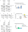
Figure 1.. pS13 HTT levels are regulated by inhibitor of nuclear factor kappa B kinase subunit beta (IKBKB) and okadaic acid–sensitive phosphatases.
(A, B) Treatment with okadaic acid (OA) of HEK293T cells overexpressing N571 Q55 HTT, with or without IKBKB or its kinase-dead mutant IKBKB KD. (A) Western blotting showing robustly increased pS13 HTT levels and increased auto-phosphorylation of IKBKB at residues S177/S181 upon IKBKB but not IKBKB KD overexpression in combination with OA treatment. Kinase expression and HTT levels were assessed using anti-FLAG and mAb 2166 antibodies, respectively. (A, B) Normalized pS13 signal on total HTT levels measured by Singulex immunoassay performed on the same lysates as in (A), confirming the Western blotting results. (Means and SDs were calculated on three biological replica. One-way analysis of variance, Dunnett’s test [***P < 0.0001]). (C, D) Western blotting of HEK293T cells overexpressing N571 Q55 HTT with or without IKBKB treated with IKBKB-inhibiting compound BAY-65-1942 at 0.1, 1, and 10 μM, without OA (C) and in combination with OA (D), demonstrating the capacity of IKBKB to increase pS13 HTT levels only in the presence of OA, and of BAY-65-1942 compound to inhibit IKBKB’s activity on pS13 in a dose-dependent way. IKBKB expression and its auto-phosphorylation were assessed using anti-FLAG and anti-pIKBKB antibodies, respectively. Protein loading and HTT levels were assessed using anti-GAPDH and mAb 2166 antibodies, respectively. (C, D, E, F) Same experimental conditions as in (C, D) evaluated on endogenous HTT in HEK293T cells confirming the overexpression results without OA (E) or in presence of OA (F). IKBKB expression and its auto-phosphorylation were assessed using anti-FLAG and anti-pIKBKB antibodies, respectively. Protein loading and HTT levels were assessed using anti-GAPDH and mAb 2166 antibodies, respectively. (C, D, E, F, G, H) Singulex immunoassay on same lysates as in (C, D, E, F) (means and SDs of n = 2 overexpressed HTT merged with n = 2 endogenous HTT experiments) without OA (G) and in combination with OA (H), confirming Western blotting results. (Means and SDs were calculated on four biological replica. One-way analysis of variance, Dunnett’s test [**P < 0.001; ***P < 0.0001]).
Figure 2.
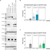
Figure 2.. Protein phosphatase 2A (PP2A) interferes on inhibitor of nuclear factor kappa B kinase subunit beta’s (IKBKB’s) capacity to phosphorylate HTT S13.
(A, B) Endogenous PP2A knockdown consequent to specific siRNA treatment in HEK293T cells transiently co-transfected with N571 Q55 HTT and IKBKB produced an increased response to IKBKB compared with siRNA control. (A) Western blotting showing that IKBKB overexpression leads to higher phosphorylation on HTT S13 when PP2A is silenced (as shown by anti-PP2A Ab). Increased auto-phosphorylation of IKBKB on S177/S181 as a result of endogenous PP2A knockdown. Protein loading, kinase and phosphatase expression, and HTT levels were assessed using anti-GAPDH, anti-FLAG, anti-PP2A, and mAb 2166 antibodies, respectively. (A, B) Normalized pS13 signal on total HTT levels measured by Singulex immunoassay performed on the same lysates as in (A) confirming Western blotting results. (Means and SDs were calculated on three biological replica. One-way analysis of variance, Dunnett’s test [*P < 0.05]). (C, D) Endogenous PP2A in HEK293T cells interacts with IKBKB. (C) Western blotting of HEK293T cells lysates overexpressing IKBKB FLAG–tagged (INPUT) pulled-down by anti-FLAG antibody (CO-IP) or unrelated anti-glial fibrillary acidic protein antibody (MOCK), and revealed by anti-FLAG and anti-PP2A antibodies. Anti-FLAG and anti-PP2A signals in CO-IP at the expected molecular weights demonstrate the PP2A–IKBKB interaction. (D) ELISA assay using anti-PP2A as capture antibody and anti-FLAG as detection antibody performed on HEK293T cell lysates transfected with/without IKBKB FLAG–tagged confirms the interaction between endogenous PP2A and IKBKB. (Means and SDs were calculated on three biological replica. T test [*P < 0.05]). (E, F) IKBKB’s capacity to influence pS13 HTT levels is affected by PP2A. (E) Western blotting of HEK293T cells overexpressing N571 Q55 HTT with or without IKBKB FLAG–tagged, PP2A FLAG–tagged or the combination of both. The lack of up-regulation of pS13 HTT levels by IKBKB when PP2A is overexpressed demonstrates that PP2A can regulate IKBKB’s activity (as confirmed by the absence of IKBKB auto-phosphorylation in the same condition) rather than dephosphorylating HTT S13 (as shown by the lack of modulation of pS13 HTT levels in the presence of PP2A). Protein loading, kinase and phosphatase expression, and HTT levels were assessed using anti-GAPDH, anti-FLAG, and mAb 2166 antibodies, respectively. (E, F) Normalized pS13 signal on total HTT levels measured by Singulex immunoassay, performed on the same lysates as in (E) confirming the Western blotting results. (Means and SDs were calculated on three biological replica. One-way analysis of variance, Dunnett’s test [*P < 0.05]).
Figure 3.
Figure 3.. Inhibitor of nuclear factor kappa B kinase subunit beta (IKBKB) effects on pS13 HTT levels are independent of IKBKB’s activity in the canonical IKK complex.
(A) Schematic representation of IKBKB mutant constructs used here. (B, C) Western blotting of HEK293T cells overexpressing N571 Q55 HTT with or without IKBKB WT and IKBKB mutants, in presence or absence of OA treatment. pS13 HTT levels increased only in the presence of the IKBKB WT, the IKBKB NBD, and the IKBKB LZ mutants when cells were treated with OA, indicating that the canonical IKK pathway is not involved in regulation of pS13 by IKBKB. Protein loading, kinases expression and activity, and HTT levels were assessed using anti-GAPDH, anti-FLAG, anti-pIKBKB, and mAb 2166 antibodies, respectively. (B, C) Normalized pS13 signal on total HTT levels measured by Singulex immunoassay, performed on the same lysates as in (B) confirming the Western blotting results. (Means and SDs were calculated on three biological replica. One-way analysis of variance, Dunnett’s test [***P < 0.0001]).
Figure 4.
Figure 4.. Inhibitor of nuclear factor kappa B kinase subunit beta (IKBKB) regulation of endogenous pS13 HTT levels is associated with an interferon regulatory factor 3 (IRF3) mediated, non-canonical IKK pathway.
(A, B) HEK293T cells overexpressing IKBKB WT and IKBKB mutants, IKBKA, IKBKG, and inhibitor of nuclear factor kappa-B kinase subunit epsilon (IKBKE) in absence of OA. (A) Western blotting showing a modest increase of endogenous pS13 HTT levels upon expression of IKBKB WT, IKBKB NBD mutant, IKBKB LZ mutant and of the non-canonical IKBKE kinase. The non-canonical IRF3 pathway is involved in pS13 HTT regulation as demonstrated by the increase of endogenous phosphor-IRF3. Kinases expression, HTT levels, and endogenous IRF3 expression were assessed using anti-FLAG, mAb 2166, and anti-IRF3 antibodies, respectively. (A, B) Normalized pS13 signal on total HTT levels measured by Singulex immunoassay, performed on the same lysates as in (A) confirming the Western blotting results. (Means and SDs were calculated on three biological replica. One-way analysis of variance, Dunnett’s test [*P < 0.05; **P < 0.001]). (C, D) HEK293T cells overexpressing IKBKB WT and IKBKB mutants, IKBKA, IKBKG, and IKBKE in the presence of OA. (A, B, C) Western blotting showing that the presence of OA greatly enhances the effects described in (A) and (B), coherent with effects on pS13 HTT being dependent on the catalytic activity of IKBKB. (D) Normalized pS13 signal on total HTT levels measured by Singulex immunoassay, performed on the same lysates as in (C) confirming the Western blotting results. (Means and SDs were calculated on three biological replica. One-way analysis of variance, Dunnett’s test [***P < 0.0001]).
Figure 5.
Figure 5.. Inhibitor of nuclear factor kappa B kinase subunit beta (IKBKB) interacts with HTT in cells in a manner dependent on its catalytic activity but independent of HTT phosphorylation at S13.
(A, B, C) Western blotting of HEK293T cells overexpressing N571 Q55 HTT WT or S13A/S16A phosphor-abrogative mutant with or without IKBKB FLAG–tagged. pS13 HTT levels, IKBKB expression, and HTT levels were assessed using anti-pS13, anti-FLAG, and mAb 2166 antibodies, respectively. (B) TR-FRET protein–protein interaction assay using 2B7-Tb/anti-FLAG–D2 antibody pair showing a strong interaction of N571 Q55 HTT WT not only with IKBKB but also with the phosphor-abrogative N571 Q55 HTT S13A/S16A mutant. (B, C) ELISA protein–protein interaction assay using 2B7 as capture and anti-FLAG as detection antibodies used as orthogonal readout confirming the data in (B). (D) ELISA protein–protein interaction assay of HEK293T cells overexpressing N571 Q55 HTT WT and IKBKB WT or IKBKB KD, IKBKB S/E, IKBKB NBD, IKBKB LZ, or IKBKA, showing that the interaction of N571 Q55 HTT with IKBKB is robustly affected only in the presence of the kinase-dead mutation and therefore this interaction is dependent on the integrity of IKBKB’s catalytic domain. (Means and SDs were calculated on three biological replica. One-way analysis of variance, Dunnett’s test [***P < 0.0001]).
Figure 6.
Figure 6.. HTT N-terminal phosphor-abrogative/mimetic mutations affect mutant HTT aggregation.
(A) Immunofluorescence of HEK293T cells overexpressing HTT Ex1 Q72-EGFP WT or phosphor-mutants (S13A/S16A, S13D/S16D, T3A, T3D, T3A/S13A/S16A, T3D/S13D/S16D), showing increased HTT aggregates in the presence of the phosphor-abrogative S13A/S16A mutation and decreased aggregation in the presence of the phosphor-mimetic S13D/S16D mutation. T3A and T3D mutations produced a comparable decrease in HTT aggregation. Expression of a triple phosphor-abrogative mutant T3A/S13A/S16A resulted in increased aggregation to a comparable degree to the double-S13A/S16A mutation, whereas the respective phosphor-mimetic triple mutant (T3D/S13D/S16D) led to the opposite result. Nuclear signal, aggregates signal, and HTT levels were assessed by DAPI staining, GFP-signal acquisition, and 2B7 antibody staining, respectively. (Representative images of n = 3 independent experiments. Scale bars represent 50 μm). B Quantitative analysis of images in A. (Means and SDs were calculated on three biological replica. One-way analysis of variance, Dunnett’s test [*P < 0.05; **P < 0.001; ***P < 0.0001]).
Figure S1.
Figure S1.. Negative controls for immunofluorescence analysis. Immunofluorescence of HEK293T cells untransfected (NT) or transfected with eGFP or Ex1 Q16-EGFP constructs reported as negative controls for high-content imaging analysis.
The overexpression of Ex1 Q16-EGFP is properly detected by 2B7 antibody staining and GFP-signal acquisition, without showing aggregates signal. Nuclear signal was assessed by DAPI staining. (Representative images of n = 3 independent experiments. Scale bars represent 50 μm).
Figure 7.
Figure 7.. Inhibitor of nuclear factor kappa B kinase subunit beta (IKBKB) influences pS13 HTT levels through cross-talk between T3 and S13.
(A) Western blotting of HEK293T cells overexpressing HTT Ex1 Q72-EGFP WT or phosphor-abrogative mutants (T3A or S13A/S16A) with or without IKBKB. Protein loading, pS13 HTT levels, pT3 HTT levels, IKBKB expression, and HTT levels were assessed using anti-GAPDH, anti-pS13, anti-pT3, anti-FLAG, and 4C9 antibodies, respectively. (A, B, C) Densitometry analysis of Western blotting in (A) showing normalized pS13 HTT levels on total HTT (B) and normalized pT3 HTT levels on total HTT (C), demonstrating that the presence of T3A mutation increases basal pS13 HTT levels and enhances the effects of IKBKB on pS13 HTT levels. (Means and SDs were calculated on three biological replica. One-way analysis of variance, Dunnett’s test [*P < 0.05; **P < 0.001]).
Figure 8.
Figure 8.. Inhibitor of nuclear factor kappa B kinase subunit beta (IKBKB) influences mutant HTT aggregation dependent on an intact catalytic site and on the presence of S13/S16 residues.
(A, B) HEK293T cells overexpressing HTT Ex1 Q72-EGFP WT and IKBKB WT or IKBKB KD, IKBKB S/E or IKBKA. (A) Immunofluorescence showing decreased mutant HTT aggregation only in the presence of WT IKBKB kinase. Aggregation levels are not modulated in the presence of the KD mutation, S/E mutation, or in the presence of IKBKA. Nuclear signal, aggregates signal, HTT levels, and kinases expression were assessed by DAPI staining, GFP-signal acquisition, 2B7 antibody, and anti-FLAG antibody staining, respectively. (Representative images of n = 3 independent experiments. Scale bars represent 50 μm). (A, B) Quantitative analysis of images in (A). (Means and SDs were calculated on at least three biological replica. One-way analysis of variance, Dunnett’s test [***P < 0.0001]). (C, D) HEK293T cells overexpressing HTT Ex1 Q72-EGFP WT or phosphor-abrogative mutants (S13A/S16A, T3A, T3A/S13A/S16A) with or without IKBKB. (C) Immunofluorescence showing decreased mutant HTT aggregation levels in the presence of IKBKB when S13/S16 residues are intact, with T3A modification further enhancing the capacity of IKBKB to reduce mutant HTT aggregation. Aggregate levels were assessed by GFP-signal acquisition and HTT levels by 2B7 antibody staining. (Representative images of n = 3 independent experiments. (A) For the HTT Ex1 Q72-EGFP WT ± IKBKB, the same images of (A) were reported. Scale bars represent 50 μm). (C, D) Quantitative analysis of images in (C). The fold change of each bar was calculated with respect to its own control without IKBKB. (Means and SDs were calculated on three biological replica. One-way analysis of variance, Dunnett’s test [*P < 0.05]).

References

    1. Aiken CT, Steffan JS, Guerrero CM, Khashwji H, Lukacsovich T, Simmons D, Purcell JM, Menhaji K, Zhu YZ, Green K, et al. (2009) Phosphorylation of threonine 3: Implications for huntingtin aggregation and neurotoxicity. J Biol Chem 284: 29427–29436. 10.1074/jbc.M109.013193 - DOI - PMC - PubMed
    1. Alpaugh M, Galleguillos D, Forero J, Morales LC, Lackey SW, Kar P, Di Pardo A, Holt A, Kerr BJ, Todd KG, et al. (2017) Disease-modifying effects of ganglioside GM1 in Huntington's disease models. EMBO Mol Med 9: 1537–1557. 10.15252/emmm.201707763 - DOI - PMC - PubMed
    1. Ansaloni A, Wang ZM, Jeong JS, Ruggeri FS, Dietler G, Lashuel HA (2014) One-pot semisynthesis of exon 1 of the huntingtin protein: New tools for elucidating the role of posttranslational modifications in the pathogenesis of Huntington's disease. Angew Chem Int Ed Engl 53: 1928–1933. 10.1002/anie.201307510 - DOI - PubMed
    1. Atwal RS, Xia J, Pinchev D, Taylor J, Epand RM, Truant R (2007) Huntingtin has a membrane association signal that can modulate huntingtin aggregation, nuclear entry and toxicity. Hum Mol Genet 16: 2600–2615. 10.1093/hmg/ddm217 - DOI - PubMed
    1. Atwal RS, Desmond CR, Caron N, Maiuri T, Xia J, Sipione S, Truant R (2011) Kinase inhibitors modulate huntingtin cell localization and toxicity. Nat Chem Biol 7: 453–460. 10.1038/nchembio.582 - DOI - PubMed

Publication types