. 2023 Aug 8;14(1):4755.
doi: 10.1038/s41467-023-40154-8.

Long-term ex situ normothermic perfusion of human split livers for more than 1 week

Affiliations

Long-term ex situ normothermic perfusion of human split livers for more than 1 week

Ngee-Soon Lau et al. Nat Commun. .

Abstract

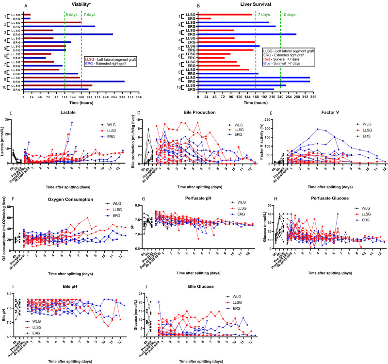
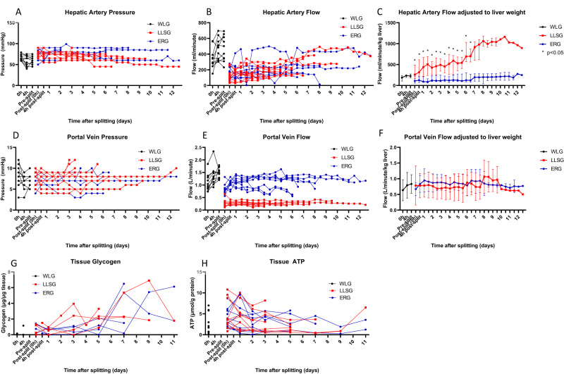
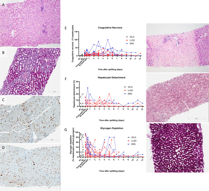
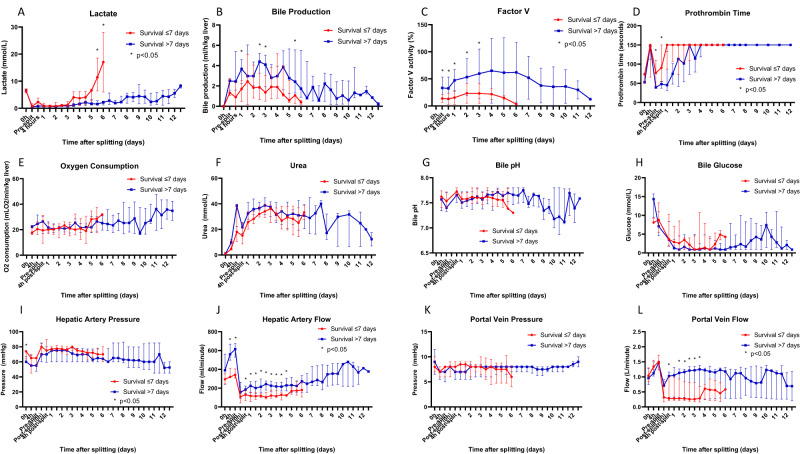
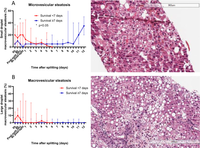
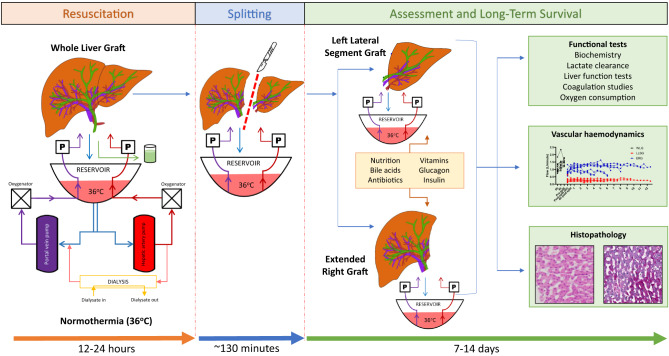
Current machine perfusion technology permits livers to be preserved ex situ for short periods to assess viability prior to transplant. Long-term normothermic perfusion of livers is an emerging field with tremendous potential for the assessment, recovery, and modification of organs. In this study, we aimed to develop a long-term model of ex situ perfusion including a surgical split and simultaneous perfusion of both partial organs. Human livers declined for transplantation were perfused using a red blood cell-based perfusate under normothermic conditions (36 °C) and then split and simultaneously perfused on separate machines. Ten human livers were split, resulting in 20 partial livers. The median ex situ viability was 125 h, and the median ex situ survival was 165 h. Long-term survival was demonstrated by lactate clearance, bile production, Factor-V production, and storage of adenosine triphosphate. Here, we report the long-term ex situ perfusion of human livers and demonstrate the ability to split and perfuse these organs using a standardised protocol.


Conflict of interest statement

The authors declare no competing interests.

Figures

Fig. 1
Fig. 1. Biochemical and functional evidence of the long-term function of human split livers.
Organ viability was continuously assessed until partial livers no longer fulfilled viability criteria* (A). Perfusion continued until organs failed (B), characterised by a lactate >10 mmol/L with a lack of bile production or unresponsive hypoglycaemia. All livers demonstrated lactate clearance (C), bile production (D), production of Factor-V (E), and evidence of oxygen consumption (F) until the point of organ failure. Perfusate pH and glucose were typically stable during perfusion until organ failure, which resulted in refractory acidosis and unresponsive hypoglycaemia (G, H). Bile pH was typically alkalotic and bile glucose was typically in the hypoglycaemic range during perfusion (I, J). *Viability according to the criteria proposed by the VITTAL clinical trial (≤2.5 mmol/L, and two or more of: bile production, pH ≥ 7.30, glucose metabolism, hepatic arterial flow ≥150 ml/min and portal vein flow ≥500 ml/min, or homogeneous perfusion).
Fig. 2
Fig. 2. Monitored haemodynamic indices and tissue ATP and glycogen during long-term perfusion of human split livers.
Using a pressure-controlled system with a goal of 60 mmHg for the hepatic artery, the ERG and LLSG typically achieved 200 ml/min of blood flow (A, B). After adjustment for liver weight, the LLSG achieved significantly higher flows/min/kg of liver than the ERG (median 316 ml/min [IQR 224–613 ml/min] vs 126 ml/min [IQR 72–209 ml/min], p = 0.003, at 4 h after splitting, Mann–Whitney U Test) (C) (n = 20 partial livers; 10 ERGs, 10 LLSGs, expressed as median (IQR)). With a goal of 8 mmHg for the portal vein, the ERG and LLSG typically achieved 1.0 L/min and 300–400 ml/min, respectively (D, E). After adjustment for liver weight, the portal vein flow was similar between ERG and LLSGs (F) (n = 20 partial livers; 10 ERGs, 10 LLSGs, expressed as median (IQR)). Hepatic tissue ATP and glycogen levels remained stable or increased during perfusion in both ERGs and LLSGs compared to baseline (G, H). ATP adenosine triphosphate, ERG extended right graft, LLSG left lateral segment graft, *p < 0.05.
Fig. 3
Fig. 3. Histopathology analysis of liver core biopsies taken throughout long-term perfusion of human split livers.
Slides were stained with haematoxylin and eosin to assess architectural integrity (A), Periodic acid-Schiff for glycogen depletion (B) and ki67 for cellular proliferation (C, D). The amount of cellular proliferation decreased from early in perfusion (C) to later in perfusion (D). Assessment of each slide was performed by a blinded specialist pathologist for coagulative necrosis (E), hepatocyte detachment (F) and glycogen depletion (G). Levels of coagulative necrosis and hepatocyte detachment remained low until the point of organ failure (E, F). Glycogen deposition within hepatocytes increased with long-term perfusion (G).
Fig. 4
Fig. 4. Factors related to the long-term survival of human split livers.
Perfusate lactate levels were not significantly different between livers that survived >7 days or ≤7 days (A). Bile production and Factor-V levels were significantly higher in the livers that survived >7 days (bile: median 3.674 ml/h/kg liver [IQR 2.247–4.576 ml/h/kg liver] vs 1.714 ml/h/kg liver [IQR 0.478–2.516 ml/h/kg liver], p = 0.008 at 24 h, Mann–Whitney U Test; Factor-V: mean 47.3 ± 19.9% vs 15.4 ± 12.7%, p < 0.001 at 24 h, unpaired two-sided t-test) (B, C). Prothrombin time was significantly shorter for livers that survived >7 days immediately before and 4 h after splitting (median 54 s [IQR 38–48 s] vs 150 s [IQR 55–91 s] at 4 h, p = 0.015, Mann–Whitney U Test) (D). Oxygen consumption, perfusate urea, bile pH and bile glucose did not demonstrate significant differences between the two groups (EH). Hepatic artery flow was significantly higher in the livers that survived >7 days for the same hepatic artery pressure (median 615 ml/min [IQR 530–674 ml/min] vs 342 ml/min [IQR 308–405 ml/min], p = 0.002, just before splitting, Mann–Whitney U Test) (I, J). Portal venous pressure was not significantly different between the two groups (K). Portal venous flow was significantly higher in the livers that survived >7 days between days 1–3 after splitting (median 1.030 ml/min [IQR 0.320–1.310 ml/min] vs 0.280 ml/min [IQR 0.220–0.970 ml/min], p = 0.049, 1 day after splitting, Mann–Whitney U Test) (L). All grouped data are presented as median (IQR) except for Factor-V, which was normally distributed and presented as mean (standard deviation), n = 20 partial livers, 9 survived >7 days, 11 survived ≤7 days. Normally distributed data and non-normally distributed data were compared at each grouped time point using an unpaired two-sided t-test and a Mann–Whitney U Test, respectively. *p < 0.05.
Fig. 5
Fig. 5. Steatosis analysis of liver core biopsies taken throughout long-term perfusion of human split livers.
Slides were stained with haematoxylin and eosin and assessed by a blinded specialist pathologist for microvesicular (A) and macrovesicular (B) steatosis. Microvesicular steatosis seen on core biopsies taken before splitting was significantly less severe in livers that survived >7 days (median 5% [IQR 0–7.5%] vs 20% [IQR 5–35%], p = 0.041 at 0 h, Mann–Whitney U Test) (A). All grouped data are presented as median (IQR), n = 20 partial livers, 9 survived >7 days, 11 survived ≤7 days, *p < 0.05.
Fig. 6
Fig. 6. Schematic diagram of our machine perfusion setup.
A commercially available organ perfusion system (Liver assist, Xvivo, Groningen, Netherlands) that uses a dual-pump system (P) and an open venous reservoir was modified for long-term perfusion by adding long-term oxygenators (A), a gas blender (B) and a dialysis filter (C).
Fig. 7
Fig. 7. Summary of experimental design for the long-term perfusion of human split livers.
Donated human livers are resuscitated under normothermic conditions for 12–24 h before a conventional split is performed. The left lateral segment graft and extended right graft are then perfused on separate perfusion machines for long-term assessment of liver function using functional tests, haemodynamic evaluation and histopathology.

References

    1. Lascaris B, de Meijer VE, Porte RJ. Normothermic liver machine perfusion as a dynamic platform for regenerative purposes What does the future have in store for us? J. Hepatol. 2022;77:825–836. doi: 10.1016/j.jhep.2022.04.033. - DOI - PubMed
    1. Mergental H, et al. Transplantation of discarded livers following viability testing with normothermic machine perfusion. Nat. Commun. 2020;11:2939. doi: 10.1038/s41467-020-16251-3. - DOI - PMC - PubMed
    1. Brüggenwirth IMA, de Meijer VE, Porte RJ, Martins PN. Viability criteria assessment during liver machine perfusion. Nat. Biotechnol. 2020;38:1260–1262. doi: 10.1038/s41587-020-0720-z. - DOI - PubMed
    1. Eshmuminov D, et al. An integrated perfusion machine preserves injured human livers for 1 week. Nat. Biotechnol. 2020;38:189–198. doi: 10.1038/s41587-019-0374-x. - DOI - PMC - PubMed
    1. Lascaris B, et al. Long-term normothermic machine preservation of human livers: what is needed to succeed? Am. J. Physiol. Gastrointest. Liver Physiol. 2022;322:G183–G200. doi: 10.1152/ajpgi.00257.2021. - DOI - PubMed

Publication types