. 2023 Aug 8;13(1):12879.
doi: 10.1038/s41598-023-40025-8.

Macular superficial vascular density on optical coherence tomography angiography in children with unilateral anisometropic and bilateral hyperopic amblyopia

Affiliations

Macular superficial vascular density on optical coherence tomography angiography in children with unilateral anisometropic and bilateral hyperopic amblyopia

Yeon Woong Chung et al. Sci Rep. .

Abstract

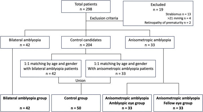
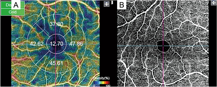
We analyzed whether macular superficial vascular density (SVD) and foveal vascular zone (FAZ) on optical coherence tomography angiography (OCTA) can distinguish between bilateral ametropic and anisometropic amblyopia. We included 42, 33, and 50 eyes in the bilateral ametropic amblyopia, anisometropic amblyopia, and normal control groups, respectively. Using macular swept-source optical coherence tomography angiography, we measured and analyzed the superficial FAZ areas and five sectoral macular SVDs after magnification correction. The anisometropic amblyopic eye group showed significantly increased foveal SVDs (p < 0.001) and significantly decreased superficial FAZ areas (p < 0.001), compared with the remaining groups. Additionally, the bilateral ametropic amblyopia group had significantly decreased nasal SVDs. SVDs and superficial FAZ areas differed among hyperopic amblyopia subtypes. These findings may reflect vascular distribution differences and macular changes in hyperopic amblyopia subtypes compared with normal eyes.


Conflict of interest statement

The authors declare no competing interests.

Figures

Figure 1
Figure 1
The sequential flow for group selection.
Figure 2
Figure 2
Representative image of (A) superficial vascular density and (B) superficial foveal avascular zone area in the ocular coherence tomography angiography.
Figure 3
Figure 3
Comparison of the foveal superficial vascular density among groups. ***P < 0.001 on post hoc analysis.
Figure 4
Figure 4
Comparison of parafoveal superficial vascular density (SVD) among groups. (A) superior, (B) inferior, (C) nasal, and (D) temporal SVDs. Only the bilateral amblyopia group had significantly decreased nasal superficial vascular density compared to the normal control group (p = 0.006), the amblyopic eye (p < 0.001), and the fellow eye group (p = 0.002) in the anisometropic amblyopia. **P < 0.01 and ***P < 0.001 when the post hoc analysis compared the two groups.
Figure 5
Figure 5
Comparison of the superficial foveal avascular zone area among groups. The area was significantly decreased in the anisometropic amblyopia group, compared with other groups. ***P < 0.001 when the post hoc analysis compared the two groups.

Similar articles

References

    1. Friedman DS, et al. Prevalence of amblyopia and strabismus in white and African American children aged 6 through 71 months the Baltimore Pediatric Eye Disease Study. Ophthalmology. 2009;116(2128–2134):e2121–2122. doi: 10.1016/j.ophtha.2009.04.034. - DOI - PMC - PubMed
    1. Williams C, et al. Prevalence and risk factors for common vision problems in children: Data from the ALSPAC study. Br. J. Ophthalmol. 2008;92:959–964. doi: 10.1136/bjo.2007.134700. - DOI - PubMed
    1. Simons K. Amblyopia characterization, treatment, and prophylaxis. Surv. Ophthalmol. 2005;50:123–166. doi: 10.1016/j.survophthal.2004.12.005. - DOI - PubMed
    1. Gopal SKS, Kelkar J, Kelkar A, Pandit A. Simplified updates on the pathophysiology and recent developments in the treatment of amblyopia: A review. Indian J. Ophthalmol. 2019;67:1392–1399. doi: 10.4103/ijo.IJO_11_19. - DOI - PMC - PubMed
    1. Martin PR, Lee BB, White AJ, Solomon SG, Ruttiger L. Chromatic sensitivity of ganglion cells in the peripheral primate retina. Nature. 2001;410:933–936. doi: 10.1038/35073587. - DOI - PubMed