

A stromal lineage maintains crypt structure and villus homeostasis in the intestinal stem cell niche
- PMID: 37553612
- PMCID: PMC10408166
- DOI: 10.1186/s12915-023-01667-2
A stromal lineage maintains crypt structure and villus homeostasis in the intestinal stem cell niche
Abstract
Background: The nutrient-absorbing villi of small intestines are renewed and repaired by intestinal stem cells (ISCs), which reside in a well-organized crypt structure. Genetic studies have shown that Wnt molecules secreted by telocytes, Gli1+ stromal cells, and epithelial cells are required for ISC proliferation and villus homeostasis. Intestinal stromal cells are heterogeneous and single-cell profiling has divided them into telocytes/subepithelial myofibroblasts, myocytes, pericytes, trophocytes, and Pdgfralow stromal cells. Yet, the niche function of these stromal populations remains incompletely understood.
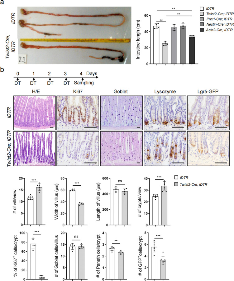
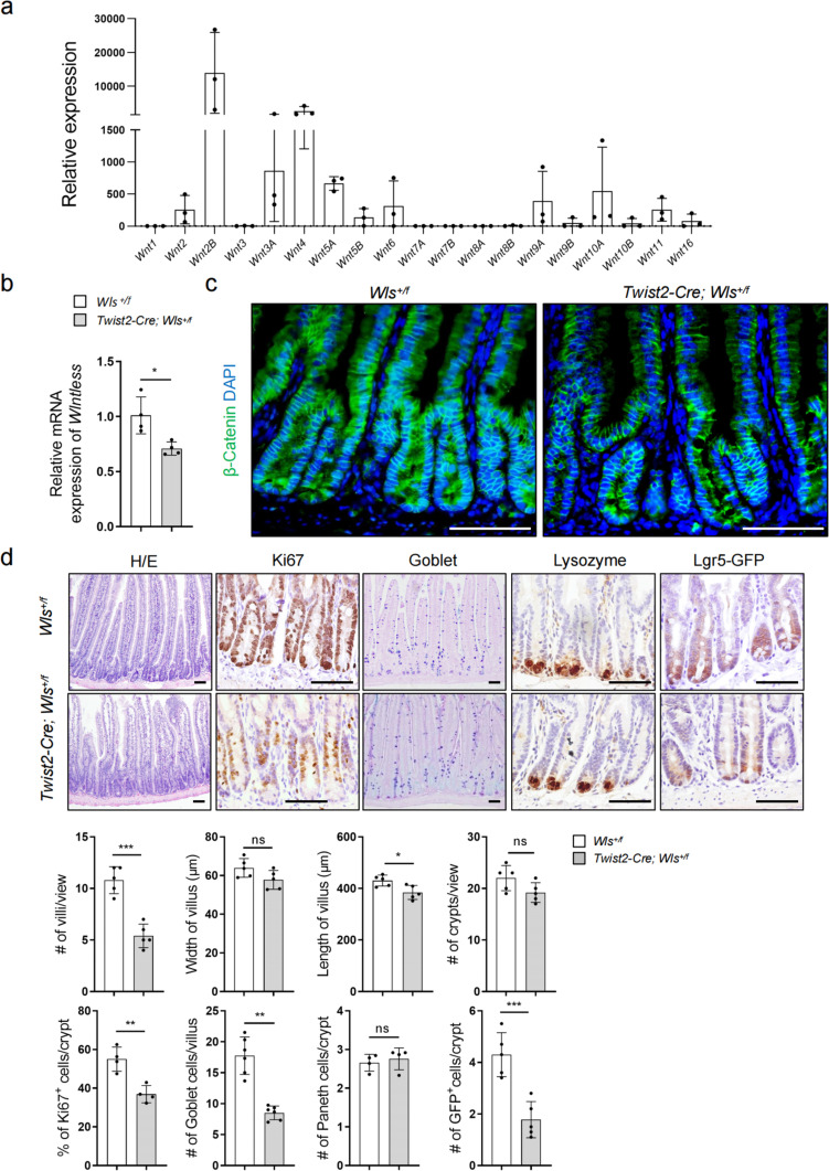
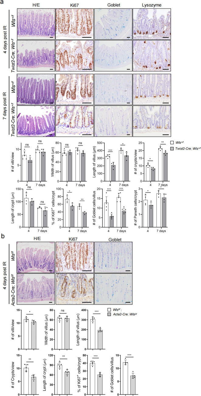
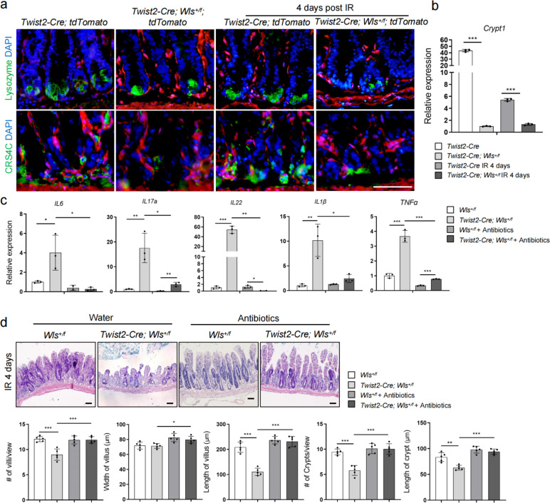
Results: We show here that a Twist2 stromal lineage, which constitutes the Pdgfralow stromal cell and trophocyte subpopulations, maintains the crypt structure to provide an inflammation-restricting niche for regenerating ISCs. Ablating Twist2 lineage cells or deletion of one Wntless allele in these cells disturbs the crypt structure and impairs villus homeostasis. Upon radiation, Wntless haplo-deficiency caused decreased production of anti-microbial peptides and increased inflammation, leading to defective ISC proliferation and crypt regeneration, which were partially rescued by eradication of commensal bacteria. In addition, we show that Wnts secreted by Acta2+ subpopulations also play a role in crypt regeneration but not homeostasis.
Conclusions: These findings suggest that ISCs may require different niches for villus homeostasis and regeneration and that the Twist2 lineage cells may help to maintain a microbe-restricted environment to allow ISC-mediated crypt regeneration.
Keywords: ISC; Inflammation; Mesenchymal; Niche; Paneth cell; Wntless.
© 2023. BioMed Central Ltd., part of Springer Nature.
Conflict of interest statement
The authors declare that they have no competing interests.
Figures


Similar articles
-
Time-resolved fate mapping identifies the intestinal upper crypt zone as an origin of Lgr5+ crypt base columnar cells.Cell. 2024 Jun 6;187(12):3039-3055.e14. doi: 10.1016/j.cell.2024.05.001. Cell. 2024. PMID: 38848677 Free PMC article.
-
Paneth cells inhibit intestinal stem cell proliferation through the bone morphogenic protein 7 pathway under rotavirus-mediated intestinal injury.World J Gastroenterol. 2025 Jul 14;31(26):107044. doi: 10.3748/wjg.v31.i26.107044. World J Gastroenterol. 2025. PMID: 40678707 Free PMC article.
-
Intestinal stem cell niche: An upcoming area of immense importance in gastrointestinal disorders.Indian J Gastroenterol. 2025 Feb;44(1):8-23. doi: 10.1007/s12664-024-01699-8. Epub 2024 Nov 8. Indian J Gastroenterol. 2025. PMID: 39514159 Review.
-
Enteroendocrine Cells Protect the Stem Cell Niche by Regulating Crypt Metabolism in Response to Nutrients.Cell Mol Gastroenterol Hepatol. 2023;15(6):1293-1310. doi: 10.1016/j.jcmgh.2022.12.016. Epub 2023 Jan 4. Cell Mol Gastroenterol Hepatol. 2023. PMID: 36608902 Free PMC article.
-
Crosstalk between hippo and Wnt pathways in intestinal stem cells regeneration.Cell Signal. 2025 Nov;135:112023. doi: 10.1016/j.cellsig.2025.112023. Epub 2025 Jul 24. Cell Signal. 2025. PMID: 40714276 Review.
Cited by
-
Circulating lung cancer exosomes damage the niche of intestinal stem cells.Transl Lung Cancer Res. 2025 Mar 31;14(3):718-735. doi: 10.21037/tlcr-24-758. Epub 2025 Mar 10. Transl Lung Cancer Res. 2025. PMID: 40248740 Free PMC article.
-
Impact of Enterococcus faecium AL41 on growth performance, immune parameters, morphology, and tight junction proteins in intestine of chicks.Poult Sci. 2025 Aug;104(8):105361. doi: 10.1016/j.psj.2025.105361. Epub 2025 May 28. Poult Sci. 2025. PMID: 40451071 Free PMC article.
-
Dietary probiotic based on a dual-strain Bacillus subtilis improves immunity, intestinal health, and growth performance of broiler chickens.J Anim Sci. 2024 Jan 3;102:skae183. doi: 10.1093/jas/skae183. J Anim Sci. 2024. PMID: 39022917 Free PMC article.
-
Fibroblast-induced mammary epithelial branching depends on fibroblast contractility.PLoS Biol. 2024 Jan 10;22(1):e3002093. doi: 10.1371/journal.pbio.3002093. eCollection 2024 Jan. PLoS Biol. 2024. PMID: 38198514 Free PMC article.
-
Skin Telocytes Could Fundament the Cellular Mechanisms of Wound Healing in Platelet-Rich Plasma Administration.Cells. 2024 Aug 8;13(16):1321. doi: 10.3390/cells13161321. Cells. 2024. PMID: 39195210 Free PMC article. Review.
References
-
- Bjerknes M, Cheng H. The stem-cell zone of the small intestinal epithelium. I. Evidence from Paneth cells in the adult mouse. Am J Anatomy. 1981;160(1):51–63. - PubMed
-
- Clevers HC, Bevins CL. Paneth cells: maestros of the small intestinal crypts. Annu Rev Physiol. 2013;75(1):289–311. - PubMed
-
- Sato T, Vries RG, Snippert HJ, van de Wetering M, Barker N, Stange DE, van Es JH, Abo A, Kujala P, Peters PJ, et al. Single Lgr5 stem cells build crypt–villus structures in vitro without a mesenchymal niche. Nature. 2009;459(7244):262–265. - PubMed
-
- El Aidy S, van den Bogert B, Kleerebezem M. The small intestine microbiota, nutritional modulation and relevance for health. Curr Opin Biotech. 2015;32:14–20. - PubMed
Publication types
MeSH terms
Substances
LinkOut - more resources
Full Text Sources
Medical
Molecular Biology Databases
Miscellaneous
