. 2024 Apr;65 Suppl 1(Suppl 1):14-24.
doi: 10.1002/em.22569. Epub 2023 Aug 25.

Genome-wide impact of cytosine methylation and DNA sequence context on UV-induced CPD formation

Affiliations

Genome-wide impact of cytosine methylation and DNA sequence context on UV-induced CPD formation

Hannah E Wilson et al. Environ Mol Mutagen. 2024 Apr.

Abstract

Exposure to ultraviolet (UV) light is the primary etiological agent for skin cancers because UV damages cellular DNA. The most frequent form of UV damage is the cyclobutane pyrimidine dimer (CPD), which consists of covalent linkages between neighboring pyrimidine bases in DNA. In human cells, the 5' position of cytosine bases in CG dinucleotides is frequently methylated, and methylated cytosines in the TP53 tumor suppressor are often sites of mutation hotspots in skin cancers. It has been argued that this is because cytosine methylation promotes UV-induced CPD formation; however, the effects of cytosine methylation on CPD formation are controversial, with conflicting results from previous studies. Here, we use a genome-wide method known as CPD-seq to map UVB- and UVC-induced CPDs across the yeast genome in the presence or absence in vitro methylation by the CpG methyltransferase M.SssI. Our data indicate that cytosine methylation increases UVB-induced CPD formation nearly 2-fold relative to unmethylated DNA, but the magnitude of induction depends on the flanking sequence context. Sequence contexts with a 5' guanine base (e.g., GCCG and GTCG) show the strongest induction due to cytosine methylation, potentially because these sequence contexts are less efficient at forming CPD lesions in the absence of methylation. We show that cytosine methylation also modulates UVC-induced CPD formation, albeit to a lesser extent than UVB. These findings can potentially reconcile previous studies, and define the impact of cytosine methylation on UV damage across a eukaryotic genome.

Keywords: 5‐methylcytosine; DNA sequence specificity; UV‐induced damage; cyclobutane pyrimidine dimers (CPDs); genome‐wide damage mapping.


Figures

Figure 1.
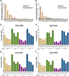
Figure 1.. CPD Formation in Non-Methylated Yeast DNA.
A. CPD-seq read counts for non-methylated wild-type yeast genomic DNA irradiated with 500J/m2 of UVB light or No UV control. B. Same as (A), but for DNA irradiated with 90J/m2 of UVC light. C-D. Normalized CPDs at different tetranucleotide contexts flanking CPD lesions at (C) TT or (D) TC dinucleotides in UVB-irradiated non-methylated yeast genomic DNA. CPD-seq read density across both UVB replicates was normalized to the tetranucleotide sequence context frequencies across the yeast genome. E-F. Same as (C-D), but for UVC-irradiated samples.
Figure 2.
Figure 2.. Validation of Methylated Yeast DNA.
A. Agarose gel electrophoresis of yeast genomic DNA samples digested with McrBC, which specifically cleaves methylcytosine-containing DNA. M.SssI is a CpG methyltransferase (MT). B. Same as panel A, except using HpaII to digest the DNA. HpaII will only cleave sites that are not methylated (i.e., no 5-methylcytosine).
Figure 3.
Figure 3.. Effect of cytosine methylation on UVB-induced CPD formation.
A. Schematic showing protocol of how the effects of CpG methylation by M.SssI CpG methyltransferase on UVB- or UVC-induced CPD formation was analyzed across the yeast genome using CPD-seq. CPD-seq schematic adapted from [30]. B. Number of CPD-seq reads associated with putative lesions at the indicated dinucleotides in UVB-irradiated methylated DNA (5mC) versus UVB-irradiated unmethylated DNA and non-UV (and unmethylated) DNA controls. UVB-irradiated unmethylated DNA data is from Fig. 1A. C. Plot showing number of CPD-seq reads in each tetranucleotide sequence context (centered on a CPD-forming dipyrimidine) in the UVB-irradiated 5-methylcytosine (5mC) DNA relative to the UVB-irradiated unmethylated (No Methyl) DNA control. Red dots represent tetranucleotide sequences that match a NYCG pattern, as these are targets for 5mC methylation by M.SssI methyltransferase. D-E. Normalized ratio of CPD-seq reads in UVB-irradiated 5-methylcytosine (5mC) DNA relative to the UVB-irradiated unmethylated (No Methyl) DNA control. Ratio was normalized so that the number of CPD-seq reads at TT dinucleotides would be the same between the two samples. D. Shows normalized ratios for lesions at TC and CC dipyrimidines, which when flanked by a 3’ guanine are targeted for methylation; E. shows normalized ratios for lesions at TT and CT dipyrimidines, which would not be methylated. The color of the bar indicates the 3’ flanking base.
Figure 4.
Figure 4.. Effect of cytosine methylation on UVB-induced CPD formation using optimized methylation protocol.
A. Agarose gel electrophoresis showing that optimized methylation protocol using M.SssI CpG methyltransferase (MT) blocks cleavage of methylated yeast genomic DNA by BstBI, which only cleaves unmethylated TTCGAA sites. CPD-seq schematic adapted from [30]. B. CPD-seq reads associated with putative lesions at the indicated dinucleotides in repeat experiment (replicate #2) of UVB-irradiated methylated DNA (5mC) versus non-UV (and unmethylated) DNA control using optimized methylation protocol. C. Plot showing number of CPD-seq reads in UVB-irradiated methylated DNA (5mC) relative to UVB-irradiated unmethylated (No Methyl) DNA control for each tetranucleotide sequence context. Red dots indicate tetranucleotides containing NYCG sequence, which can be methylated. D-E. Normalized ratio of CPD lesions in UVB-irradiated methylated DNA (5mC) relative to UVB-irradiated unmethylated (No Methyl) DNA control for each tetranucleotide sequence context. CPD counts were normalized so number of CPDs at TT dinucleotides (which cannot be methylated) were the same between the two samples. Color of the bar indicates the flanking 3’ base.
Figure 5.
Figure 5.. CPD-seq analysis of methylated DNA following UVC irradiation.
A. Plot of dinucleotide counts of putative lesions giving rise to CPD-seq reads for methylated yeast genomic DNA (5mC) irradiated with 90J/m2 of UVC light relative to unmethylated DNA that is not UV irradiated (No UV). B. Plot of CPD-seq reads associated with each tetranucleotide sequence context (centered on a dipyrimidine) for UVC-irradiated methylated DNA (5mC) relative to UVC-irradiated unmethylated (No Methyl) DNA. Red dots represent tetranucleotide sequences containing a NYCG sequence, which are targets for methylation by M.SssI methyltransferase. C-D. Normalized ratio of CPD-seq reads in UVC-irradiated methylated DNA (5mC) relative to UVC-irradiated unmethylated DNA. Normalization was performed using the number of TT CPD-seq reads in each CPD-seq library, since these should not be affected by methylation. Color of the bar indicates the flanking 3’ base.
Figure 6.
Figure 6.. Magnitude of CPD induction due to methylation depends on efficiency of CPD formation in unmethylated DNA.
A-B. Plot of methylation-dependent CPD induction (i.e., normalized ratio of CPDs in 5mC DNA relative to No methyl control) relative to frequency of CPD-seq reads in unmethylated DNA for NYCG sequence contexts in (A) UVB-irradiated (both replicates combined) or (B) UVC-irradiated yeast genomic DNA. R2 value calculated by linear regression analysis: **P < 0.001; *P < 0.05.

Similar articles

Cited by

References

    1. Alexandrov LB, Jones PH, Wedge DC, Sale JE, Campbell PJ, Nik-Zainal S and Stratton MR (2015) Clock-like mutational processes in human somatic cells. Nat Genet 47:1402–7. - PMC - PubMed
    1. Alexandrov LB, Nik-Zainal S, Wedge DC, Aparicio SA, Behjati S, Biankin AV, Bignell GR, Bolli N, Borg A, Borresen-Dale AL, Boyault S, Burkhardt B, Butler AP, Caldas C, Davies HR, Desmedt C, Eils R, Eyfjord JE, Foekens JA, Greaves M, Hosoda F, Hutter B, Ilicic T, Imbeaud S, Imielinsk M, Jager N, Jones DT, Jones D, Knappskog S, Kool M, Lakhani SR, Lopez-Otin C, Martin S, Munshi NC, Nakamura H, Northcott PA, Pajic M, Papaemmanuil E, Paradiso A, Pearson JV, Puente XS, Raine K, Ramakrishna M, Richardson AL, Richter J, Rosenstiel P, Schlesner M, Schumacher TN, Span PN, Teague JW, Totoki Y, Tutt AN, Valdes-Mas R, van Buuren MM, van 't Veer L, Vincent-Salomon A, Waddell N, Yates LR, Zucman-Rossi J, Futreal PA, McDermott U, Lichter P, Meyerson M, Grimmond SM, Siebert R, Campo E, Shibata T, Pfister SM, Campbell PJ and Stratton MR (2013) Signatures of mutational processes in human cancer. Nature 500:415–21. - PMC - PubMed
    1. Banyasz A, Esposito L, Douki T, Perron M, Lepori C, Improta R and Markovitsi D (2016) Effect of C5-Methylation of Cytosine on the UV-Induced Reactivity of Duplex DNA: Conformational and Electronic Factors. J Phys Chem B 120:4232–42. - PubMed
    1. Bohm KA, Hodges AJ, Czaja W, Selvam K, Smerdon MJ, Mao P and Wyrick JJ (2021) Distinct roles for RSC and SWI/SNF chromatin remodelers in genomic excision repair. Genome Res 31:1047–1059. - PMC - PubMed
    1. Bohm KA, Morledge-Hampton B, Stevison S, Mao P, Roberts SA and Wyrick JJ (2023) Genome-wide maps of rare and atypical UV photoproducts reveal distinct patterns of damage formation and mutagenesis in yeast chromatin. Proc Natl Acad Sci U S A 120:e2216907120. - PMC - PubMed