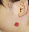
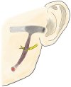
First Branchial Cleft Fistula Piercing through the Main Trunk of the Facial Nerve
- PMID: 37554142
- PMCID: PMC10406015
- DOI: 10.1097/GOX.0000000000005173
First Branchial Cleft Fistula Piercing through the Main Trunk of the Facial Nerve
Abstract
First branchial cleft fistulas are congenital malformations that result from the incomplete closure of the ectodermal portion of the first branchial cleft. These fistulas typically appear as small pits or subcutaneous masses in the upper neck and cheek and can cause pain due to infection and inflammation. Surgical excision is the most effective treatment, but special attention is necessary to avoid facial nerve injury due to the proximity of the lesion to the nerve and variations in their arrangement. Here, we report the successful treatment of a first branchial cleft fistula piercing through the main trunk of the facial nerve in a 3-year-old girl. Intraoperative findings revealed that the fistula in the parotid gland opened into the cheek area from the ear canal. Identification of the facial nerve trunk was challenging due to the malformation of the lower end of the auricular cartilage, which is an anatomical landmark of the facial nerve. The trunk of the facial nerve was divided proximally by the fistula and merged just past the fistula. Preoperative magnetic resonance is important for determining the fistula location, surrounding anatomical variations, and fistula-facial nerve arrangement. Furthermore, early surgical treatment should be considered to prevent tissue scarring and adhesion due to infection, which can lead to facial nerve injury.
Copyright © 2023 The Authors. Published by Wolters Kluwer Health, Inc. on behalf of The American Society of Plastic Surgeons.
Conflict of interest statement
The authors have no financial interest to declare in relation to the content of this article.
Figures


References
-
- Chen Z, Wang Z, Dai C. An effective surgical technique for the excision of first branchial cleft fistula: make-inside-exposed method by tract incision. Eur Arch Otorhinolaryngol. 2010;267:267–271. - PubMed
-
- Olsen KD, Maragos NE, Weiland LH. First branchial cleft anomalies. Laryngoscope. 1980;90:423–436. - PubMed
-
- Belenky WM, Medina JE. First branchial cleft anomalies. Laryngoscope. 1980;90:28–39. - PubMed
-
- Crymble B, Braithwaite F. Anomalies of the first branchial cleft. Brit J Surg. 1964;51:420–423. - PubMed
Publication types
LinkOut - more resources
Full Text Sources
Miscellaneous
