Review
. 2022 Aug 17;10(3):326-357.
doi: 10.1080/23328940.2022.2109931. eCollection 2023.

Cardiovascular responses to hot skin at rest and during exercise

Affiliations
Review

Cardiovascular responses to hot skin at rest and during exercise

Ting-Heng Chou et al. Temperature (Austin). .

Abstract

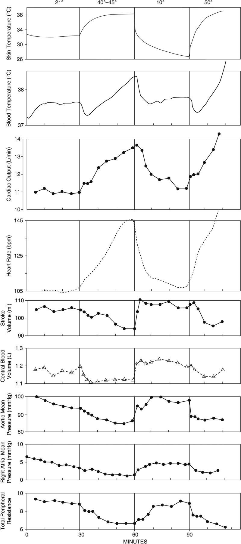
Integrative cardiovascular responses to heat stress during endurance exercise depend on various variables, such as thermal stress and exercise intensity. This review addresses how increases in skin temperature alter and challenge the integrative cardiovascular system during upright submaximal endurance exercise, especially when skin is hot (i.e. >38°C). Current evidence suggests that exercise intensity plays a significant role in cardiovascular responses to hot skin during exercise. At rest and during mild intensity exercise, hot skin increases skin blood flow and abolishes cutaneous venous tone, which causes blood pooling in the skin while having little impact on stroke volume and thus cardiac output is increased with an increase in heart rate. When the heart rate is at relatively low levels, small increases in heart rate, skin blood flow, and cutaneous venous volume do not compromise stroke volume, so cardiac output can increase to fulfill the demands for maintaining blood pressure, heat dissipation, and the exercising muscle. On the contrary, during more intense exercise, hot skin does not abolish exercise-induced cutaneous venoconstriction possibly due to high sympathetic nerve activities; thus, it does not cause blood pooling in the skin. However, hot skin reduces stroke volume, which is associated with a decrease in ventricular filling time caused by an increase in heart rate. When the heart rate is high during moderate or intense exercise, even a slight reduction in ventricular filling time lowers stroke volume. Cardiac output is therefore not elevated when skin is hot during moderate intensity exercise.

Keywords: Skin temperature; cardiovascular control; cutaneous venous volume; heat stress; hemodynamic; hyperthermia; skin blood flow.


Conflict of interest statement

No potential conflict of interest was reported by the author(s).

Figures

Figure 1.
Figure 1.
Schematic illustration of the cardiovascular responses to hot skin at rest and during submaximal dynamic exercise. (a) At rest and during mild intensity exercise, hot skin increases SBF and abolishes cutaneous venous tone which raises CVV. The extra blood to the skin is provided by an increase in CO which is achieved by an increase in HR and no changes in SV. Hot skin also causes visceral vasoconstriction which redistributes some blood from visceral vascular beds to the skin. At rest and during mild exercise (when core body temperature is relatively low), the extent of the increases in HR and CVV might be not enough to affect SV when TPR (cardiac afterload) is reduced by cutaneous vasodilation. MAP is maintained by an increase in CO while TPR is lowered. (b) During more intense exercise, hot skin increases SBF, but SBF levels off when the core body temperature is above ~38°C. Hot skin does not abolish cutaneous venoconstriction induced by intense exercise, and thus there is no significant blood pooling in the skin. Visceral vasoconstriction is higher during moderate exercise relative to mild exercise as HR is higher. HR can be raised to very high levels by hot skin which decreases ventricular filling time and SV; therefore, CO does not change when skin is hot during moderate intensity exercise. MAP maintains or only slightly decreases. CO, cardiac output; CVV, cutaneous venous volume; HR, heart rate; MAP, mean arterial pressure; SBF, skin blood flow; SV, stroke volume; TPR, total peripheral resistance.
Figure 2.
Figure 2.
Influence of skin temperature on forearm (skin) blood flow during dynamic exercise. Increasing skin temperature (Tsk) shifts the threshold of core temperature for cutaneous vasodilation to lower levels during 30–50% VO2max cycling exercise. Therefore, at a given core temperature, increasing Tsk raises skin blood flow during dynamic exercise at least before skin blood flow reaches the plateau level (i.e. when core temperature is above 38°C). Redrawn with permission from Ref [23].
Figure 3.
Figure 3.
Weighted mean skin temperature (a), esophageal temperature (b), and core-to-skin temperature gradient (C) responses during exercise with different water temperatures in the suit.
Figure 4.
Figure 4.
Cardiovascular responses during exercise with different water temperatures in the suit. (a) Cardiac output; *significant increase in 20°C. (b) Heart rate; all trials were significantly different from each other at every time point (P < 0.05). (c) Stroke volume. (d) Cutaneous blood flow; *15 min and 25 min were significantly higher than 5 min of the same trial in 20°C, 30°C, and 40°C trials (P < 0.05). (e) Forearm venous volume; the resting forearm venous volumes were significantly higher compared to exercising values of the same trial in 30°C, 40°C, and 50°C trials (P < 0.05) indicating exercise caused venoconstriction which was maintained throughout exercise bouts in all trials. Values are means ± SE (n = 8). *Significantly different from 5–10 min of the same trial; †significantly different from 15–20 min of the same trial; ‡significantly different from the 20°C trial at the same time point; §significantly different from the 30°C trial at the same time point, P < 0.05. Used with permission from Chou et al. [15].
Figure 5.
Figure 5.
Cardiovascular responses during exercise with cool skin and warm skin after precooling of core temperature. Warm skin significantly increased heart rate, cutaneous blood flow, and cardiac output while did not impair stroke volume relative to cool skin when core is pre-cooled (~35.8°C) during moderate intensity exercise. Values are means ± SE (n = 8). *Warm skin condition significantly higher than cool skin condition, P < 0.05. Used with permission from Lee et al. [45].
Figure 6.
Figure 6.
Cardiovascular responses during exercise with cool skin and warm skin after preheating of core temperature. Warm skin significantly increased heart rate and decreased stroke volume while did not affect cardiac output and cutaneous blood flow relative to cool skin when core is preheated (37.3°C) during moderate intensity exercise. Values are means ± SE (n = 8). *Warm skin condition significantly higher than cool skin condition, P < 0.05. †Main effect of skin temperature condition, P < 0.05. Used with permission from Lee et al. [45].
Figure 7.
Figure 7.
Schematic of the effect of thermal stress on the Frank. Starling relations.
Figure 8.
Figure 8.
The relationships between splanchnic and renal blood flow, heart rate, and sympathetic nervous activity. The relationships between splanchnic blood flow (SBF) or renal blood flow (RBF) presented as a percent of resting values vs. heart rate, plasma norepinephrine (NE) concentration vs. heart rate, and plasma renin activity (PRA) vs. heart rate are illustrated. Note that NE and PRA begin to rise and SBF (RBF) begins to fall at the same heart rates (i.e. at 50–60 bpm when stresses are applied at rest and at 90–100 bpm when the stress is exercising under a variety of conditions including heat stress) indicating an overall increase in sympathetic nervous activity under stress which increases heart rate and decreases SBF and RBF. Redrawn with permission from Ref [144].
Figure 9.
Figure 9.
Schematic description of the effect of dynamic exercise on skin blood flow response to hyperthermia. Exercise lowers skin blood flow relative to resting conditions at a given core body temperature by three ways: (a) an initial adrenergic cutaneous vasoconstriction at the onset of exercise, (b) an increase in core body temperature threshold at which cutaneous vasodilation begins, and (c) a plateau of skin blood flow at 50–60% maximal resting level above a core body temperature of 38°C. Redrawn with permission from Ref [14].
Figure 10.
Figure 10.
Average cardiovascular and temperature data from six subjects during mild exercise (0% grade, 2.7 or 3.5 mph) when heating and cooling the skin. During heating periods, right atrial mean pressure, central blood volume, aortic mean pressure, total peripheral resistance, and stroke volume fell suggesting that there was a displacement of blood from the central to the peripheral circulations. Blood volume was most likely redistributed from the central to the skin during heating, but skin blood flow and volume were not measured. Redrawn with permission from Ref [148].
Figure 11.
Figure 11.
Average cardiovascular and temperature data from four subjects during moderate intensity exercise (7.5% grade, 3.5 mph) when heating and cooling the skin. In contrast to mild exercise, aortic mean pressure and central blood volume increased while total peripheral resistance was only slightly reduced during heating. The right atrial mean pressure was not measured. Therefore, it seems that blood was not redistributed from the central to the skin while stroke volume still declined when heating the skin during moderate intensity exercise. Redrawn with permission from Ref [148].
Figure 12.
Figure 12.
Temperature and cardiovascular responses during moderate intensity exercise (62% VO2peak) when heating and cooling the skin with or without β-blocker ingestion. Skin temperature (Tsk) was preheated to 38°C or 33°C before exercise bouts and was rapidly cooled toward 28°C at 20 min of exercise by a water-perfused suit. Subjects ingested either β-blocker (βB) or placebo (PL) before exercise in two hot skin trials. (a) Skin temperature, (b) esophageal temperature (c) cardiac output, (d) heart rate, (e) stroke volume, (f) cutaneous blood flow, and (g) forearm venous volume responses during exercise. *Tsk 38°C-PL significantly different from Tsk 33°C-PL and Tsk 38°C-βB at the same time point, P < 0.05; †Tsk 38°C-βB significantly different from Tsk 33°C-PL at the same time point, P < 0.05; ‡significantly different from 5–10 min within trials, P < 0.05; §significantly different from 15–20 min within trials, P < 0.05; #Tsk 33°C-PL significantly different from Tsk 38°C-PL and Tsk 38°C-βB at the same time point. The shaded areas represent skin cooling from min 20 to min 40. Values are means ± SE (n = 9). Used with permission from Chou et al. [16].

Similar articles

Cited by

References

    1. Montain SJ, Coyle EF.. Influence of graded dehydration on hyperthermia and cardiovascular drift during exercise. J Appl Physiol (1985). 1992;73(4):1340–1350. - PubMed
    1. Gonzalez-Alonso J, Mora-Rodriguez R, Below PR, et al. Dehydration reduces cardiac output and increases systemic and cutaneous vascular resistance during exercise. J Appl Physiol (1985). 1995;79(5):1487–1496. - PubMed
    1. Gonzalez-Alonso J, Mora-Rodriguez R, Below PR, et al. Dehydration markedly impairs cardiovascular function in hyperthermic endurance athletes during exercise. J Appl Physiol (1985). 1997;82(4):1229–1236. - PubMed
    1. Trangmar SJ, Gonzalez-Alonso J. New insights into the impact of dehydration on blood flow and metabolism during exercise. Exerc Sport Sci Rev. 2017;45(3):146–153. - PubMed
    1. Rowell LB. Human cardiovascular adjustments to exercise and thermal stress. Physiol Rev. 1974;54(1):75–159. - PubMed

LinkOut - more resources