. 2023 Jul 15;26(8):107403.
doi: 10.1016/j.isci.2023.107403. eCollection 2023 Aug 18.

Soluble prefusion-closed HIV-envelope trimers with glycan-covered bases

Affiliations

Soluble prefusion-closed HIV-envelope trimers with glycan-covered bases

Adam S Olia et al. iScience. .

Abstract

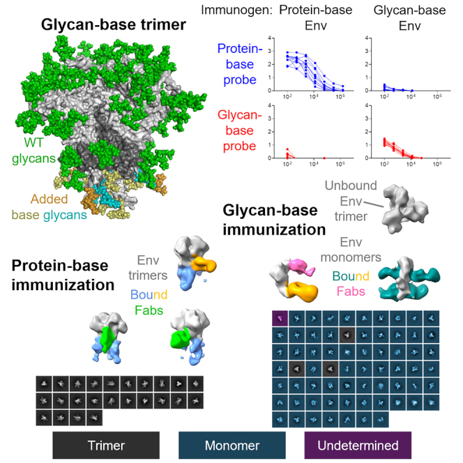
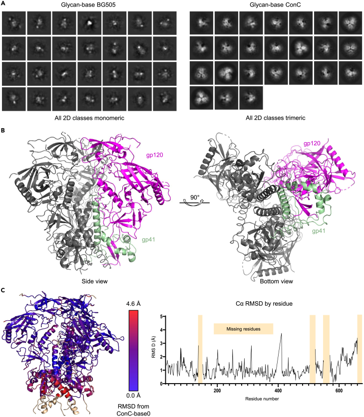
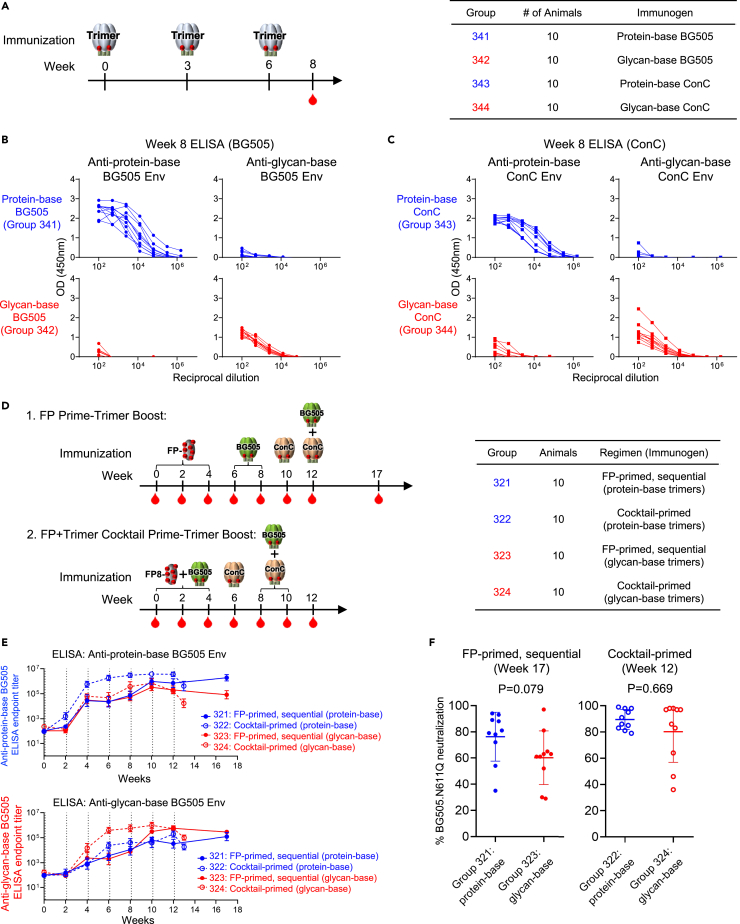
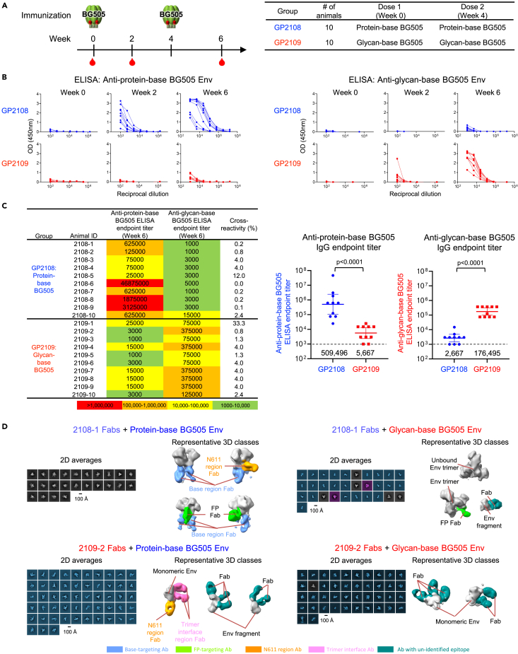
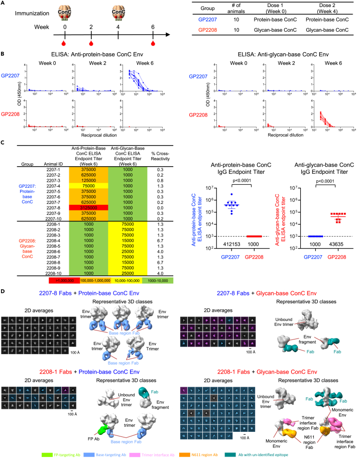
Soluble HIV-1-envelope (Env) trimers elicit immune responses that target their solvent-exposed protein bases, the result of removing these trimers from their native membrane-bound context. To assess whether glycosylation could limit these base responses, we introduced sequons encoding potential N-linked glycosylation sites (PNGSs) into base-proximal regions. Expression and antigenic analyses indicated trimers bearing six-introduced PNGSs to have reduced base recognition. Cryo-EM analysis revealed trimers with introduced PNGSs to be prone to disassembly and introduced PNGS to be disordered. Protein-base and glycan-base trimers induced reciprocally symmetric ELISA responses, in which only a small fraction of the antibody response to glycan-base trimers recognized protein-base trimers and vice versa. EM polyclonal epitope mapping revealed glycan-base trimers -even those that were stable biochemically- to elicit antibodies that recognized disassembled trimers. Introduced glycans can thus mask the protein base but their introduction may yield neo-epitopes that dominate the immune response.

Keywords: Molecular structure; Virology.


Conflict of interest statement

NIH has submitted a PCG patent application (PCT/US2023/065009) for Env trimers described in this manuscript on which A.S.O., C.C., T.Z., D.H.R., A.C., R.R., Y.Y., and P.D.K. are co-inventors. The other authors declare no competing interests.

Figures

None
Graphical abstract
Figure 1
Figure 1
Design and selection of glycan base-covered Env trimers (A) BG505 DS-SOSIP Env trimer model highlighting sequence segments that comprise the exposed protein base. (B) Close-up view and sequence information showing location of N-linked glycans introduced in each sequence segment. Italicized numbers denote sequence insertions, and “BG505” refers to protein-base BG505 trimer. (C) Matrix of 16 Env variants and antigenic criteria used for selection. The first half of the table describes the glycan sequons that were added to each construct with + or – notation. The second half of the table summarizes the antibody binding of each construct, denoted with Yes or No binding, or in red with the number of base-directed antibodies blocked from binding. Constructs in green were selected for further characterization. (D) Glycan-base BG505 trimer, modeled with six-introduced N-linked glycans. See also Table S1.
Figure 2
Figure 2
Physical properties of glycan base-covered Env trimers (A) SDS-PAGE of affinity purified glycan-base constructs from BG505 (left panel), and ConC (right panel). (B) ELISA response of sera from trimer-only or fusion peptide primed NHP immunizations assayed against selected glycan-base candidates. (C) Table showing the number of additional protein N-linked glycosylation (PNG) sites for each construct along with the percentage of furin cleavage, as determined by densitometric of the Coomassie gel in panel (A). Constructs highlighted in red were selected as the top candidates to move on for further characterization. (D) Size exclusion chromatography profiles of the two selected constructs with blue shading showing trimer containing fractions. (E) Differential scanning calorimetry scans of the two constructs, with calculated melting temperatures of 67.3°C and 76.5°C for the BG505 and ConC versions respectively. (F) Negative stain EM 2D class averages of BG505 and ConC glycan-base constructs. See also Figure S1.
Figure 3
Figure 3
Glycosylation and antigenic profiles of BG505 and ConC base-covered trimers (A) Mass spectrometry glycosylation analysis of protein-base and glycan-base constructs of BG505 and ConC. (B) Surface plasmon resonance analysis of soluble CD4 binding to glycan-base BG505 and ConC, as compared to protein-base BG505. (C) MSD antibody binding analysis of glycan-base BG505 and ConC as compared to their protein-base counterparts, all stabilized by DS-SOSIP-2G-3Mut-RnS7Mut. See also Figures S2 and S3.
Figure 4
Figure 4
Cryo-EM of ConC Glycan-Base Trimer (A) Cryo-EM 2D class averages for BG505 and ConC glycan-base constructs. The classification for the BG505 glycan-base did not reveal any trimeric classes with all particles appearing as monomers, whereas ConC showed a nearly fully trimeric appearance. (B) Cryo-EM reconstruction of the ConC glycan-base trimer at 4.1 Å resolution. (C) Color-coded representation of RMSD of glycan-base ConC from protein-base ConC (PDB: 6CK9). Increasing RMSD are colored in red, and residues that could not be resolved in the glycan-base trimer reconstruction have been modeled and colored in wheat. See also Figures S4 and S5; Table S2.
Figure 5
Figure 5
Trimer immunogenicity in mice reveals reciprocally symmetric responses (A) Immunization scheme and groups are shown for mice immunized with protein-base (blue) or glycan-base (red) variants of BG505 and ConC Env trimers. (B) Anti-trimer ELISA responses at week 8 for BG505 trimer-immunized groups and (C) ConC trimer-immunized groups. (D) Immunization schema and groups are shown for mice immunized with protein-base or glycan-base trimers using FP-primed, sequential or FP + trimer cocktail-primed vaccine regimens. (E) Longitudinal trimer-specific serum immunogenicity is shown for each group, as assessed by ELISA against protein-base BG505 trimer (top) or glycan-base BG505 trimer (bottom). Immunization timepoints are indicated by vertical dotted lines. (F) Percent neutralization against BG505.N611Q virus is shown after the final immunization using a 1:50 serum dilution for FP-primed, sequential groups (left) and cocktail-primed groups (right). Mean ± SD are shown. See also Figure S6 and Table S4.
Figure 6
Figure 6
Glycan-base BG505 trimer elicit responses capable of dissembling trimer (A) Immunization scheme and groups are shown for guinea pigs immunized with protein-base (blue) or glycan-base (red) BG505 trimer variants. (B) Anti-trimer ELISA responses at weeks 0, 2 and 6 are shown for each group against protein-base BG505 Env (left) and glycan-base BG505 Env (right). (C) Week 6 ELISA endpoint titers using a starting dilution of 1:1000 and cross-reactivity (%) of responses are listed for each animal (left) and group comparison shown for week 6 titers against each trimer (right). (D) Electron microscopy of polyclonal epitope mapping (EMPEM) of antibodies elicited by immunization. Fabs were generate from whole plasma IgG and incubated with indicated HIV-1 Env. Negative stain EM were carried out using Env-Fab complexes purified by size exclusion chromatography, 2D averages were shown to the left of each panel, false colored in gray for trimeric particles, cyan for monomeric particles, and purple for undetermined. Representative 3D reconstructions were shown to the right with HIV-1 Env or Env fragments colored in gray, and Fabs colored by potential targeting sites. See also Tables S3 and S4.
Figure 7
Figure 7
ConC trimer immunogenicity in guinea pigs reveals reciprocally symmetric responses (A) Immunization scheme and groups shown for guinea pigs immunized with protein-base (blue) or glycan-base (red) ConC Env trimers. (B) Anti-trimer ELISA responses at weeks 0, 2 and 6 are shown for each group against protein-base ConC Env (left) and glycan-base ConC Env (right). (C) Week 6 ELISA endpoint titers using a starting dilution of 1:1000 and cross-reactivity (%) of responses are listed for each animal (left) and group comparison shown for week 6 titers against each ConC trimer (right). (D) Electron microscopy of polyclonal epitope mapping (EMPEM) of antibodies elicited by immunization. Fabs were generate from whole plasma IgG and incubated with indicated HIV-1 Env. Negative stain EM were carried out using Env-Fab complexes purified by size exclusion chromatography, 2D averages were shown to the left of each panel, false colored in gray for trimeric particles, cyan for monomeric particles, and purple for undetermined. Representative 3D reconstructions were shown to the right with HIV-1 Env or Env fragments colored in gray, and Fabs colored by potential targeting sites. See also Tables S3 and S4.

References

    1. Kwong P.D., Doyle M.L., Casper D.J., Cicala C., Leavitt S.A., Majeed S., Steenbeke T.D., Venturi M., Chaiken I., Fung M., et al. HIV-1 evades antibody-mediated neutralization through conformational masking of receptor-binding sites. Nature. 2002;420:678–682. doi: 10.1038/nature01188. - DOI - PubMed
    1. McKeating J.A., McKnight A., Moore J.P. Differential loss of envelope glycoprotein gp120 from virions of human immunodeficiency virus type 1 isolates: Effects on infectivity and neutralization. J. Virol. 1991;65:852–860. doi: 10.1128/JVI.65.2.852-860.1991. - DOI - PMC - PubMed
    1. Moore J.P., McKeating J.A., Weiss R.A., Sattentau Q.J. Dissociation of gp120 from HIV-1 virions induced by soluble CD4. Science. 1990;250:1139–1142. doi: 10.1126/science.2251501. - DOI - PubMed
    1. Hraber P., Seaman M.S., Bailer R.T., Mascola J.R., Montefiori D.C., Korber B.T. Prevalence of broadly neutralizing antibody responses during chronic HIV-1 infection. AIDS. 2014;28:163–169. doi: 10.1097/QAD.0000000000000106. - DOI - PMC - PubMed
    1. Kwon Y.D., Pancera M., Acharya P., Georgiev I.S., Crooks E.T., Gorman J., Joyce M.G., Guttman M., Ma X., Narpala S., et al. Crystal structure, conformational fixation and entry-related interactions of mature ligand-free HIV-1 Env. Nat. Struct. Mol. Biol. 2015;22:522–531. doi: 10.1038/nsmb.3051. - DOI - PMC - PubMed

LinkOut - more resources