

Fetus in Fetu: A Rare Congenital Anomaly Diagnosed Postnatally by Ultrasonography and MRI
- PMID: 37554598
- PMCID: PMC10405024
- DOI: 10.7759/cureus.41550
Fetus in Fetu: A Rare Congenital Anomaly Diagnosed Postnatally by Ultrasonography and MRI
Abstract
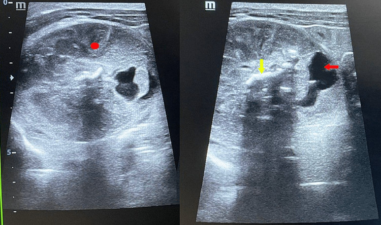
Fetus in fetu (FIF) is a rare congenital anomaly with two controversial theories regarding its embryogenesis. Although it is an extremely rare condition, it should be kept in mind as a differential diagnosis while evaluating children with abdominal calcification. Radiological findings on postnatal days 2 and 5 of a neonate with an antenatal scan showing an abdominal mass in the fetus are described here. Ultrasonography and magnetic resonance imaging (MRI) revealed the mass in which the contents favored a diagnosis of the FIF. Characteristic features of FIF on MRI have been less explored and knowledge regarding the same will be of immense help to the radiologist. Complete surgical excision followed by histopathology confirmed the diagnosis.
Keywords: abdominal ultrasonography; fetus-in-fetu; mri - magnetic resonance imaging; pediatric teratoma; preoperative diagnosis.
Copyright © 2023, M. et al.
Conflict of interest statement
The authors have declared that no competing interests exist.
Figures


Similar articles
-
Fetus-in-fetu: mimicking teratoma on antenatal ultrasound.Case Rep Perinat Med. 2023 Mar 10;12(1):20220024. doi: 10.1515/crpm-2022-0024. eCollection 2023 Jan. Case Rep Perinat Med. 2023. PMID: 40041259 Free PMC article.
-
Preoperative diagnosis of a "humanoid" fetus in fetu using multimode ultrasound: a case report.BMC Pediatr. 2020 Oct 19;20(1):483. doi: 10.1186/s12887-020-02389-y. BMC Pediatr. 2020. PMID: 33076884 Free PMC article.
-
Diagnostic dilemma in a neglected case of fetus-in-fetu solved with Magnetic Resonance Imaging and MDCT--a case report and review of literature.J Radiol Case Rep. 2011;5(10):29-37. doi: 10.3941/jrcr.v5i10.833. Epub 2011 Oct 1. J Radiol Case Rep. 2011. PMID: 22470767 Free PMC article. Review.
-
Fetus-in-Fetu: A Differential Diagnosis of Neonatal Fetiform Encysted Abdominal Mass.Cureus. 2023 Jan 12;15(1):e33725. doi: 10.7759/cureus.33725. eCollection 2023 Jan. Cureus. 2023. PMID: 36793819 Free PMC article.
-
Fetus in fetu: two case reports and literature review.BMC Pediatr. 2014 Apr 2;14:88. doi: 10.1186/1471-2431-14-88. BMC Pediatr. 2014. PMID: 24693883 Free PMC article. Review.
Cited by
-
An uncommon presentation of abdominal mass in an infant: A case report of fetus in fetu.Radiol Case Rep. 2025 Apr 17;20(7):3331-3334. doi: 10.1016/j.radcr.2025.03.073. eCollection 2025 Jul. Radiol Case Rep. 2025. PMID: 40297260 Free PMC article.
References
-
- Foetus-in-foetu. Grant P, Pearn JH. Med J Aust. 1969;1:1016–1019. - PubMed
-
- Fetus in fetu removal in a 47-year-old man. Dagradi AD, Mangiante GL, Serio GE, Musajo FG, Menestrina FV. https://www.surgjournal.com/article/0039-6060(92)90266-3/fulltext. Surgery. 1992;112:598–602. - PubMed
Publication types
LinkOut - more resources
Full Text Sources