Review
. 2023 Jul 25;9(8):e18653.
doi: 10.1016/j.heliyon.2023.e18653. eCollection 2023 Aug.

Caveolin-1 in endothelial cells: A potential therapeutic target for atherosclerosis

Affiliations
Review

Caveolin-1 in endothelial cells: A potential therapeutic target for atherosclerosis

Yan Shu et al. Heliyon. .

Abstract

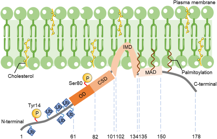
Atherosclerosis (AS) is a chronic vascular disease characterized by lipid accumulation and the activation of the inflammatory response; it remains the leading nation-wide cause of death. Early in the progression of AS, stimulation by pro-inflammatory agonists (TNF-α, LPS, and others), oxidized lipoproteins (ox-LDL), and biomechanical stimuli (low shear stress) lead to endothelial cell activation and dysfunction. Consequently, it is crucial to investigate how endothelial cells respond to different stressors and ways to alter endothelial cell activation in AS development, as they are the earliest cells to respond. Caveolin-1 (Cav1) is a 21-24-kDa membrane protein located in caveolae and highly expressed in endothelial cells, which plays a vital role in regulating lipid transport, inflammatory responses, and various cellular signaling pathways and has atherogenic effects. This review summarizes recent studies on the structure and physiological functions of Cav1 and outlines the potential mechanisms it mediates in AS development. Included are the roles of Cav1 in the regulation of endothelial cell autophagy, response to shear stress, modulation of the eNOS/NO axis, and transduction of inflammatory signaling pathways. This review provides a rationale for proposing Cav1 as a novel target for the prevention of AS, as well as new ideas for therapeutic strategies for early AS.

Keywords: Atherosclerosis; Autophagy; Caveolin-1; Endothelial cells; Inflammation; eNOS/NO axis.


Conflict of interest statement

The authors declare that they have no known competing financial interests or personal relationships that could have appeared to influence the work reported in this paper.

Figures

Fig. 1
Fig. 1
Schematic representation of Cav1 with membrane topology. Cav1 is a membrane protein of 178aa in length with an IMD (102-134aa) inserted into the plasma membrane, and both the N and C termini are expressed in the cytoplasm to form a unique hairpin loop structure. Cav1 contains six lysine sites (Lys5/26/30/39/47/57) at the N terminus that can be modified for ubiquitination; it is modified by phosphorylation at the tyrosine 14 and serine 80 sites, with three palmitoylation sites (Cys133/143/156) in the C-terminal structural domain and MAD (135-150aa) for membrane insertion. Its OD (61-101aa) is an oligomeric structural domain directing the homo-zwitterionization of Cav1 molecules and hetero-oligomerization of cholesterol, fatty acids, and others. The CSD (82-101aa) is located within the OD structural domain and Cav1 exerts different biological functions by binding to different proteins through the CSD. OD, oligomerization domain; CSD, caveolin scaffolding domain; IMD, intramembrane domain; MAD, membrane attachment domain.
Fig. 2
Fig. 2
Mechanism of Cav1 induced endothelial dysfunction leading to AS. The main features of atherosclerosis are lipid accumulation and chronic inflammatory activation. When endothelial cells sense nutrient depletion, they activate mTORC1 signaling and inhibit AMPK signaling, thereby inhibiting the formation of ULK1 and Class III PI3K complexes, preventing the formation of autophagic phagocytic vesicles and thus inhibiting autophagy, resulting in reduced levels of protective intracellular autophagy to degrade excess cholesterol and damaged organelles, and reduced degradation of Cav1 through reduced autophagy, leading to increased LDL transcytosis across the endothelial cells. When endothelial cells respond to shear stress stimulation leading to a decrease in glycocalyx and a decrease in caveolae, the co-localization of caveolae with eNOS-pS1177 is impaired thereby attenuating NO signaling. Activation of TLR4 by LSS and thus NOX2 induces an increase in Cav1 phosphorylation leading to eNOS inactivation, while inflammatory cytokines induce inflammation in endothelial cells upregulating eNOS signaling leading to a massive accumulation of NO leading to a state of oxidative stress in endothelial cells. Activation of the TLR4-MyD88 axis by LPS increases Cav1 expression, promoting the downstream NF-κB inflammatory signaling pathway. At the same time, Cav1 also mediates the activation of the JNK signaling pathway after Ox-LDL treatment, and the increased release of inflammatory cytokines such as IL-6, ICAM-1, and TNF-α leads to an inflammatory state in endothelial cells. Ox-LDL disrupts the balance between eNOS and inflammation-induced iNOS in endothelial cells and increases NO overproduction in response to superoxide anion (O2•-) to produce peroxynitrite (ONOO-); meanwhile, Ox-LDL can activate Cav1 expression through TLR4 signaling to induce NF-κB ectopic entry into the nucleus to regulate iNOS transcription, leading to increased endothelial cell injury and apoptosis. In conclusion, endothelial cell dysplasia induces endothelial cell dysfunction, promoting early atherosclerosis formation. mTORC1: mechanistic target of rapamycin complex 1; AMPK: AMP-activated protein kinase; PI3K: phosphoinositide 3-kinase; ULK1: unc-51 like autophagy activating kinase 1; eNOS: endothelial nitric oxide synthase; iNOS: inducible nitric oxide synthase; TLR4: Toll-like receptor 4; LSS: low shear stress; NOX2: NADPH oxidase 2; MYD88: myeloid differentiation primary response 88; LPS: lipopolysaccharide; JNK: c-Jun N-terminal kinase; ox-LDL: oxidized LDL; IL-6: Interleukin 6; ICAM, intercellular cell adhesion molecule; TNF-α: tumor necrosis factor alpha; NF-κB, nuclear factor kappa-B.

References

    1. Libby P. The changing landscape of atherosclerosis. Nature. 2021;592(7855):524–533. doi: 10.1038/s41586-021-03392-8. - DOI - PubMed
    1. Tsao C.W., Aday A.W., Almarzooq Z.I., Alonso A., Beaton A.Z., Bittencourt M.S., Boehme A.K., Buxton A.E., Carson A.P., Commodore-Mensah Y., Elkind M.S.V., Evenson K.R., Eze-Nliam C., Ferguson J.F., Generoso G., Ho J.E., Kalani R., Khan S.S., Kissela B.M., Knutson K.L., Levine D.A., Lewis T.T., Liu J., Loop M.S., Ma J., Mussolino M.E., Navaneethan S.D., Perak A.M., Poudel R., Rezk-Hanna M., Roth G.A., Schroeder E.B., Shah S.H., Thacker E.L., VanWagner L.B., Virani S.S., Voecks J.H., Wang N.Y., Yaffe K., Martin S.S. Heart disease and stroke statistics-2022 update: a report from the American heart association. Circulation. 2022;145(8):e153–e639. doi: 10.1161/cir.0000000000001052. - DOI - PubMed
    1. Björkegren J.L.M., Lusis A.J. Atherosclerosis: recent developments. Cell. 2022;185(10):1630–1645. doi: 10.1016/j.cell.2022.04.004. - DOI - PMC - PubMed
    1. Arora S., Stouffer G.A., Kucharska-Newton A.M., Qamar A., Vaduganathan M., Pandey A., Porterfield D., Blankstein R., Rosamond W.D., Bhatt D.L., Caughey M.C. Twenty year trends and sex differences in young adults hospitalized with acute myocardial infarction. Circulation. 2019;139(8):1047–1056. doi: 10.1161/circulationaha.118.037137. - DOI - PMC - PubMed
    1. Collaborators GMaCoD Global, regional, and national age-sex specific all-cause and cause-specific mortality for 240 causes of death, 1990-2013: a systematic analysis for the Global Burden of Disease Study 2013. Lancet (London, England) 2015;385(9963):117–171. doi: 10.1016/s0140-6736(14)61682-2. - DOI - PMC - PubMed