. 2023 Oct;10(29):e2302658.
doi: 10.1002/advs.202302658. Epub 2023 Aug 9.

Topical Drug Delivery of Concentrated Cabazitaxel in an α-Tocopherol and DMSO Solution

Affiliations

Topical Drug Delivery of Concentrated Cabazitaxel in an α-Tocopherol and DMSO Solution

Boyang Sun et al. Adv Sci (Weinh). 2023 Oct.

Abstract

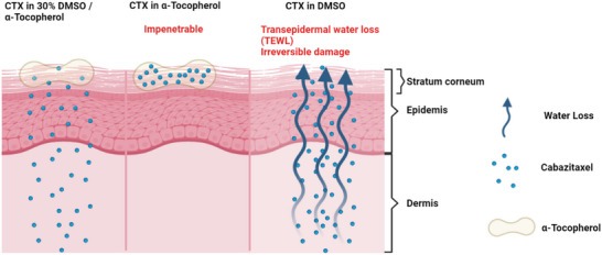
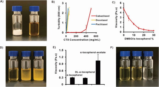
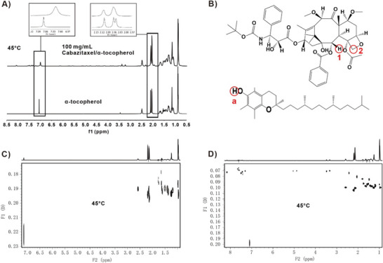
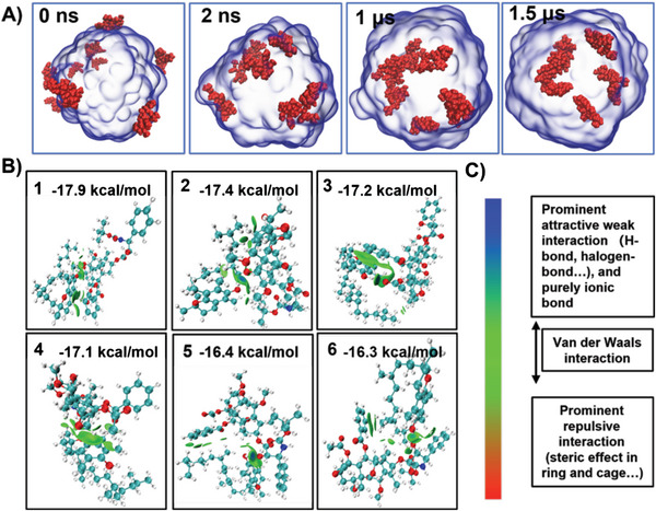
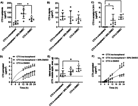
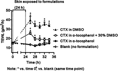
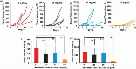
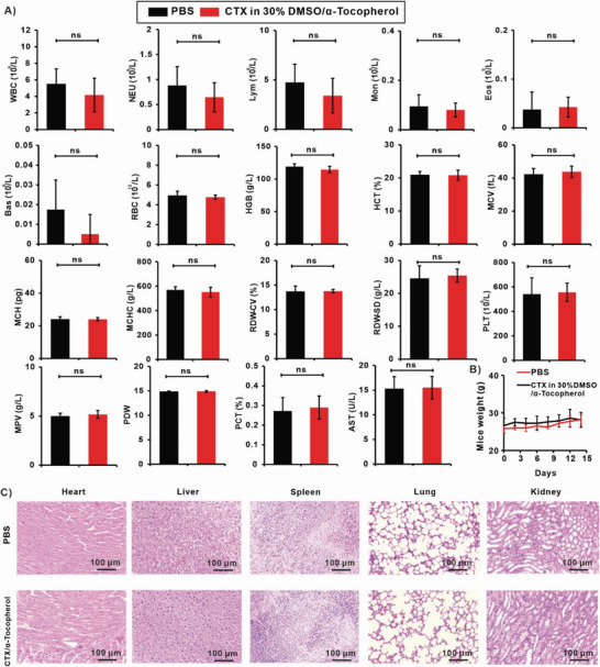
Topical chemotherapy approaches are relevant for certain skin cancer treatments. This study observes that cabazitaxel (CTX), a broad-spectrum second-generation taxane cytotoxic agent, can be dissolved in α-tocopherol at high concentrations exceeding 100 mg mL-1 . 2D nuclear magnetic resonance (NMR) analysis and molecular dynamics (MD) are used to study this phenomenon. The addition of 30% dimethyl sulfoxide (DMSO) to the α-tocopherol/CTX solution improves its working viscosity and enhances CTX permeation through human skin in vitro (over 5 µg cm-2 within 24 h), while no detectable drug permeates when CTX is dissolved in α-tocopherol alone. In a transepidermal water loss assay, the barrier impairment induced by CTX in 30% DMSO in α-tocopherol, but not in pure DMSO, is reversible 8 h after the formulation removal from the skin surface. Antitumor efficacy of the topical CTX formulation is evaluated in nude mice bearing A431 human squamous carcinoma skin cancer xenografts. With topical application of concentrated CTX solutions (75 mg mL-1 ), tumor growth is significantly suppressed compared to lower concentration groups (0, 25, or 50 mg mL-1 CTX). Taken together, these findings show that topical delivery of CTX using a DMSO and α-tocopherol solvent warrants further study as a treatment for skin malignancies.

Keywords: cabazitaxel; skin cancer; transdermal delivery; α-tocopherol.


Conflict of interest statement

The authors declare no conflict of interest.

Figures

Figure 1
Figure 1
Schematic illustration of the therapeutic effect of CTX in α‐tocopherol, CTX in 30% DMSO/α‐tocopherol and CTX in DMSO, respectively. CTX in α‐tocopherol is unable to penetrate the skin layer while CTX in DMSO causes the transepidemal water loss (TEWL) in the skin.
Figure 2
Figure 2
CTX can be dissolved in α‐tocopherol (Vitamin E) at high concentrations and solution viscosity can be adjusted with DMSO. A) Photograph of 100 mg of CTX powder (left) and 100 mg CTX powder dissolved in 1 mL α‐tocopherol (right). B) The turbidity (optical absorbance at 600 nm) of taxanes (CTX, DTX, PTX) dissolved in α‐tocopherol. C) The viscosity α‐tocopherol with varying amounts of DMSO added. D) Photograph of 100 mg of CTX (left) DTX (middle) PTX (right) in 30% DMSO/ α‐tocopherol. E) Viscosity or F) photographs of 100 mg CTX dissolved in 30% DMSO/α‐tocopherol, 30% DMSO/ DL‐α‐tocopherol, 30% DMSO/ α‐tocopherol‐Acetate. Data shows mean ± S.D. for n = 3.
Figure 3
Figure 3
2‐D DOSY Nuclear Magnetic Resonance analysis of CTX/α‐tocopherol interaction. A) Stacked 1H‐NMR spectra of α‐tocopherol (bottom) and 100 mg mL−1 CTX/ α‐tocopherol (top) in 30% DMSO‐d 6 at 45 °C. B) Chemical structure of CTX and α‐tocopherol. C) 2D‐DOSY spectra of α‐tocopherol in 30% DMSO‐d 6 (500 MHz, 45 °C). D) 2D‐DOSY spectra of 100 mg mL−1 CTX / α‐tocopherol in 30% DMSO‐d 6 (500 MHz, 45 °C).
Figure 4
Figure 4
A) Molecular dynamics simulations of CTX dissolving in α‐tocopherol at 0, 2, 1000, and 1500 ns. The drug molecules (red) gradually dissolve into the α‐tocopherol solvent (blue). B) The six conformations in terms of free energy of binding are mostly green at the interaction interface. By combining the colorimetric card and the description in C) Intermolecular interactions are found to be mainly van der Waals interactions.
Figure 5
Figure 5
CTX permeation through human skin and retention in skin layers in vitro. CTX detected in A) human epidermis, B) dermis, and C) acceptor compartment after the application of 40 µL of 100 mg mL−1 CTX in α‐tocopherol, α‐tocopherol with 30% DMSO, and DMSO for 24 h (n = 6, * significant at p < 0.05, ***p < 0.001). (40 µL of the formulation applied on 1 cm2 skin); D) CTX permeation kinetics profiles from α‐tocopherol (n = 3), α‐tocopherol with 30% DMSO (n = 5), and DMSO (n = 6, means and S.D.). E) Cumulative amount of CTX that permeated through the skin in 24 h. F) CTX permeation profiles from a second permeation experiment with different skin donor and sampling intervals.
Figure 6
Figure 6
The effects of the formulations on TEWL and the irreversibility of that effect. Means and S.D., *p < 0.05 versus time 0; + p < 0.05 versus blank at the same time point; n = 5 (blank), n = 3 (α‐tocopherol), n = 5 (30% DMSO), n = 6 (DMSO).
Figure 7
Figure 7
In vivo tumor inhibition efficacy on mice bearing subcutaneous A431 human skin cancer xenografts. A) Tumor volume change after received 20 µL of topical 0, 25, 50, and 75 mg mL−1 CTX in 30% DMSO/ α‐tocopherol on day 3 and 10. Arrow indicates drug administration dates. Statistically significant difference (ANOVA) was test for the tumors on B). day 7 and C). day 14 with ANOVA and Turkey's post‐test (*p < 0.5, **p < 0.05.).
Figure 8
Figure 8
Systemic toxicity analysis of CTX formulations: CD‐1 Mice received transdermal administration of 20 µL 75 mg mL−1 CTX in 30% DMSO/α‐tocopherol at day 0 and 7 for toxicity analysis experiments (n = 5), at day 14 mice were sacrificed for toxicity analysis. A) Mice blood routine blood test and serum chemistry profile. B) Mice weight was monitored and recorded for 14 days. C) The H&E staining analysis of major organs. Statistical significance was analyzed via one‐way ANOVA with Tukey's post‐test: ns p > 0.05.

Similar articles

Cited by

References

    1. Rembielak A., Ajithkumar T., Clin. Oncol. 2019, 31, 735. - PubMed
    1. Ciążyńska M., Kamińska‐Winciorek G., Lange D., Lewandowski B., Reich A., Sławińska M., Pabianek M., Szczepaniak K., Hankiewicz A., Ułańska M., Morawiec J., Błasińska‐Morawiec M., Morawiec Z., Piekarski J., Nejc D., Brodowski R., Zaryczańska A., Sobjanek M., Nowicki R. J., Owczarek W., Słowińska M., Wróbel K., Bieniek A., Woźniacka A., Skibińska M., Narbutt J., Niemczyk W., Ciążyński K., Lesiak A., Sci. Rep. 2021, 11, 4337. - PMC - PubMed
    1. Simões M. C. F., Sousa J. J. S., Pais A., Cancer Lett. 2015, 357, 8. - PubMed
    1. Pfeifer G. P., Besaratinia A., Photochem. Photobiol. Sci. 2012, 11, 90. - PMC - PubMed
    1. Khan M. A., Pandit J., Sultana Y., Sultana S., Ali A., Aqil M., Chauhan M., Drug Deliv. 2015, 22, 795. - PubMed

LinkOut - more resources