Review
. 2023 Sep;483(3):281-298.
doi: 10.1007/s00428-023-03590-x. Epub 2023 Aug 9.

Emerging entities: high-grade/large B-cell lymphoma with 11q aberration, large B-cell lymphoma with IRF4 rearrangement, and new molecular subgroups in large B-cell lymphomas. A report of the 2022 EA4HP/SH lymphoma workshop

Affiliations
Review

Emerging entities: high-grade/large B-cell lymphoma with 11q aberration, large B-cell lymphoma with IRF4 rearrangement, and new molecular subgroups in large B-cell lymphomas. A report of the 2022 EA4HP/SH lymphoma workshop

Leticia Quintanilla-Martinez et al. Virchows Arch. 2023 Sep.

Abstract

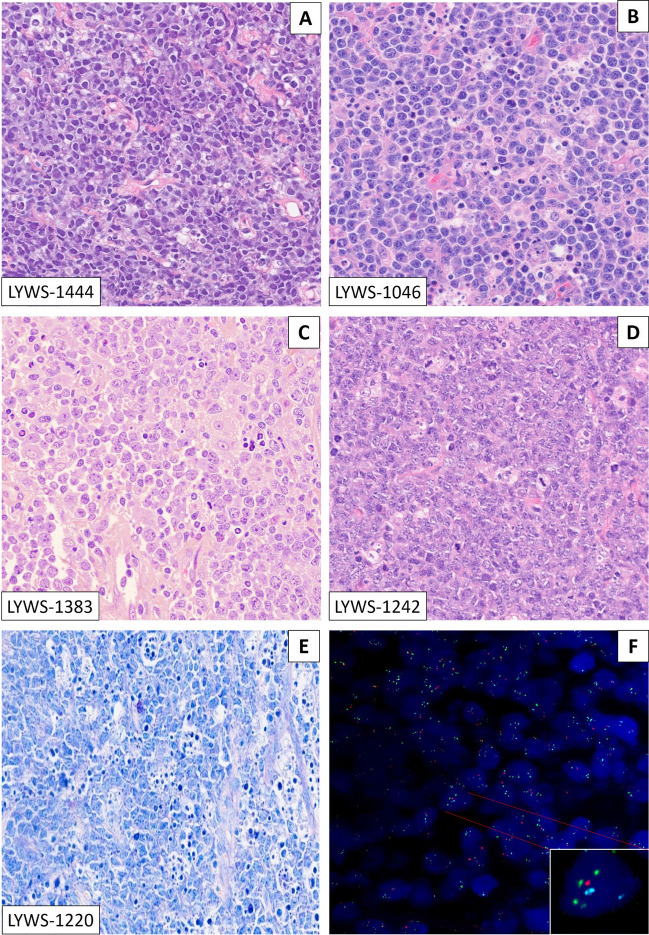
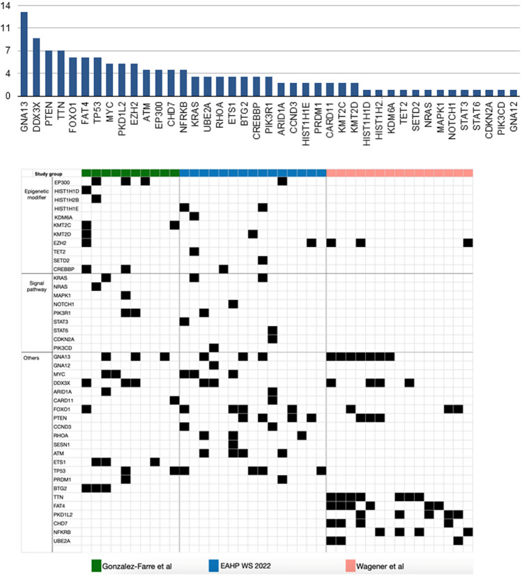
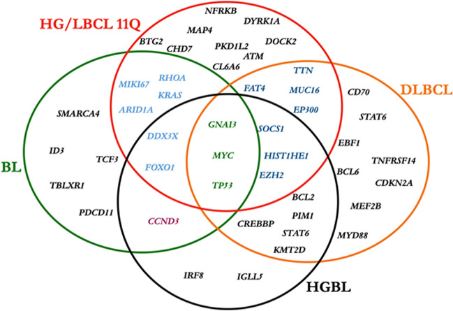
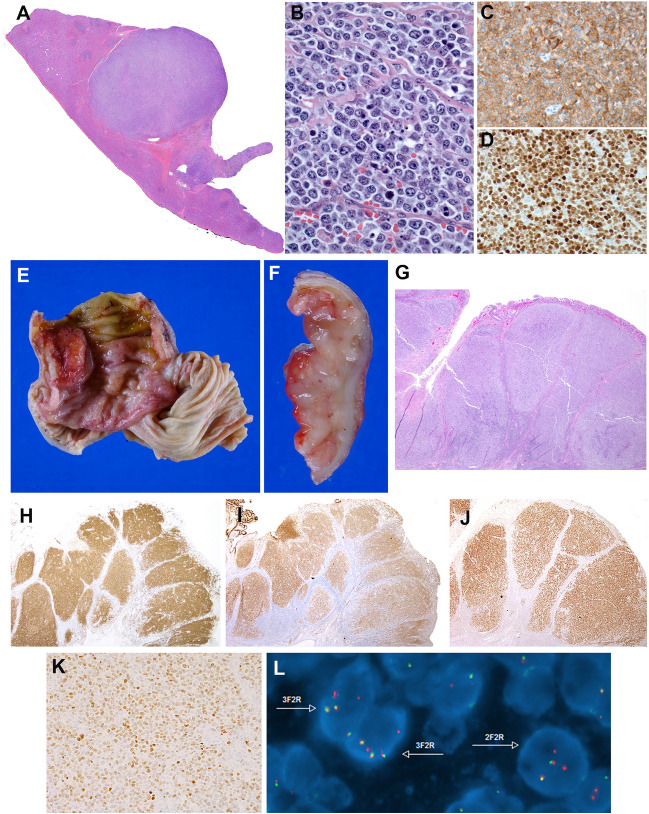
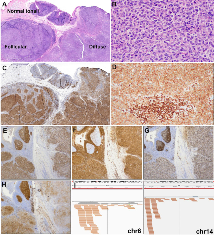
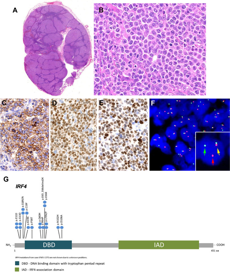
Emerging entities and molecular subgroups in large B-cell lymphomas (LBCLs) were discussed during the 2022 European Association for Haematopathology/Society for Hematopathology workshop in Florence, Italy. This session focused on newly recognized diseases and their diagnostic challenges. High-grade/large B-cell lymphoma with 11q aberration (HG/LBCL-11q) is defined by chromosome 11q-gains and telomeric loss. FISH analysis is recommended for the diagnosis. HG/LBCL-11q can occur in the setting of immunodeficiency, including ataxia-telangiectasia, and predominates in children. The morphological spectrum of these cases is broader than previously thought with often Burkitt-like morphology and coarse apoptotic bodies. It has a Burkitt-like immunophenotype (CD10+, BCL6+, BCL2-) but MYC expression is weak or negative, lacks MYC rearrangement, and is in contrast to Burkitt lymphoma 50% of the cases express LMO2. LBCL with IRF4 rearrangement (LBCL-IRF4) occurs mainly in the pediatric population but also in adults. LBCL-IRF4 has an excellent prognosis, with distinguishing molecular findings. IRF4 rearrangements, although characteristic of this entity, are not specific and can be found in association with other chromosomal translocations in other large B-cell lymphomas. Other molecular subgroups discussed included primary bone diffuse large B-cell lymphoma (PB-DLBCL), which has distinctive clinical presentation and molecular findings, and B-acute lymphoblastic leukemia (B-ALL) with IGH::MYC translocation recently segregated from Burkitt lymphoma with TdT expression. This latter disorder has molecular features of precursor B-cells, often tetrasomy 1q and recurrent NRAS and KRAS mutations. In this report, novel findings, recommendations for diagnosis, open questions, and diagnostic challenges raised by the cases submitted to the workshop will be discussed.

Keywords: 11q aberration; B-ALL with MYC-R; CCND1-R in DLBCL; High-grade/large B-cell lymphoma; IRF4-rearrangement; Plasmablastic transformation; Primary bone lymphoma.


Conflict of interest statement

The authors declare no competing interests.

Figures

Fig. 1
Fig. 1
The morphological spectrum of high-grade/large B-cell lymphoma with 11q aberration: the morphological spectrum varied from blastoid (A) to cases with intermediate morphology, ranging from cases very similar to BL (B) to other more similar to DLBCL (C); only one case was submitted with typical large cell morphology (D). Starry-sky pattern with conspicuous, coarse apoptotic debris was detected in 5 cases. E FISH analysis showed 11q gain/loss in all cases (F). AD H&E stain; E Giemsa stain; F FISH analysis with ZytoLight® SPEC 11q gain/loss Triple Color Probe
Fig. 2
Fig. 2
Mutational profile of HG/LBCL-11q cases submitted to the workshop. The mutational landscape of the workshop cases showed similarities but also difference with previous studies. Mutations in GNA13, DDX3X, MYC, KRAS, EZH2, CREBBP, and FOXO1 were confirmed and new mutations in ATM, CCND3, and RHOA were identified. Typical Burkitt lymphoma driver mutations (ID3 or TCF3 were not detected). The comparison was performed with the studies of Gonzalez-Farre et al. [21] and Wagener et al. [22]
Fig. 3
Fig. 3
Comparison of the mutational patterns of Burkitt lymphoma (BL), high-grade/large B-cell lymphoma (HG/LBCL)-11q, high-grade B-cell lymphoma (HGBL) wit double/triple hit, and diffuse large B-cell lymphoma (DLBCL). Mutations in GNA13, MYC, and TP53 are shared by BL, HG/LBCL-11q, HGBL, and DLBCL. BTG2 and DYRK1A gene mutations are considered typical for HG/LBCL-11q
Fig. 4
Fig. 4
Histologic and immunophenotypic features of large B-cell lymphoma with IRF4-rearrangement presenting in extranodal sites. AD Case LYWS-1163 courtesy of E. Shuyu. A Spleen section showing a well-circumscribed nodular infiltration. B The tumor is composed of large centroblastic lymphoid cells with open chromatin, several large nuclei, and abundant cytoplasm. C The tumor cells are CD10 and D IRF4/MUM1 strongly positive. EL Case LYWS-1112 courtesy of D. Wang. E Terminal ileum with a 2.2-cm large polypoid mass. F The intestinal crossed section shows a white soft mass infiltrating the mucosa, submucosa, and the muscularis propria. G H&E section reveals a follicular lymphoid infiltrate with large, back-to-back follicles. The follicles are composed of medium to large-sized centroblasts. H The tumor cells are positive for CD79a, I CD10, J BCL6, and K IRF4/MUM1. L Interphase FISH analysis using break apart probes for IRF4. Most cells have 3 fusion signals (yellow) and 2 red signals with loss of the green signals indicating an IRF4 translocation
Fig. 5
Fig. 5
Histologic and immunophenotypic features of large B-cell lymphoma with IRF4-rearrangement presenting in the tonsil. Case LYWS-1279 courtesy of R. Mariani. A Panoramic view of a tonsil showing residual normal tonsil areas and lymphoma infiltration with follicular and diffuse pattern. B Higher magnification shows a lymphoid infiltrate composed of large-sized centroblasts. C IRF4/MUM1 stain shows in the normal residual lymphoid tissue few positive plasma cells. The left side shows a follicular growth pattern whereas the left side reveals a diffuse growth pattern. D The tumor cells show an aberrant CD5 expression. Note the strong CD5 expression of the reactive T cells. E The tumor cells are positive for BCL6, and F BCL2. G The MIB1 stain shows the normal polarization of residual germinal centers, whereas the tumor shows a proliferation of approximately 80% in both the follicular and in the diffuse areas. H The CD10 stain is strong and homogeneous positive in the residual germinal center whereas the stain is weak in the follicular areas and partially lost in the diffuse areas. I IGV screenshots of chimeric pairs in chromosomes 6 and 14 supporting the presence of IGH::IRF4 juxtaposition
Fig. 6
Fig. 6
Histologic and immunophenotypic features of large B-cell lymphoma with IRF4-rearrangement in an adult patient. Case LYWS-1370 courtesy of B. Bisig. A Panoramic view of a lymph node with preserved capsule and effacement of the architecture by the presence of multiple, large confluent nodules. No diffuse areas or necrosis are observed. B Higher magnification demonstrates that the nodules are composed of large cells with fine chromatin, multiple nucleolei, abundant cytoplasm typical of centroblasts. Some apoptotic figures and some mitosis are observed. C The tumor cells were CD10 positive as well as D BCL6 and E IRF4/MUM1. F Interphase FISH analysis using break apart probes for IRF4 revealed 1 fusion signal (yellow arrow), one red signal (red arrow), and one green signal (green arrow) indicative of an IRF4 rearrangement. G Diagram of the relative positions of IRF4 driver mutations. The approximate location of somatic mutations identified is indicated. The analysis was performed by next generation sequencing. IRF4 mutations are mainly in the DNA binding domain (DBD). Domains of the protein are represented according to the Uniprot database (www.uniprot.org)
Fig. 7
Fig. 7
Histologic, immunophenotype, and cytogenetic features of a B-cell acute lymphoblastic leukemia with IGH::MYC according to the 2022 ICC. Case LYWS-1026 courtesy of G. Caponetti. A CT scan demonstrates supradiaphragmatic lymphadenopathy (red arrow). B CT scan reveals a soft tissue lesion expanding the posterior aspect of the left iliacus muscle (red arrow) C PET scan shows intense FDG uptake by the soft tissue mass (red arrow). D The bone marrow aspirate shows 81% blasts with vacuolated cytoplasm typical of Burkitt lymphoma. E The bone marrow biopsy is hypercellular for age and shows a diffuse infiltrate with relatively large blastoid cells. F The tumor cells are positive for CD19. G CD10, H TdT, and I CD34 are negative. J CD10 is strongly positive in the tumor cells. K BCL2 is negative, whereas L MYC is strongly positive. M The MIB1 stain shows a 100% proliferation. N The karyotype analysis reveals an abnormal male karyotype with a supernumerary isochromosome for the entire long arm of chromosome 1, which results in tetrasomy 1q (black arrow head). Additionally, a balanced translocation between chromosome 8 and 14 is observed with breakpoints at bands 8q24.2 and 14q32 resulting in a IGH::MYC translocation. O Interphase FISH analysis using break apart probes for MYC, BCL2, and BCL6 reveals in MYC 1 fusion signal (yellow arrow), one red signal (red arrow), and one green signal (green arrow) indicative of MYC rearrangement. In contrast, the analyses of BCL2 and BCL6 demonstrate two normal fusion signals (yellow arrows)
Fig. 8
Fig. 8
Primary diffuse large B-cell lymphoma of the bone (AB). Case LYWS-1231 courtesy of Y-Ch. Liu. AB Imaging studies (MRI) of the left femur shows a lobulated mass extended from the mid diaphysis into the distal epiphysis including both the medial and lateral femoral condyles. B The bone biopsy showed a polymorphic infiltrate predominantly of large cells confirming the diagnosis of diffuse large B-cell lymphoma. CL Diffuse large B-cell lymphoma with CCND1 rearrangement. Case LYWS-1380 courtesy of K.S. Kurz. C Lymph node biopsy with effaced architecture by a diffuse infiltrate of predominantly medium-sized to large blast cells. D Giemsa stain reveals that the tumor cells have basophilic cytoplasm and round to oval nuclei with open chromatin and prominent nucleolus. E Cyclin D1 is positive in the tumor cells. F In other areas of the lymph node the tumor cells were cyclin D1 negative. G CD5 stain is positive in the reactive T cells but negative in the tumor cells. H SOX11 remains negative. I MUM1 is positive in the majority of tumor cells whereas CD10 remained negative (not shown). J Ki-67 demonstrate a high proliferation rate. KL Interphase FISH analysis using break apart probes for CCND1 and MYC reveals 1 fusion signal (yellow arrow), one red signal (red arrow), and one green signal (green arrow) indicative of CCND1 (K) and MYC (L) rearrangements

References

    1. Gascoyne RD, Campo E, Jaffe ES, et al. et al. Diffuse large B-cell lymphoma, NOS. In: Swerdlow SH, Campo E, Harris NL, et al.et al., editors. WHO classification of tumours of haematopoetic and lymphoid tissues. Lyon: International Agency for Research on Cancer; 2017.
    1. Swerdlow SH, Campo E, Pileri SA, et al. The 2016 revision of the World Health Organization classification of lymphoid neoplasms. Blood. 2016;127(20):2375–2390. - PMC - PubMed
    1. Song JY, Dirnhofer S, Piris MA, Quintanilla-Martinez L, Pileri S, Campo E. Diffuse large B-cell lymphomas, not otherwise specified, and emerging entities. Virchows Arch. 2023;482(1):179–192. - PubMed
    1. Alizadeh AA, Eisen MB, Davis RE, et al. Distinct types of diffuse large B-cell lymphoma identified by gene expression profiling. Nature. 2000;403(6769):503–511. - PubMed
    1. Pedersen MO, Gang AO, Poulsen TS, et al. MYC translocation partner gene determines survival of patients with large B-cell lymphoma with MYC- or double-hit MYC/BCL2 translocations. Eur J Haematol. 2014;92(1):42–48. - PubMed