. 2023 Oct;41(5):688-698.
doi: 10.1007/s10637-023-01386-z. Epub 2023 Aug 9.

Cytotoxicity of poly-guanidine in medulloblastoma cell lines

Affiliations

Cytotoxicity of poly-guanidine in medulloblastoma cell lines

Gabriel Gallo-Oller et al. Invest New Drugs. 2023 Oct.

Abstract

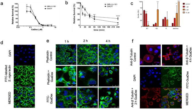
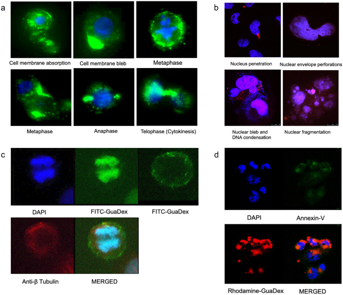
Medulloblastoma (MB) is the most common pediatric brain tumor. The therapy frequently causes serious side effects, and new selective therapies are needed. MB expresses hyper sialylation, a possible target for selective therapy. The cytotoxic efficacy of a poly guanidine conjugate (GuaDex) incubated with medulloblastoma cell cultures (DAOY and MB-LU-181) was investigated. The cells were incubated with 0.05-8 µM GuaDex from 15 min to 72 h. A fluorometric cytotoxicity assay (FMCA) measured the cytotoxicity. Labeled GuaDex was used to study tumor cell interaction. FITC-label Sambucus nigra confirmed high expression of sialic acid (Sia). Immunofluorescence microscopy was used to visualize the cell F-actin and microtubules. The cell interactions were studied by confocal and fluorescence microscopy. Annexin-V assay was used to detect apoptosis. Cell cycle analysis was done by DNA content determination. A wound-healing migration assay determined the effects on the migratory ability of DAOY cells after GuaDex treatment. IC50 for GuaDex was 223.4 -281.1 nM. FMCA showed potent growth inhibition on DAOY and MB-LU-181 cells at 5 uM GuaDex after 4 h of incubation. GuaDex treatment induced G2/M phase cell cycle arrest. S. nigra FITC-label lectin confirmed high expression of Sia on DAOY medulloblastoma cells. The GuaDex treatment polymerized the cytoskeleton (actin filaments and microtubules) and bound to DNA, inducing condensation. The Annexin V assay results were negative. Cell migration was inhibited at 0.5 µM GuaDex concentration after 24 h of incubation. GuaDex showed potent cytotoxicity and invasion-inhibitory effects on medulloblastoma cells at low micromolar concentrations. GuaDex efficacy was significant and warrants further studies.

Keywords: Cell migration; Cytoskeleton polymerization; Cytotoxicity efficacy; DAOY; DNA condensation; MB-LU-181; Poly-guanidine; Polyamines; Sialic acid.


Conflict of interest statement

GG, TDS, AA, SN, and MM-M declare no conflict of interest. ARH is the inventor of the poly-guanidine construct and the CEO of DexTech Medical (publ.), the owner of the corresponding patents.

Figures

Fig. 1
Fig. 1
FMCA cytotoxicity assay: a DAOY and MB-LU-181 MB incubated 72 h with GuaDex at 0.5–8 µM concentration. b DAOY and MB-LU-181 MB cells incubated 4 h with GuaDex at 5 µM concentration. c. Flow cytometry: GuaDex concentration-dependent arrest of DAOY cells in G2/M cell cycle phase, 24 h incubation. Confocal microscopy: d DAOY cells, Sialic acid (Sia) expression, FITC labeled S. nigra lectin. e DAOY cells incubated for 1- 4 h with GuaDex at 5 µM concentration. Stabilization of actin filaments. f Stabilization of microtubule filaments at DAOY cells after incubation 2 and 4 h with GuaDex at 5 µM concentration. DAPI staining (blue), FITC-labeled S. nigra lectin (green) FITC-GuaDex (green), Alexa Fluor® 488 phalloidin (green), Alexa Fluor® 594 Anti-β Tubulin (red), Magnification 40x
Fig. 2
Fig. 2
Fluorescent microscope images of DAOY cells. a 5 µM FITC-Guadex, 4 h incubation. FITC-Guadex cell membrane absorption, formation of blebs, binding to microtubule structures during cell cycle phases. DAPI (blue), and FITC-GuaDex (green). Magnification 63x. b Confocal microscopy images, nucleus, and DNA damage in DAOY cells incubated with Rhodamine-GuaDex at 5 µM concentration after 24 h incubation. Rhodamine-GuaDex (red) and DAPI nucleus staining (blue). Magnification 63x. c Immunofluorescent microscope images of DAOY cell in G2/M after 4 h incubation with FITC-GuaDex at 5 µM concentration. FITC-GuaDex (green) is binding with microtubule (red) and chromatin (blue). Magnification 60x. d Confocal microscope images. DAOY cells were incubated with Rhodamine-GuaDex (red) at 5 µM concentration for 1 h, Annexin-V assay (green). GuaDex appeared negative in the Annexin V apoptosis assay. Magnification 40x
Fig. 3
Fig. 3
Guadex inhibitory growth effect on DAOY medulloblastoma cell lines after 24-h incubation at 0.25 to 1 µM. a Inverted microscope images, b Cell migration measured after the scratch assay, as described by Bobadilla et al. [31]. The distance migrated was monitored, and the area was measured manually by the area method. The cell migration rate was indirectly evaluated as the percentage of wound area at a specific time

References

    1. Millard NE, De Braganca KC. Medulloblastoma. J Child Neurol. 2016;31(12):1341–1353. doi: 10.1177/0883073815600866. - DOI - PMC - PubMed
    1. Louis DN, Perry A, Wesseling P, et al. The 2021 WHO Classification of Tumors of the Central Nervous System: a summary. Neuro Oncol. 2021;23(8):1231–1251. doi: 10.1093/neuonc/noab106. - DOI - PMC - PubMed
    1. Rolland A, Aquilina K. Surgery for recurrent medulloblastoma: a review. Neurochirurgie. 2021;67(1):69–75. doi: 10.1016/j.neuchi.2019.06.008. - DOI - PubMed
    1. Northcott PA, Robinson GW, Kratz CP, et al. Medulloblastoma Nat Rev Dis Primers. 2019;5(1):11. doi: 10.1038/s41572-019-0063-6. - DOI - PubMed
    1. Packer RJ, Zhou T, Holmes E, et al. Survival and secondary tumors in children with medulloblastoma receiving radiotherapy and adjuvant chemotherapy: results of Children’s Oncology Group trial A9961. Neuro Oncol. 2012;15:97–103. doi: 10.1093/neuonc/nos267. - DOI - PMC - PubMed

Publication types

LinkOut - more resources