. 2023 Dec;46(6):2386-2401.
doi: 10.1007/s10753-023-01885-4. Epub 2023 Aug 9.

Regulating NLRP3 Inflammasome-Induced Pyroptosis via Nrf2: TBHQ Limits Hyperoxia-Induced Lung Injury in a Mouse Model of Bronchopulmonary Dysplasia

Affiliations

Regulating NLRP3 Inflammasome-Induced Pyroptosis via Nrf2: TBHQ Limits Hyperoxia-Induced Lung Injury in a Mouse Model of Bronchopulmonary Dysplasia

Minrong Wang et al. Inflammation. 2023 Dec.

Abstract

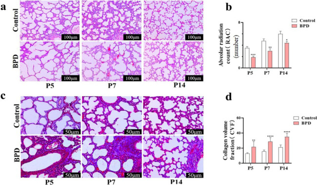
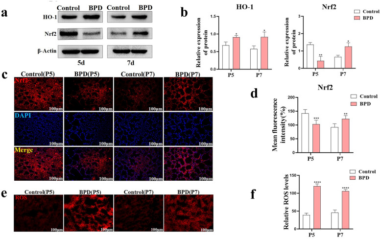
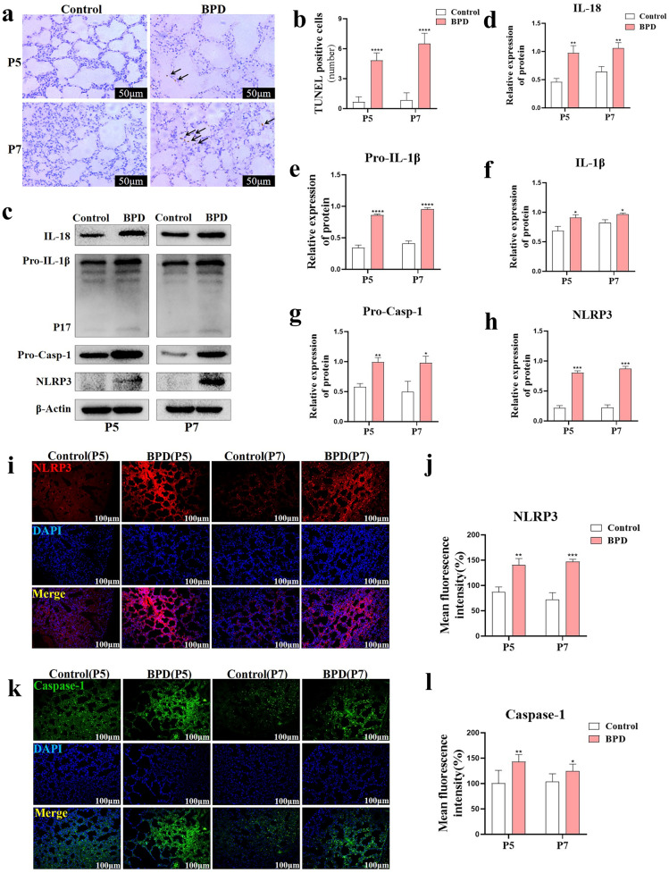
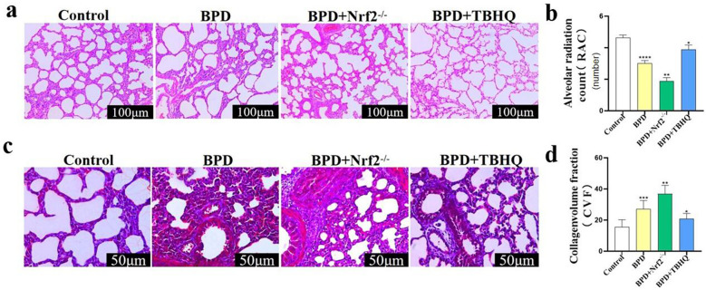
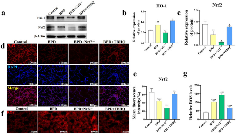
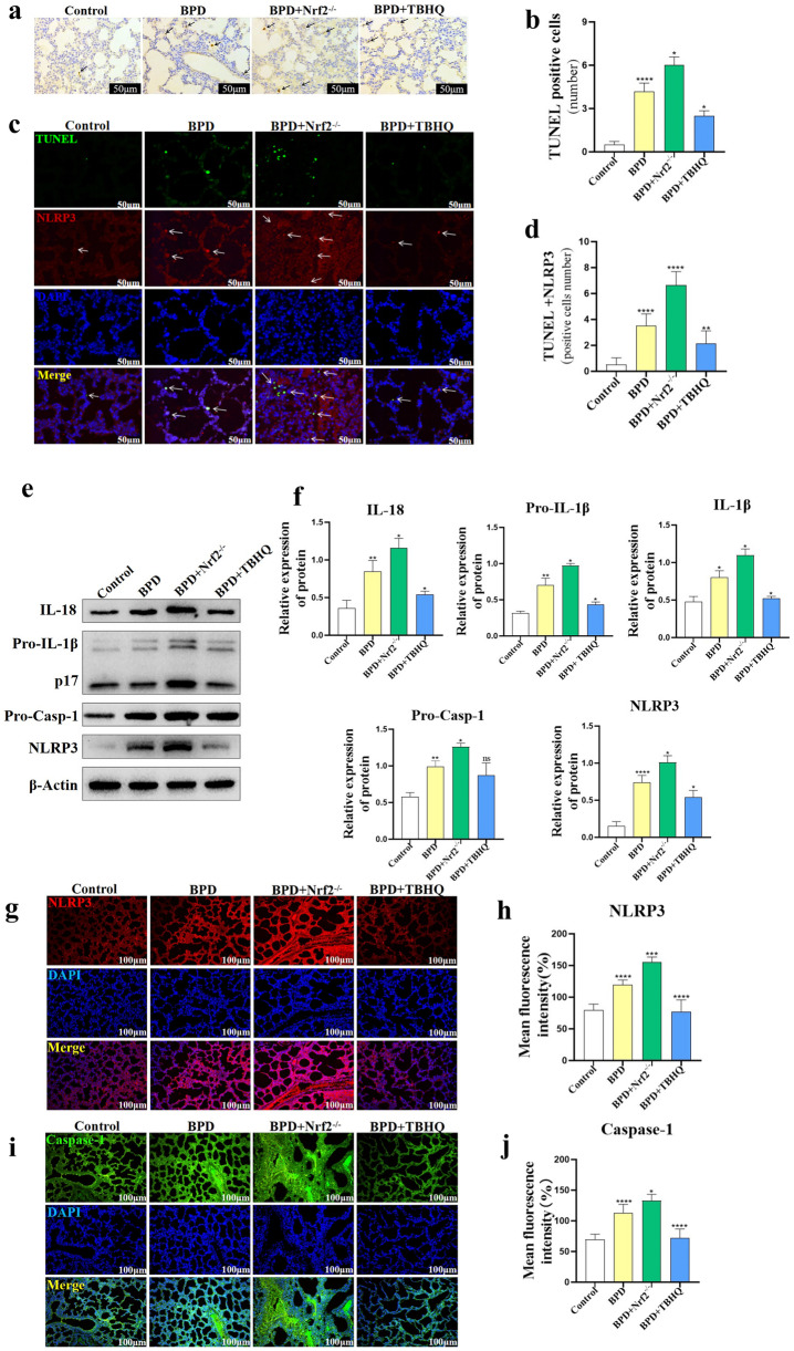
Nuclear factor e2-related factor 2 (Nrf2) plays a key role in cellular resistance to oxidative stress injury. Oxidative stress injury, caused by Nrf2 imbalance, results in increased pyroptosis, DNA damage, and inflammatory activation, which may lead to the arrest of alveolar development and bronchopulmonary dysplasia (BPD) in premature infants under hyperoxic conditions. We established a BPD mouse model to investigate the effects of tert-butylhydroquinone (TBHQ), an Nrf2 activator, on oxidative stress injury, pyroptosis, NLRP3 inflammasome activation, and alveolar development. TBHQ reduced abnormal cell death in the lung tissue of BPD mice and restored the number and normal structure of the alveoli. TBHQ administration activated the Nrf2/heme oxygenase-1 (HO-1) signaling pathway, resulting in the decrease in the following: reactive oxygen species (ROS), activation of the NOD-like receptor pyrin domain containing 3 (NLRP3) inflammasome, and IL-18 and IL-1β expression and activation, as well as inhibition of pyroptosis. In contrast, after Nrf2 gene knockout in BPD mice, there was more severe oxidative stress injury and cell death in the lungs, there were TUNEL + and NLRP3 + co-positive cells in the alveoli, the pyroptosis was significantly increased, and the development of alveoli was significantly blocked. We demonstrated that TBHQ may promote alveolar development by enhancing Nrf2-induced antioxidation in the lung tissue of BPD mice and that the decrease in the NLRP3 inflammasome and pyroptosis caused by Nrf2 activation may be the underlying mechanism. These results suggest that TBHQ is a promising treatment for lung injury in premature infants with hyperoxia.

Keywords: NLRP3; Nrf2; ROS; TBHQ; bronchopulmonary dysplasia; pyroptosis.


Conflict of interest statement

The authors declare no competing interests.

Figures

Fig. 1
Fig. 1
The hyperoxia-induced BPD neonatal mouse model was established. a, b The pathological changes of lung tissue at P5, P7, and P14 in the control and BPD groups were evaluated with H&E staining, and RAC statistical analysis was performed. Data are expressed as the mean ± SEM for n = 6 mice/group (one asterisk denotes P < 0.05; two asterisks denote P < 0.01; three asterisks denote P < 0.001). Scale bars = 100 μm. c, d The changes in lung collagen fibers at P5, P7, and P14 in the control and BPD groups were evaluated by Masson’s staining, and CVF quantitative analysis was performed. Data are expressed as mean ± SEM for n = 6 mice/group (one asterisk denotes P < 0.05; three asterisks denote P < 0.001). Scale bars = 50 μm.
Fig. 2
Fig. 2
Nrf2 antioxidant factor and oxidative stress imbalance in the BPD group. a, b The expression of Nrf2 and HO-1 after hyperoxia treatment was detected by western blot with β-actin as the reference, and quantitative analysis was performed. Data are expressed as the mean ± SEM for n = 6 mice/group (one asterisk denotes P < 0.05; two asterisks denote P < 0.01). c, d Immunofluorescence was used to detect the expression of Nrf2 (Cy3 red) in lung tissues of the control and BPD groups at P5 and P7, and the difference in average fluorescence intensity was analyzed. The nuclei were stained with DAPI (blue). Data are expressed as the mean ± SEM for n = 6 mice/group (two asterisks denote P < 0.01; three asterisks denote P < 0.001). Scale bars = 100 μm. e, f DHE-ROS was used to detect and quantify ROS content in lung tissue of the control and BPD groups at P5 and P7. Data are expressed as the mean ± SEM for n = 6 mice/group (two asterisks denote P < 0.01; three asterisks denote P < 0.001; four asterisks denote P < 0.0001). Scale bars = 100 μm.
Fig. 3
Fig. 3
Hyperoxia-induced BPD mice have increased pyroptosis induced by the NLRP3 inflammasome. a, b TUNEL in situ staining was used to observe and analyze the effect of hyperoxia on TUNEL-positive cells in the lung tissue of neonatal mice. Data are expressed as the mean ± SEM for n = 6 mice/group (four asterisks denote P < 0.0001). ch Western blotting was used to detect NLRP3, Caspase-1, IL-18, and IL-1β in the lung tissues of the control and BPD groups at P5 and P7. Data are expressed as the mean ± SEM for n = 6 mice/group (one asterisk denotes P < 0.05; two asterisks denote P < 0.01; three asterisks denote P < 0.001; four asterisks denote P < 0.0001). il In situ immunofluorescence detection of NLRP3 and Caspase-1, and the average fluorescence intensity. Data are expressed as the mean ± SEM for n = 6 mice/group (one asterisk denotes P < 0.05; two asterisks denote P < 0.01; three asterisks denote P < 0.001). Scale bars = 100 μm.
Fig. 4
Fig. 4
Effect of TBHQ on lung development in the BPD group. a, b The RAC values of the lung tissue at P7 in the control, BPD, BPD + Nrf2−/−, and BPD + TBHQ groups were evaluated by pathological staining. Data are expressed as the mean ± SEM for n = 6 mice/group (two asterisks denote P < 0.01; four asterisks denote P < 0.0001). Scale bars = 100 μm. c, d The changes in collagen fibers in the lung tissue at P7 in the control, BPD, BPD + Nrf2−/−, and BPD + TBHQ groups were evaluated by Masson’s staining, and CVF quantitative analysis was performed. Data are expressed as the mean ± SEM for n = 6 mice/group (one asterisk denotes P < 0.05; two asterisks denote P < 0.01; three asterisks denote P < 0.001). *P represents the statistical data values of the control and BPD groups, the BPD and BPD + Nrf2−/− groups, and the BPD and BPD + TBHQ groups in turn. Scale bars = 50 μm.
Fig. 5
Fig. 5
Effect of TBHQ on oxidative stress in the lung tissue of the BPD group. ac Immunoblotting was used to detect the protein expression of Nrf2 and HO-1 in the lung tissues of the control, BPD, BPD + Nrf2−/−, and BPD + TBHQ groups at P5. Data are expressed as the mean ± SEM for n = 6 mice/group (one asterisk denotes P < 0.05; two asterisks denote P < 0.01). d, e The average immunofluorescence intensity of Nrf2 in the lung tissues of the control, BPD, BPD + Nrf2−/−, and BPD + TBHQ groups was compared. Data are expressed as the mean ± SEM for n = 6 mice/group (three asterisks denote P < 0.001; four asterisks denote P < 0.0001). Scale bars = 100 μm. f, g DHE-ROS was used to detect and quantify ROS content in the lung tissues of the control, BPD, BPD + Nrf2−/−, and BPD + TBHQ groups. Data are expressed as the mean ± SEM for n = 6 mice/group (four asterisks denote P < 0.0001). *P represents the statistical data values of the control and BPD groups, the BPD and BPD + Nrf2−/− groups, and the BPD and BPD + TBHQ groups in turn. Scale bars = 100 μm.
Fig. 6
Fig. 6
Effect of TBHQ on pyroptosis in lung tissue of newborn mice in the BPD group. a, b TUNEL staining was used to analyze the degree of cell death in lung tissue of newborn mice at P5 in the control, BPD, BPD + Nrf2−/−, and BPD + TBHQ groups. Data are expressed as mean ± SEM for n = 6 mice/group (one asterisk denotes P < 0.05; two asterisks denote P < 0.01; four asterisks denote P < 0.0001). Scale bars = 50 μm. c, d TUNEL + and NLRP3 + co-staining were used to analyze the number of TUNEL + and NLRP3 + co-positive cells in lung tissue of neonatal mice in the control group, the BPD group, the BPD + Nrf2−/− group, and the BPD + TBHQ group at P5. Data are expressed as mean ± SEM of n = 6 mice/group (two asterisks denote P < 0.01; four asterisks denote P < 0.0001). Scale bar = 50 μm. e, f Immunoblotting method was used to detect NLRP3, Caspase-1, IL-18, and IL-1β in lung tissue of the control group, the BPD group, the BPD + Nrf2−/− group, and the BPD + TBHQ group Protein expression and activation level. Data are expressed as the mean ± SEM for n = 6 mice/group (one asterisk denotes P < 0.05; two asterisks denote P < 0.01; four asterisks denote P < 0.0001). gj The protein contents of NLRP3 and Caspase-1 in lung tissues of the control group, the BPD group, the BPD + Nrf2−/− group, and the BPD + TBHQ group were determined by immunofluorescence staining. Data are expressed as mean ± SEM for n = 6 mice/group (one asterisk denotes P < 0.05; three asterisks denote P < 0.001; four asterisks denote P < 0.0001). *P represents the statistical data values of the control and BPD groups, the BPD and BPD + Nrf2−/− groups, and the BPD and BPD + TBHQ groups in turn. Scale bars = 100 μm.
Fig. 7
Fig. 7
In the model of hyperoxia (85% O2)-induced BPD in newborn mice, TBHQ can reduce the production of ROS, oxidative stress injury, and the degree of pyroptosis in lung tissue. This is achieved by regulating the Nrf2/NLRP3/Caspase-1 signaling pathway. The expression of the antioxidant protein Nrf2 was unbalanced in newborn mice under hyperoxia stimulation. Simultaneously, the NLRP3 protein was overregulated in the lung tissue, which promoted the activation and expression of Caspase-1, resulting in significant upregulation of pyroptosis proteins and inflammatory factors (IL-18 and IL-1β). Post-administration of the Nrf2 agonist drug TBHQ, pyroptosis in the lung tissue of BPD was reduced, and the number and normal structure of the alveoli was restored. Furthermore, mice with targeted knockout (KO) of the Nrf2 gene obtained opposing results.

References

    1. Principi N, Di Pietro GM, Esposito S. Bronchopulmonary dysplasia: Clinical aspects and preventive and therapeutic strategies. Journal of Translational Medicine. 2018;16(1):36. doi: 10.1186/s12967-018-1417-7. - DOI - PMC - PubMed
    1. Bhattacharya, S., J.A. Mereness, A.M. Baran, R.S. Misra, D.R. Peterson, R.M. Ryan, et al. 2021. Lymphocyte-specific biomarkers associated with preterm birth and bronchopulmonary dysplasia. Frontiers in Immunology 11: 563473. 10.3389/fimmu.2020.563473. (PubMed: 33552042, PubMedCentral: PMC7859626). - PMC - PubMed
    1. Williams E, Greenough A. Advances in treating bronchopulmonary dysplasia. Expert Review of Respiratory Medicine. 2019;13(8):727–735. doi: 10.1080/17476348.2019.1637738. - DOI - PubMed
    1. Bourbon JR, Boucherat O, Boczkowski J, Crestani B, Delacourt C. Bronchopulmonary dysplasia and emphysema: In search of common therapeutic targets. Trends in Molecular Medicine. 2009;15(4):169–179. doi: 10.1016/j.molmed.2009.02.003. - DOI - PubMed
    1. Shukla VV, Ambalavanan N. Recent advances in bronchopulmonary dysplasia. Indian Journal of Pediatrics. 2021;88(7):690–695. doi: 10.1007/s12098-021-03766-w. - DOI - PubMed

Substances

LinkOut - more resources