

Multiparametric Orthogonal Characterization of Extracellular Vesicles by Liquid Chromatography Combined with In-Line Light Scattering and Fluorescence Detection
- PMID: 37556360
- PMCID: PMC10448444
- DOI: 10.1021/acs.analchem.3c02108
Multiparametric Orthogonal Characterization of Extracellular Vesicles by Liquid Chromatography Combined with In-Line Light Scattering and Fluorescence Detection
Abstract
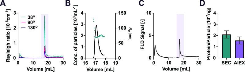
Extracellular vesicles (EVs) are membrane-enclosed biological nanoparticles with potential as diagnostic markers and carriers for therapeutics. Characterization of EVs poses severe challenges due to their complex structure and composition, requiring the combination of orthogonal analytical techniques. Here, we demonstrate how liquid chromatography combined with multi-angle light scattering (MALS) and fluorescence detection in one single apparatus can provide multiparametric characterization of EV samples, including concentration of particles, average diameter of the particles, protein amount to particle number ratio, presence of EV surface markers and lipids, EV shape, and sample purity. The method requires a small amount of sample of approximately 107 EVs, limited handling of the sample and data analysis time in the order of minutes; it is fully automatable and can be applied to both crude and purified samples.
Conflict of interest statement
The authors declare no competing financial interest.
Figures


Similar articles
-
Usefulness of Size-Exclusion Chromatography-Multi-Angle Light Scattering to Assess Particle Composition and Protein Impurities for Quality Control of Therapeutic Exosome Preparations.Pharmaceutics. 2024 Nov 27;16(12):1526. doi: 10.3390/pharmaceutics16121526. Pharmaceutics. 2024. PMID: 39771505 Free PMC article.
-
Measuring particle concentration of multimodal synthetic reference materials and extracellular vesicles with orthogonal techniques: Who is up to the challenge?J Extracell Vesicles. 2021 Jan;10(3):e12052. doi: 10.1002/jev2.12052. Epub 2021 Jan 12. J Extracell Vesicles. 2021. PMID: 33473263 Free PMC article.
-
An ultracentrifugation - hollow-fiber flow field-flow fractionation orthogonal approach for the purification and mapping of extracellular vesicle subtypes.J Chromatogr A. 2021 Feb 8;1638:461861. doi: 10.1016/j.chroma.2020.461861. Epub 2020 Dec 29. J Chromatogr A. 2021. PMID: 33472105
-
Chromatography and its hyphenation to mass spectrometry for extracellular vesicle analysis.J Chromatogr A. 2016 Mar 25;1439:26-41. doi: 10.1016/j.chroma.2016.01.017. Epub 2016 Jan 11. J Chromatogr A. 2016. PMID: 26830636 Review.
-
Preparation and characterization of extracellular vesicles.Am J Reprod Immunol. 2021 Feb;85(2):e13367. doi: 10.1111/aji.13367. Epub 2020 Nov 16. Am J Reprod Immunol. 2021. PMID: 33118232 Review.
Cited by
-
Inline Raman Spectroscopy Provides Versatile Molecular Monitoring for Platelet Extracellular Vesicle Purification with Anion-Exchange Chromatography.Int J Mol Sci. 2024 Jul 25;25(15):8130. doi: 10.3390/ijms25158130. Int J Mol Sci. 2024. PMID: 39125704 Free PMC article.
-
Microfluidics-Driven Manufacturing and Multiscale Analytical Characterization of Nanoparticle-Vesicle Hybrids.Adv Healthc Mater. 2025 Feb;14(4):e2403264. doi: 10.1002/adhm.202403264. Epub 2024 Dec 25. Adv Healthc Mater. 2025. PMID: 39722148 Free PMC article.
-
Extracellular Vesicles for Clinical Diagnostics: From Bulk Measurements to Single-Vesicle Analysis.ACS Nano. 2025 Aug 12;19(31):28021-28109. doi: 10.1021/acsnano.5c00706. Epub 2025 Jul 28. ACS Nano. 2025. PMID: 40720603 Free PMC article. Review.
-
Usefulness of Size-Exclusion Chromatography-Multi-Angle Light Scattering to Assess Particle Composition and Protein Impurities for Quality Control of Therapeutic Exosome Preparations.Pharmaceutics. 2024 Nov 27;16(12):1526. doi: 10.3390/pharmaceutics16121526. Pharmaceutics. 2024. PMID: 39771505 Free PMC article.
-
Affinity-Based Isolation and One-Pot Analysis of Extracellular Vesicles from Biofluids Using Phase Separated Zwitterionic Coacervates.Adv Sci (Weinh). 2025 May;12(20):e2411653. doi: 10.1002/advs.202411653. Epub 2025 Apr 15. Adv Sci (Weinh). 2025. PMID: 40231809 Free PMC article.
References
-
- Yáñez-Mó M.; Siljander P. R. M.; Andreu Z.; Zavec A. B.; Borràs F. E.; Buzas E. I.; Buzas K.; Casal E.; Cappello F.; Carvalho J.; Colás E.; Cordeiro-Da Silva A.; Fais S.; Falcon-Perez J. M.; Ghobrial I. M.; Giebel B.; Gimona M.; Graner M.; Gursel I.; Gursel M.; Heegaard N. H. H.; Hendrix A.; Kierulf P.; Kokubun K.; Kosanovic M.; Kralj-Iglic V.; Krämer-Albers E. M.; Laitinen S.; Lässer C.; Lener T.; Ligeti E.; Line A.; Lipps G.; Llorente A.; Lötvall J.; Manček-Keber M.; Marcilla A.; Mittelbrunn M.; Nazarenko I.; Nolte-’t Hoen E. N. M.; Nyman T. A.; O’Driscoll L.; Olivan M.; Oliveira C.; Pállinger É.; Del Portillo H. A.; Reventós J.; Rigau M.; Rohde E.; Sammar M.; Sánchez-Madrid F.; Santarém N.; Schallmoser K.; Ostenfeld M. S.; Stoorvogel W.; Stukelj R.; Van Der Grein S. G.; Helena Vasconcelos M.; Wauben M. H. M.; De Wever O. Biological Properties of Extracellular Vesicles and Their Physiological Functions. J. Extracell. Vesicles 2015, 4, 2706610.3402/jev.v4.27066. - DOI - PMC - PubMed
Publication types
MeSH terms
LinkOut - more resources
Full Text Sources
