. 2023 Aug 9;9(32):eadg9832.
doi: 10.1126/sciadv.adg9832. Epub 2023 Aug 9.

Structural basis of histone H2A lysine 119 deubiquitination by Polycomb repressive deubiquitinase BAP1/ASXL1

Affiliations

Structural basis of histone H2A lysine 119 deubiquitination by Polycomb repressive deubiquitinase BAP1/ASXL1

Jonathan F Thomas et al. Sci Adv. .

Abstract

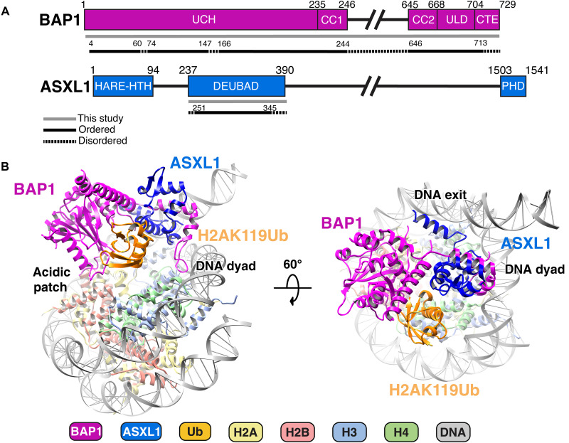
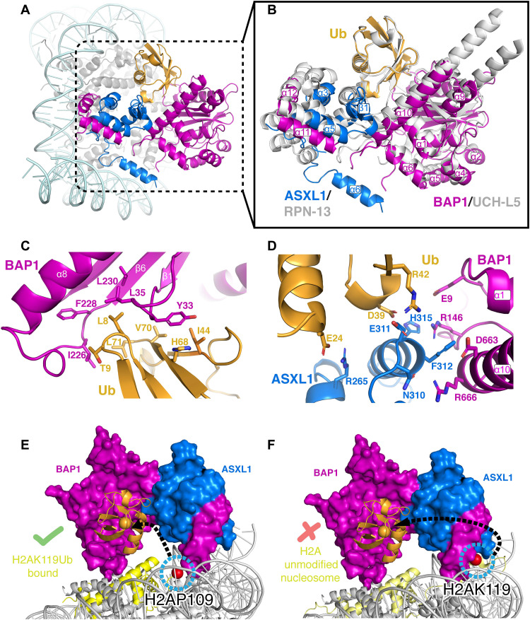
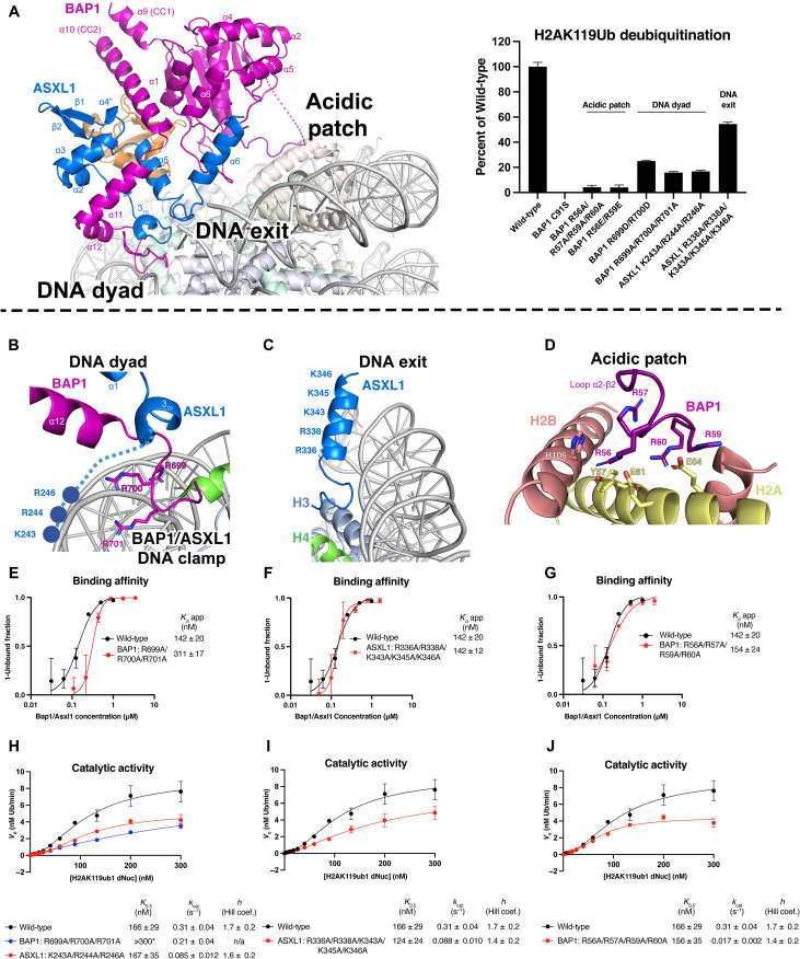
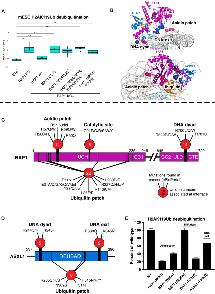
Histone H2A lysine 119 (H2AK119Ub) is monoubiquitinated by Polycomb repressive complex 1 and deubiquitinated by Polycomb repressive deubiquitinase complex (PR-DUB). PR-DUB cleaves H2AK119Ub to restrict focal H2AK119Ub at Polycomb target sites and to protect active genes from aberrant silencing. The PR-DUB subunits (BAP1 and ASXL1) are among the most frequently mutated epigenetic factors in human cancers. How PR-DUB establishes specificity for H2AK119Ub over other nucleosomal ubiquitination sites and how disease-associated mutations of the enzyme affect activity are unclear. Here, we determine a cryo-EM structure of human BAP1 and the ASXL1 DEUBAD in complex with a H2AK119Ub nucleosome. Our structural, biochemical, and cellular data reveal the molecular interactions of BAP1 and ASXL1 with histones and DNA that are critical for restructuring the nucleosome and thus establishing specificity for H2AK119Ub. These results further provide a molecular explanation for how >50 mutations in BAP1 and ASXL1 found in cancer can dysregulate H2AK119Ub deubiquitination, providing insight into understanding cancer etiology.


Figures

Fig. 1.
Fig. 1.. Overview of the structure of BAP1/ASXL1 bound to H2AK119Ub nucleosome.
(A) Bar diagram representation of BAP1 and ASXL1 domains. Protein sequences included in this study are in gray line; those resolved in the structure are in black line; and disordered regions are in dashed line. (B) Two different views of the model for the BAP1/ASXL1-H2AK119Ub nucleosome complex with key anchor points highlighted. The figure is color-coded, depicting BAP1 (purple), ASXL1 (dark blue), Ub (orange), H2A (yellow), H2B (salmon), H3 (light blue), H4 (green), and DNA (gray).
Fig. 2.
Fig. 2.. Insights into the mechanism of catalysis by BAP1/ASXL1 and conservation with other deubiquitinases.
(A) Position of Ub engaged with BAP1 and ASXL1 on the nucleosome. (B) BAP1/ASXL1/Ub aligned and superimposed with UCH-L5/RPN13/Ub (PDB ID 4uel; in gray) (22). (C) Hydrophobic interactions between BAP1 and Ub. (D) Electrostatic interactions between BAP1/ASXL1 and Ub. (E) Space-filling representation of our cryo-EM structure with the last observed residue of H2A (H2AP109; blue-circled red sphere). (F) A model of BAP1/ASXL1 from our cryo-EM structure superposed with wild-type (WT; unmodified) nucleosome (PDB ID 1kx5) (30), where the structured H2A docking domain with the unmodified H2AK119 is shown as blue-circled red sphere.
Fig. 3.
Fig. 3.. BAP1/ASXL1 interacts with DNA and acidic patch on the nucleosome.
(A) Left: Overall architecture of the BAP1/ASXL1-H2AK119Ub nucleosome complex with BAP1/ASXL1 anchor points marked. Right: Catalytic activity assays on H2AK119Ub nucleosomes (containing a DUB-cleavable native gamma-lysine isopeptide linkage) and various forms of BAP1/ASXL1 enzyme: WT, catalytically inactive (BAP1 C91S), or mutated nucleosomal anchor points (as labeled). (B) Close-up view of BAP1/ASXL1 DNA clamp contacting the nucleosome near the DNA dyad. (C) Close-up view of the ASXL1 DEUBAD α6 helix, projecting a stretch of basic residues toward a nucleosome DNA exit. (D) Close-up view of the BAP1 R-finger interacting with the acidic patch. (E to G) Quantified electromobility shift assays (EMSAs) of WT heterodimer complex versus mutants of the BAP1 DNA clamp (E), ASXL1 DNA exit (F), and BAP1 interaction with the acidic patch (G). Each data point and error bar indicate the means ± SD from three independent experiments. The SEs of dissociation constant (Kd) are indicated. (H to J) Representative kinetic curves of WT/mutant BAP1/ASXL1 enzymes with H2AK119Ub nucleosomes. Compared relative to WT are mutants of the BAP1 DNA clamp (H), ASXL1 DNA exit (I), and BAP1 interaction with the acidic patch (J). tUI-free Ub sensor was used to monitor the activity of WT BAP1/ASXL1 and its mutants (H to J). A free Ub standard curve was used to calculate the amount of enzymatically generated Ub, with initial velocities (V0) relative to H2AK119ub1 concentration used to determine H2AK119ub1 dNuc K0.5, kcat, and Hill coefficient values by applying an allosteric sigmoidal model. Kinetic constants are represented as ± SD from three independent experiments. Asterisk (*) corresponds to Michaelis-Menten fit instead of allosteric regulation fit. n/a, not applicable.
Fig. 4.
Fig. 4.. BAP1/ASXL1-nucleosome contacts are extensively mutated in cancers.
(A) Western blot analysis of H2AK119Ub levels in total protein extracts from BAP1 WT (E14) or KO mESCs, or KO after reintroduction/stable expression of BAP1 WT and various mutations (C91S, R699E/R700E; R56E/R59E; R56A/R57A/R59A/R60A). t tests were performed as indicated [non significant (n.s.), P > 0.05; *P < 0.05; **P < 0.01; ****P < 0.0001]. (B) Structure of the BAP1/ASXL1-H2AK119Ub nucleosome complex highlighting the three key nucleosome anchors (top) and Ub interaction region (bottom), and relative position of cancer mutations (colored spheres) clustered in these areas. (C and D) Bar diagrams showing cancer-associated point mutations/deletions of BAP1 (C) and ASXL1 (D) at substrate interaction surfaces (B) that can be mechanistically explained by this study. The number of unique cancer types at each interface is inside the red circles. (E) Catalytic activity assays on H2AK119Ub nucleosomes with various forms of BAP1/ASXL1 enzyme: WT or with point mutations found in cancer (from cBioPortal).

Update of

References

    1. A. P. Szczepanski, L. Wang, Emerging multifaceted roles of BAP1 complexes in biological processes. Cell Death Discov. 7, 20 (2021). - PMC - PubMed
    1. J. C. Scheuermann, A. G. de Ayala Alonso, K. Oktaba, N. Ly-Hartig, R. K. McGinty, S. Fraterman, M. Wilm, T. W. Muir, J. Müller, Histone H2A deubiquitinase activity of the Polycomb repressive complex PR-DUB. Nature 465, 243–247 (2010). - PMC - PubMed
    1. D. D. Sahtoe, W. J. Van Dijk, R. Ekkebus, H. Ovaa, T. K. Sixma, BAP1/ASXL1 recruitment and activation for H2A deubiquitination. Nat. Commun. 7, 10292 (2016). - PMC - PubMed
    1. L. Sanchez-Pulido, L. Kong, C. P. Ponting, A common ancestry for BAP1 and Uch37 regulators. Bioinformatics 28, 1953–1956 (2012). - PubMed
    1. N. A. Fursova, A. H. Turberfield, N. P. Blackledge, E. L. Findlater, A. Lastuvkova, M. K. Huseyin, P. Dobrinić, R. J. Klose, BAP1 constrains pervasive H2AK119ub1 to control the transcriptional potential of the genome. Genes Dev. 35, 749–770 (2021). - PMC - PubMed