. 2024 Jan 4;45(1):125-170.
doi: 10.1210/endrev/bnad026.

Treating Primary Aldosteronism-Induced Hypertension: Novel Approaches and Future Outlooks

Affiliations

Treating Primary Aldosteronism-Induced Hypertension: Novel Approaches and Future Outlooks

Nathan Mullen et al. Endocr Rev. .

Abstract

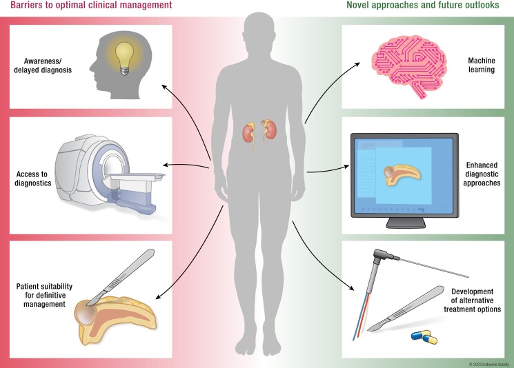
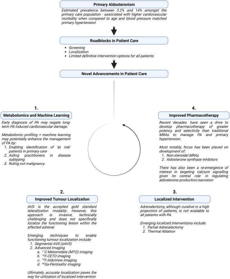
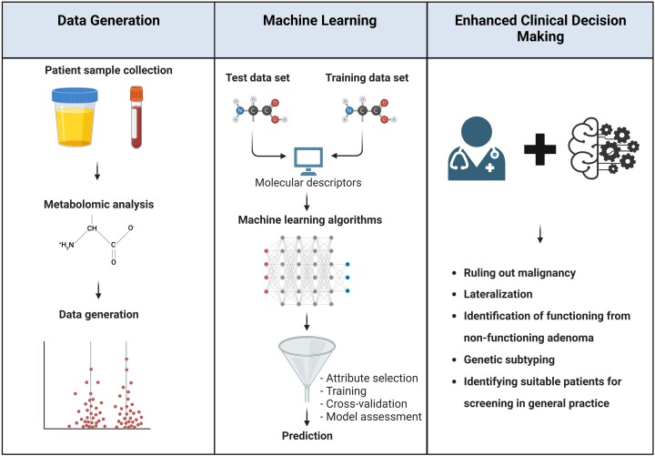
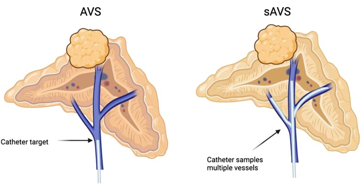
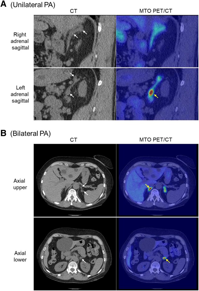
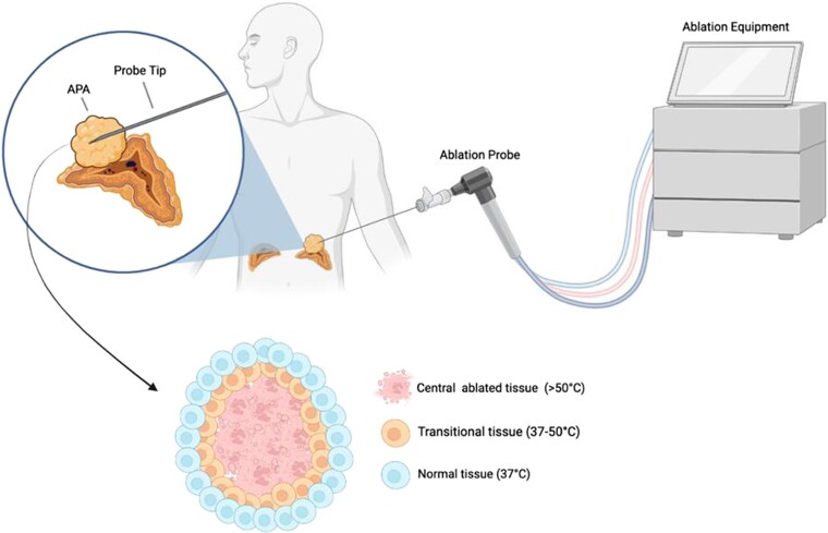
Primary aldosteronism (PA) is the most common cause of secondary hypertension and is associated with increased morbidity and mortality when compared with blood pressure-matched cases of primary hypertension. Current limitations in patient care stem from delayed recognition of the condition, limited access to key diagnostic procedures, and lack of a definitive therapy option for nonsurgical candidates. However, several recent advances have the potential to address these barriers to optimal care. From a diagnostic perspective, machine-learning algorithms have shown promise in the prediction of PA subtypes, while the development of noninvasive alternatives to adrenal vein sampling (including molecular positron emission tomography imaging) has made accurate localization of functioning adrenal nodules possible. In parallel, more selective approaches to targeting the causative aldosterone-producing adrenal adenoma/nodule (APA/APN) have emerged with the advent of partial adrenalectomy or precision ablation. Additionally, the development of novel pharmacological agents may help to mitigate off-target effects of aldosterone and improve clinical efficacy and outcomes. Here, we consider how each of these innovations might change our approach to the patient with PA, to allow more tailored investigation and treatment plans, with corresponding improvement in clinical outcomes and resource utilization, for this highly prevalent disorder.

Keywords: ablation; adrenal sparing surgery; adrenal vein sampling; functional/molecular imaging; machine learning; metabolomics; partial adrenalectomy.


Figures

None
Graphical abstract
Figure 1.
Figure 1.
The traditional approach to the diagnosis and management of patients with PA: Initial diagnosis is determined by a positive aldosterone–renin ratio (ARR) screen with at least 1 positive confirmatory test (discussed in “Screening, Diagnosis, and the Spectrum of Disease”). Following diagnosis, subtype diagnosis (ie, lateralization) is sought through use of adrenal imaging/adrenal vein sampling (AVS) (discussed in “Current Approach to Lateralization”). Unilateral disease is commonly treated with adrenalectomy of the diseased adrenal whereas in cases of bilateral disease, mineralocorticoid receptor (MR) antagonists are commonly prescribed (discussed in “Advances in Pharmacotherapy”).
Figure 2.
Figure 2.
Outline of current “roadblocks” in primary aldosteronism (PA) care, with suggested approaches to addressing these, including: 1. Improved screening for, and diagnosis of PA through application of metabolomics and machine learning; 2. Utilization of advanced lateralization techniques including molecular (functional) imaging to permit precise tumor localization; 3. Use of focal adrenal-sparing interventions (eg, adrenal-sparing surgery or thermal ablation) where appropriate; 4. Continued development of more selective pharmacological agents. Figure created with BioRender.com.
Figure 3.
Figure 3.
(A) Outline of the adrenal vein sampling (AVS) procedure. A sampling catheter is inserted in either the left or right femoral vein for sampling the left and right adrenal vein in the presence or absence of cosyntropin stimulation. Aldosterone and cortisol are sampled from locations 1-3 and, for each location, the aldosterone to cortisol ratio is calculated. Together, these values are used to determine: (1) Selectivity index: Did sampling occur from the correct location, ie, right adrenal vein (RAV), left adrenal vein (LAV), and inferior vena cava (IVC)? (2) Lateralization: Are 1 or both adrenals the source of aldosterone excess? (3) Is contralateral suppression present? (B) Example measurements demonstrating successful cannulation of both adrenal glands (location) under basal, unstimulated conditions (both locations return a selectivity index >3), with lateralization to the left adrenal and the presence of contralateral suppression. *Note, the cut-offs described in this diagram are arbitrarily defined for illustrative purposes. As is indicated by Quencher and colleagues 2021 there are marked procedural and cut-off heterogeneity between centers (64). Figure created with Biorender.com.
Figure 4.
Figure 4.
Outline of metabolomics/machine-learning workflow. Following patient sample collection, metabolomic analysis is usually carried out using liquid chromatography tandem mass spectrometry, yielding a large amount of data. Through combination with machine learning, a detailed metabolomic fingerprint can be generated of disease subtypes to streamline clinical decision making. Figure created with BioRender.com.
Figure 5.
Figure 5.
Schematic diagram outlining an AVS or segmental AVS (sAVS) procedure as described by Satani et al, 2016 (137). During a conventional AVS procedure, both central adrenal veins of the left and right adrenal are sampled to lateralize the affected adrenal. sAVS involves sampling from several adrenal tributaries to localize a section of adrenal as the source of aldosterone hypersecretion. Commonly, 3 main tributaries converge into 1 central vein. However, in some patients ≥3 tributaries may be present. Figure created with BioRender.com.
Figure 6.
Figure 6.
11C-metomidate PET/CT allows localization of aldosterone-secreting adenomas/nodules in unilateral and bilateral PA. (A) Sagittal CT demonstrating 2 discrete nodules within the left adrenal gland, but with only the inferior 1 showing high focal tracer uptake; the right adrenal gland shows normal background radiotracer uptake despite the initial suspicion of possible nodularity. Following left unilateral adrenalectomy, the patient achieved full clinical and biochemical remission and immunohistochemistry confirmed that the metomidate-avid nodule exhibited strong staining for CYP11B2; in contrast the superior nodule showed only mild CYP11B1 staining. (B) Axial CT and MTO PET/CT in a patient with bilateral PA. Both adrenal glands demonstrate focal high radiotracer uptake; within the left adrenal, the most inferior of 3 discrete nodules shows greatest metomidate uptake. The patient was managed with primary medical therapy. White arrows denote sites of suspected nodules on CT; yellow arrows identify nodules with greatest metomidate avidity.
Figure 7.
Figure 7.
Outline of adrenal ablation. During the procedure, energy in the form of radiofrequency or microwave is transferred from the ablation probe to the tumor producing lethal hyperthermia (≥50 °C) that kills cells within the targeted lesion. Outside of the core ablation zone, the transitional zone experiences various levels of sublethal hyperthermia that have differential effects on cellular viability. Figure created with BioRender.com.

Similar articles

Cited by

References

    1. Forouzanfar MH, Alexander L, Anderson HR, et al. . GBD 2013 Risk factors collaborators global, regional, and national comparative risk assessment of 79 behavioural, environmental and occupational, and metabolic risks or clusters of risks in 188 countries, 1990-2013: a systematic analysis for the global burden of disease study 2013. Lancet. 2015;386(10010):2287‐2323. - PMC - PubMed
    1. Sukor N. Endocrine hypertension—current understanding and comprehensive management review. Eur J Intern Med. 2011;22(5):433‐440. - PubMed
    1. Carretero OA, Oparil S. Essential hypertension: part I: definition and etiology. Circulation. 2000;101(3):329‐335. - PubMed
    1. Käyser SC, Dekkers T, Groenewoud HJ, et al. . Study heterogeneity and estimation of prevalence of primary aldosteronism: a systematic review and meta-regression analysis. J Clin Endocrinol Metab. 2016;101(7):2826‐2835. - PubMed
    1. Buffolo F, Monticone S, Tetti M, Mulatero P. Primary aldosteronism in the primary care setting. Curr Opin Endocrinol Diabetes Obes. 2018;25(3):155‐159. - PubMed