. 2023 Oct 23;58(20):2080-2096.e7.
doi: 10.1016/j.devcel.2023.07.011. Epub 2023 Aug 8.

Rejection of inappropriate synaptic partners in mouse retina mediated by transcellular FLRT2-UNC5 signaling

Affiliations

Rejection of inappropriate synaptic partners in mouse retina mediated by transcellular FLRT2-UNC5 signaling

Cameron L Prigge et al. Dev Cell. .

Abstract

During nervous system development, neurons choose synaptic partners with remarkable specificity; however, the cell-cell recognition mechanisms governing rejection of inappropriate partners remain enigmatic. Here, we show that mouse retinal neurons avoid inappropriate partners by using the FLRT2-uncoordinated-5 (UNC5) receptor-ligand system. Within the inner plexiform layer (IPL), FLRT2 is expressed by direction-selective (DS) circuit neurons, whereas UNC5C/D are expressed by non-DS neurons projecting to adjacent IPL sublayers. In vivo gain- and loss-of-function experiments demonstrate that FLRT2-UNC5 binding eliminates growing DS dendrites that have strayed from the DS circuit IPL sublayers. Abrogation of FLRT2-UNC5 binding allows mistargeted arbors to persist, elaborate, and acquire synapses from inappropriate partners. Conversely, UNC5C misexpression within DS circuit sublayers inhibits dendrite growth and drives arbors into adjacent sublayers. Mechanistically, UNC5s promote dendrite elimination by interfering with FLRT2-mediated adhesion. Based on their broad expression, FLRT-UNC5 recognition is poised to exert widespread effects upon synaptic partner choices across the nervous system.

Keywords: dendrite; latrophilin; retina; retinal ganglion cell; starburst amacrine cell; synaptogenesis.


Conflict of interest statement

Declaration of interests The authors declare no competing interests.

Figures
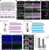
Figure 1:
Figure 1:. GAD65+ amacrine cells express UNC5C and exhibit mutual exclusion with DS circuit dendrites
A. Schematic of mouse retina (cross-section view) showing DS circuit cell types studied here. OFF starburst amacrine cells in inner nuclear layer (INL) project to inner plexiform layer (IPL) sublayer S2; ON starbursts in ganglion cell layer (GCL) project to S4. ON-OFF direction-selective ganglion cell (ooDSGC) dendrites cofasciculate with starburst dendrites in both sublayers. Amacrine cell dendrites contain both pre- and postsynaptic sites; starburst cells release GABA and acetylcholine onto ooDSGCs. B,C. Perturbation of starburst dendritic targeting using Megf10 mutants reveals mutual repulsion with GAD65+ amacrine cell dendrites. B: Retinal cross-sections stained with anti-ChAT (starburst marker) and anti-GAD65. C: Representative profile plots quantifying ChAT and GAD65 fluorescence across IPL. Controls (top; Megf10flox/flox; no Cre, n = 4) exhibit sharp boundaries between ChAT+ and GAD65+ IPL strata. In mutants (bottom, n = 4, Six3-Cre; Megf10flox/flox), starburst dendrites enter GAD65+ IPL regions, displacing GAD65 arbors (B, arrowheads; C, asterisk). Starburst dendrites also sporadically fail to innervate regions within S2 and S4; GAD65+ arbors invade these regions (arrows). D,E. Schematics of FLRT2 and UNC5C protein structure (left panels) and protein localization within IPL as per our previous study (right panels). FLRT2 is present in S2/S4 due to expression by ooDSGCs (G) and starburst cells (S). FLRT2 also localizes to S3. UNC5C is absent from S2 and S4 (E). LRR, leucine-rich repeat; Fn, Fibronectin-like domain; TM, transmembrane domain; ECD, extracellular domain; ICD, intracellular domain. Ig, immunoglobulin; Tsp, thrombospondin domains. FLRT2 binding sites for latrophilins (Lphn) and UNC5s are indicated. F,G. In situ hybridization for Unc5c and Gad65 (gene symbol Gad2) to mark GABAergic amacrine cells. Blue, Hoechst nuclear counterstain. F: Prominent Unc5c expression at P15 in GCL and INL, including amacrine subregion of INL (marked by Gad65). Unc5c labels a subset of Gad65+ amacrine cells in the INL and displaced Gad65+ amacrine cells in GCL (arrows). RGCs (Gad65-negative GCL cells) also express Unc5c. Expression is similar at P6 (Supplemental Fig. S1A). G: Representative images of Gad65+Unc5c+ double-positive cells. H. Fraction of Unc5c+ cells co-expressing Gad65, quantified from images similar to F,G. Most Unc5+ cells in GCL are GABAergic displaced amacrines rather than RGCs. N = 4 animals. Scale bars, 10 μm (B,G); 20 μm (F). Also see Supplemental Fig. S1.
Figure 2:
Figure 2:. Starburst and ooDSGC laminar targeting errors in Flrt2 and Unc5 mutants.
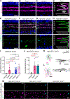
A-D. Starburst dendrite targeting errors in Flrt2Ret and Unc5d mutants. A-C, representative cross-section images; D, fluorescence profile plots (white, anti-GAD65). Magenta, anti-VAChT. Arrows (A-C) and asterisks (D) mark ectopic starburst dendrites in S1. E-H. Hb9-ooDSGC dendrite targeting errors in Flrt2Ret and Unc5c mutants. Magenta, anti-VAChT; green, anti-GFP (labeling ooDSGCs). Vertical bar, IPL neuropil. Arrows (E-G) and asterisks (H) mark ectopic ooDSGC arbors in S1 (top row) or S3 (bottom row) that are uncoupled from starburst arbors. Ax, GFP+ axons within nerve fiber layer. Blue, Hoechst nuclear counterstain. I,J: Summary of starburst (I) and ooDSGC (J) laminar targeting errors. ooDSGC phenotype strength was similar in Flrt2Ret and Unc5c−/−. Starburst phenotype strength was similar in Flrt2 whole-retina (Flrt2Ret) and starburst-specific (Flrt2SAC) mutants, as well as Unc5d−/−. Statistics (I), Kruskal-Wallis test (main effect p=1.6×10⁻⁵) with Dunn’s post-hoc test. Sample sizes (number of animals): littermate controls n = 9, Flrt2Ret n = 9, Unc5c−/− n = 5; Unc5d−/− n = 7; Flrt2SAC n = 5. Statistics (J), Kruskal-Wallis test (main effect p=9.2×10⁻⁵) with Dunn’s post-hoc test. Sample sizes (number of animals): littermate controls n = 11 (n = 6 Flrt2WT; n = 5 Unc5c+/+), Flrt2Ret n = 9, Unc5c−/− n = 5. Error bars, S.E.M. P-values (shown on graph) were corrected for multiple comparisons. K,L. Single cell reconstructions of dye-filled Hb9-ooDSGCs from Flrt2Ret mutants. F, normally-stratified ooDSGC; G, ooDSGC with ectopic S3 dendritic arbor. n = 3/12 filled cells exhibited ectopic arbors. M. En-face view of ectopic ooDSGC arbors in retinal wholemounts. To depict S1 IPL sublayer, confocal images were acquired at plane of IPL/INL border. Hb9-GFP+ arbors do not project to this location in wild-type retina (left) but are readily observed in both Unc5c−/− and Flrt2Ret mutants (arrows). Scale bars, 20 μm (A-C, E-G, M); 100 μm (K,L). Error bars, S.E.M. Also see Supplemental Figs. S2–S5.
Figure 3:
Figure 3:. Abolishing FLRT2-UNC5 binding disrupts DS circuit laminar targeting
A. FLRT2UF mutant protein. H170N mutation introduces N-glycosylation site within LRR domain at UNC5 binding surface. LPHN binding site is unaffected. Abbreviations, see Fig. 1D,E. B,C. Starburst laminar targeting errors in homozygous Flrt2UF mutants. Left, VAChT immunostaining; right, profile plots (VAChT and GA65 fluorescence). Unlike controls (B), mutants (C) exhibit ectopic dendrites within S1 (arrow, asterisk). D. Summary of starburst laminar errors in Flrt2UF mutants and Flrt2WT littermate controls. Statistics, two-tailed Mann-Whitney test; sample sizes n = 6 littermate controls; n = 6 Flrt2UF mutants. For comparison, Flrt2Ret and Unc5d−/− mutant data are replotted from Fig. 2. There was no significant difference in error frequency between Flrt2UF and the other two mutants (one-way ANOVA, F(2, 19) = 1.60; n.s., not significant). E,F. ooDSGC dendrite targeting errors are observed in Flrt2UF mutants (F) but not littermate controls (E). Arrow, ectopic ooDSGC arbor within S1. G. Summary of Hb9-ooDSGC laminar errors in Flrt2UF mutants and Flrt2WT littermate controls. Statistics, two-tailed Mann-Whitney test; sample sizes n = 6 littermate controls; n = 7 Flrt2UF mutants. For comparison, Flrt2Ret and Unc5c−/− mutant data are replotted from Fig. 2. There is a trend towards fewer errors in Flrt2UF compared to the other two mutants but the difference was not significant (one-way ANOVA, F(2, 18) = 0.85). Scale bars, 20 μm. Error bars (D,G), S.E.M.
Figure 4:
Figure 4:. Mistargeted ooDSGC arbors in Flrt2Ret and Unc5c mutants receive inhibitory synapses.
A-B. Inhibitory synapse labeling in P25 Hb9-GFP; Flrt2Ret mutant (single optical slice from representative confocal stack). A, GFP channel alone; blue arrow, ectopic ooDSGC arbor. B, GFP overlay with bassoon and gephyrin. Boxed regions are shown at high magnification (B, right). White signals indicate putative synaptic sites where bassoon and gephyrin puncta overlap. Putative synapses (white arrows) are observed along ooDSGC dendrites in normal DS circuit sublayers (S2, S4) and ectopic sublayers (S1). C. Semi-automated unbiased synapse identification in Hb9-GFP; Unc5c−/− mutant. ObjectFinder software was used to analyze images similar to B. Left, GFP channel 3D mask, demarcating ooDSGC dendrites. Right, segmentation of paired bassoon-gephyrin synaptic structures (red) within GFP mask. Synapses are evident along ectopic arbors (S1, S3, blue arrows), similar to normally targeted arbors (S2, S4). Green, non-synaptic gephyrin signal (i.e. without bassoon co-localization). Images are representative of n = 4 Unc5c−/− and n = 4 Flrt2Ret animals. Scale bars: 10 μm (A; B, left); 2 μm (B, right) 20 μm (C, left); 5 μm (D, right). Also see Supplemental Fig. S5D,E.
Figure 5:
Figure 5:. Refinement of DS circuit laminar targeting between P6 and P15.
A-D. Developmental timecourse showing starburst and Hb9-ooDSGC IPL projections at specified ages. All images from wild-type animals (i.e. Flrt2+/+; Unc5c+/+); for mutant images, see Supplemental Fig. S6. At P2 (A), starburst IPL strata are evident; ooDSGC arbors are not yet stratified. At P6 (B) and P10 (C), both cell types are largely co-stratified although uncoupled ooDSGC arbors outside S2/S4 are evident (right panels; arrows). By P15 (D) all ooDSGC dendrites are coupled with starburst scaffold. E,F. Quantification of ooDSGC (E) and starburst (F) laminar errors in Flrt2Ret mutants; Unc5c−/− mutants; and wild-type littermate controls. P15 data are replotted from Fig. 2 to facilitate comparisons with younger ages. Differences between mutant and controls do not arise until P15 (ooDSGCs) or P10 (starbursts). Statistics (E,F): Kruskal-Wallis test; P-values on graph are for main effect of genotype at each age. Sample sizes: P6 control n = 7; P6 Flrt2Ret n = 8; P6 Unc5c−/− n = 4; P10 control n = 8; P10 Flrt2Ret n = 4 (E) or n = 5 (F); P10 Unc5c−/− n = 6. Scale bars, 20 μm (bar in A also applies to B). Error bars, S.E.M. Also see Supplemental Fig. S6.
Figure 6:
Figure 6:. UNC5s function by occluding FLRT-LPHN adhesion
A. Illustration of repellent receptor model: FLRT2 transduces repulsive signals upon UNC5 binding, driving elimination of mistargeted dendrite branches (X). Model predicts cell-autonomous requirement for FLRT2 within ooDSGCs. B,C: Deletion of Flrt2 from RGCs (Flrt2RGC mutants) rules out repellent receptor model. B, Representative image (left) and profile plots (right) showing normal dendrite targeting of Hb9-ooDSGCs (anti-GFP) and starbursts (anti-VAChT) in Flrt2RGC mutants. C: quantification of mistargeted dendrites. Sample sizes: littermate controls, n = 9; Flrt2RGC n = 5. For comparison, Flrt2Ret data are replotted from Fig. 2. Statistics, Kruskal-Wallace test with post-hoc Dunn’s test. P-values were corrected for multiple comparisons. D. Illustration of adhesion-occlusion model: LPHN protein (gray) localizes throughout IPL, including starburst and ooDSGC dendrites; UNC5s are in non-DS sublayers. Dendritic contacts among DS circuit neurons are stabilized (orange +) by transcellular reciprocal FLRT2-LPHN adhesion (inset 1). Contacts with inappropriate partners also result in FLRT2-LPHN binding, but these are not stabilized (X) due to presence of UNC5 which occludes FLRT2-LPHN adhesion (inset 2). E. Co-immunoprecipitation indicates LPHN3-UNC5C-FLRT2 complex is present in P6 retina. UNC5C (left) and FLRT2 (right) are enriched in LPHN3 pull-down condition compared to input lysates or to IgG pull-down control. Upper FLRT2 band (just below 150 kDa) likely corresponds to FLRT2 dimer. F,G. Adhesion assay using primary retinal neurons co-cultured with HEK cells. F, representative images of VAChT+ starburst arbors associating with HEK cells transfected as indicated; G. quantification of VAChT fluorescence surrounding HEK cells. Starburst arbors encircle HEK cells transfected with LPHN3-GFP, but not GFP alone. Starburst-LPHN3 adhesion is blocked by co-transfection of flag-tagged wild-type (WT) UNC5C, but not UNC5CUF mutant that cannot bind FLRTs. n.d., staining was not performed. Statistics: One way ANOVA with Tukey’s post-hoc test. P-values were corrected for multiple comparisons. Sample sizes (number of cells): n = 18 EGFP; n = 32 Lphn3 alone; n = 31 Lphn3+Unc5cWT; n = 32 Lphn3+Unc5cUF. Data from three independent culture experiments. Scale bars, 20 μm (B); 10 μm (F). Error bars, S.E.M. Also see Supplemental Fig. S6.
Figure 7:
Figure 7:. Misexpression of UNC5C in starburst cells induces DS circuit laminar targeting errors.
A. Strategy for starburst-specific flex-switch AAV expression in ChatCre mice. PHP.eB, AAV serotype. B. Illustration showing confocal imaging planes used for wholemount analysis of AAV-infected retinas (C, Supplemental Fig. S7A). C. Starburst dendritic density is diminished by UNC5C misexpression. En-face views of tdTomato (tdT)+ OFF starburst dendrites in retinal wholemounts infected with specified AAVs (B shows imaging plane). Representative images of tdT-only control group (left; n = 4) and UNC5C misexpression group (right, n = 5). Lower dendrite density in UNC5C group is not explained by a difference in number of infected starburst cells (Supplemental Fig. S7A). D-F. Representative cross-sections showing IPL projections of starburst (magenta, anti-tdTomato; white, anti-ChAT) and Hb9-ooDSGC (green, anti-GFP) dendrites in retinas infected with specified AAVs. Laminar targeting is normal in retinas expressing tdTomato alone (D; n = 4 mice), but is disrupted by Chat-UNC5C misexpression (E, Hb9-DSGCs, n = 4 mice; F, starbursts, n = 5 mice). G,H. Working model for FLRT2-UNC5C function. During normal development (G), FLRT2+ DS circuit dendrites branch exuberantly as they grow. Properly stratified branches are stabilized by FLRT-LPHN adhesion (+). Branches that stray into adjacent IPL layers contact UNC5+ amacrine arbors; this blocks FLRT-LPHN adhesion leading to branch elimination (X). Upon Chat-UNC5C misexpression (H), correctly targeted arbors in S2 receive a large UNC5 signal (large X), thereby diminishing FLRT2-LPHN adhesion and suppressing growth. Ectopic UNC5 signal may be larger than endogenous UNC5 signal (small X). Survival and growth of mistargeted arbors indicates that neurons compare relative levels of UNC5 signaling – and hence relative levels of FLRT2-LPHN positive signals – across their dendritic branches to determine which branches will survive. Scale bars, 20 μm. Bar in D applies to E,F. Also see Supplemental Fig. S7.

References

    1. Sanes JR, and Zipursky SL (2020). Synaptic Specificity, Recognition Molecules, and Assembly of Neural Circuits. Cell 181, 536–556. 10.1016/j.cell.2020.04.008. - DOI - PubMed
    1. Prigge CL, and Kay JN (2018). Dendrite morphogenesis from birth to adulthood. Curr. Opin. Neurobiol. 53, 139–145. 10.1016/j.conb.2018.07.007. - DOI - PMC - PubMed
    1. Lefebvre JL, Sanes JR, and Kay JN (2015). Development of dendritic form and function. Annu. Rev. Cell. Dev. Biol. 31, 741–777. 10.1146/annurev-cellbio-100913-013020. - DOI - PubMed
    1. Pecho-Vrieseling E, Sigrist M, Yoshida Y, Jessell TM, and Arber S (2009). Specificity of sensory–motor connections encoded by Sema3e–Plxnd1 recognition. Nature 459, 842–846. 10.1038/nature08000. - DOI - PMC - PubMed
    1. Kolodkin AL, and Tessier-Lavigne M (2011). Mechanisms and molecules of neuronal wiring: a primer. Cold Spring Harbor perspectives in biology 3, 1–14. 10.1101/cshperspect.a001727. - DOI - PMC - PubMed

Publication types

Substances