. 2023 Oct:113:401-414.
doi: 10.1016/j.bbi.2023.08.005. Epub 2023 Aug 7.

Single-cell analysis of dorsal root ganglia reveals metalloproteinase signaling in satellite glial cells and pain

Affiliations

Single-cell analysis of dorsal root ganglia reveals metalloproteinase signaling in satellite glial cells and pain

Raquel Tonello et al. Brain Behav Immun. 2023 Oct.

Abstract

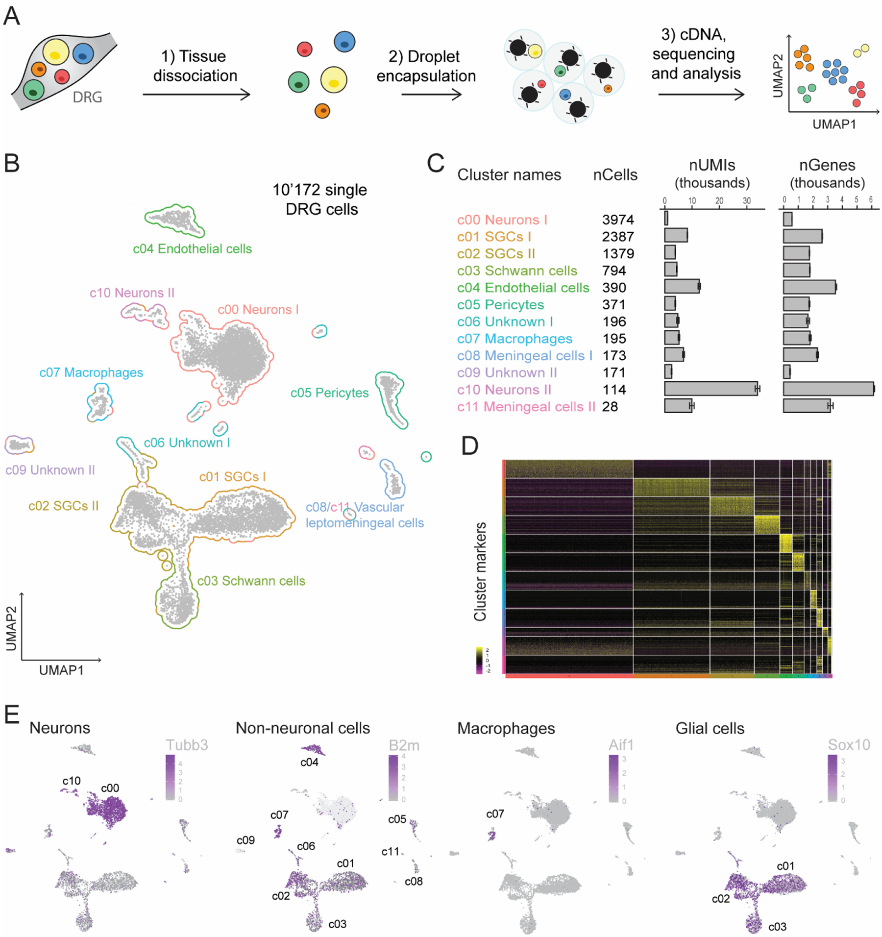
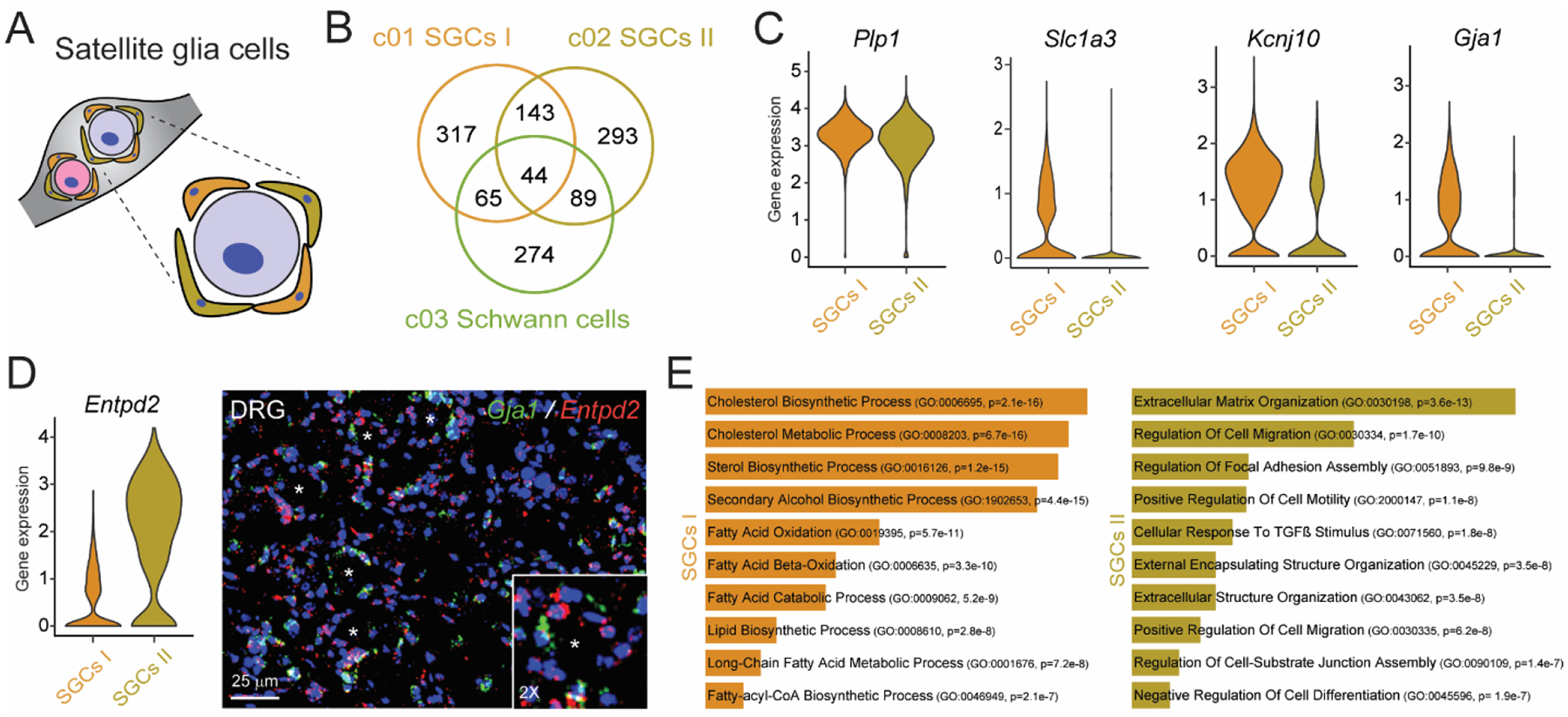
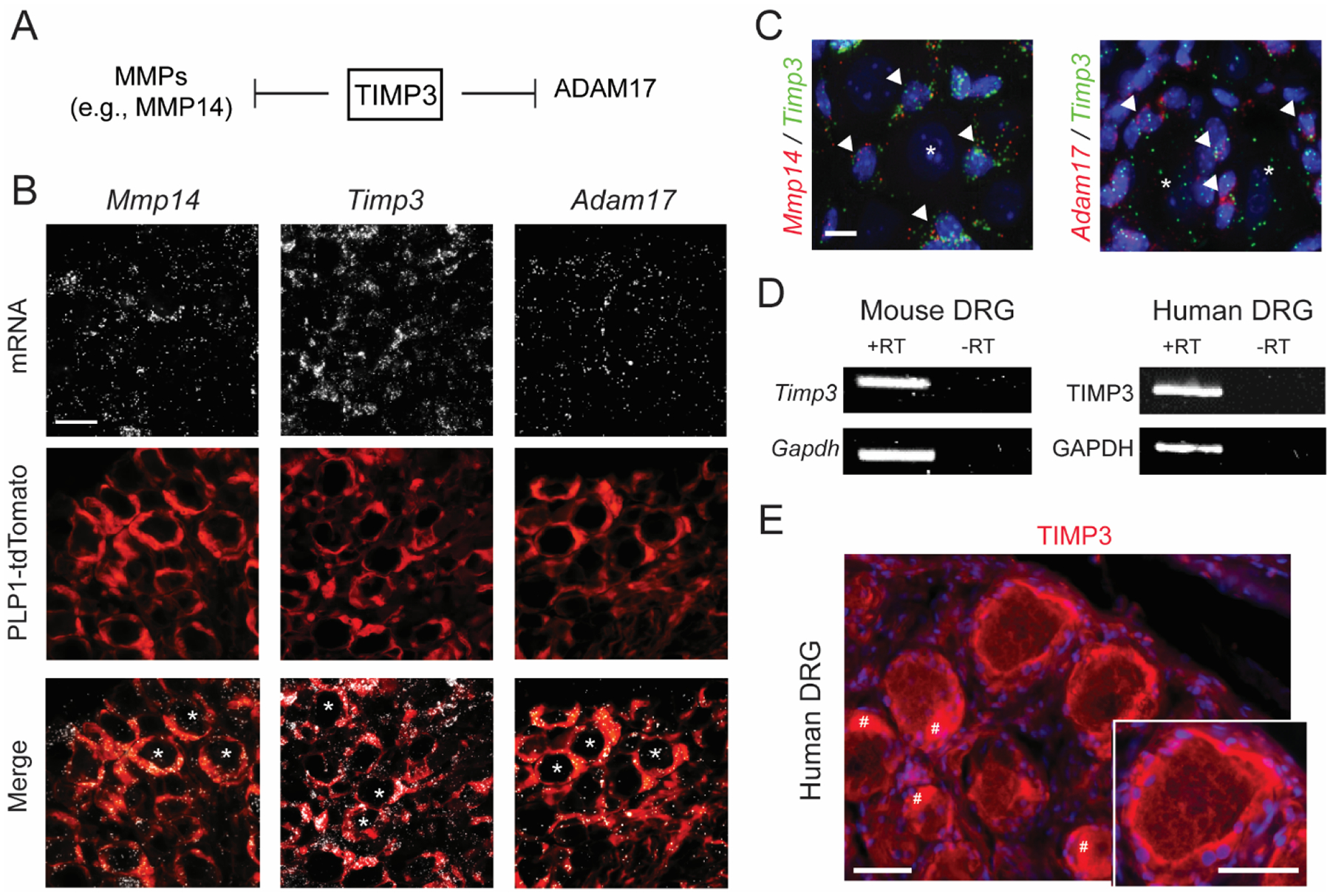
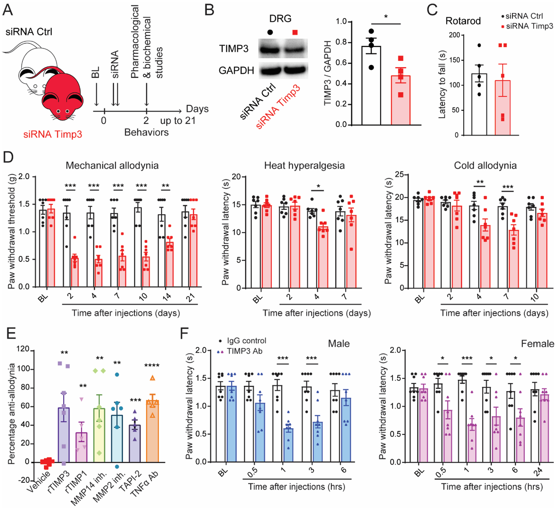
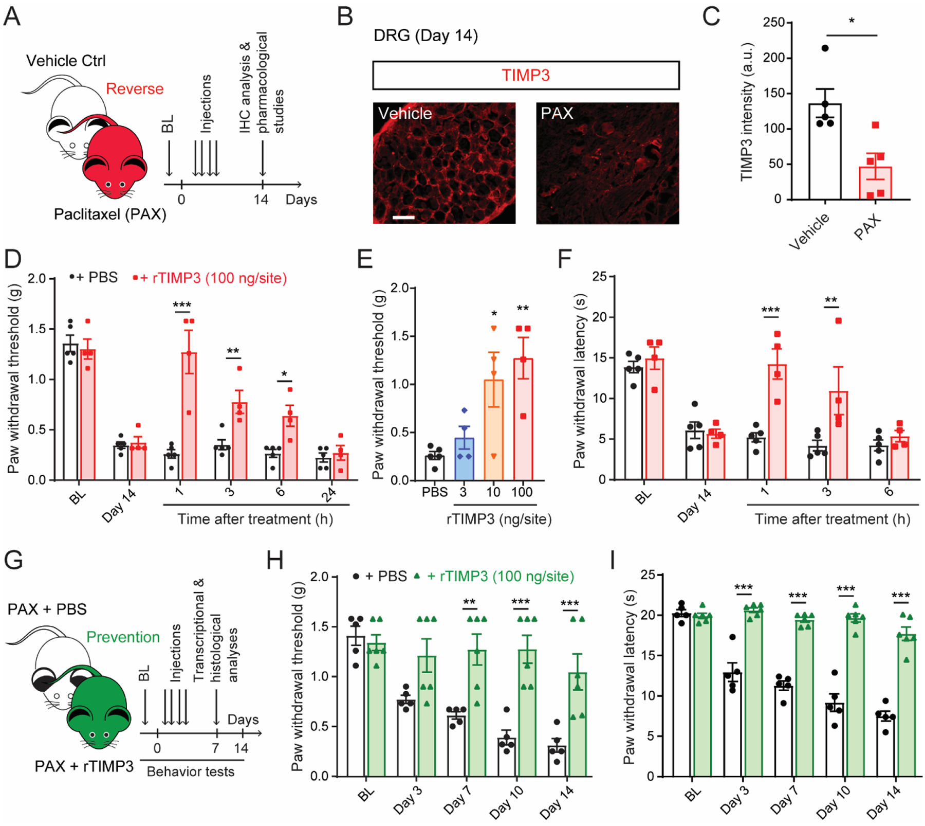
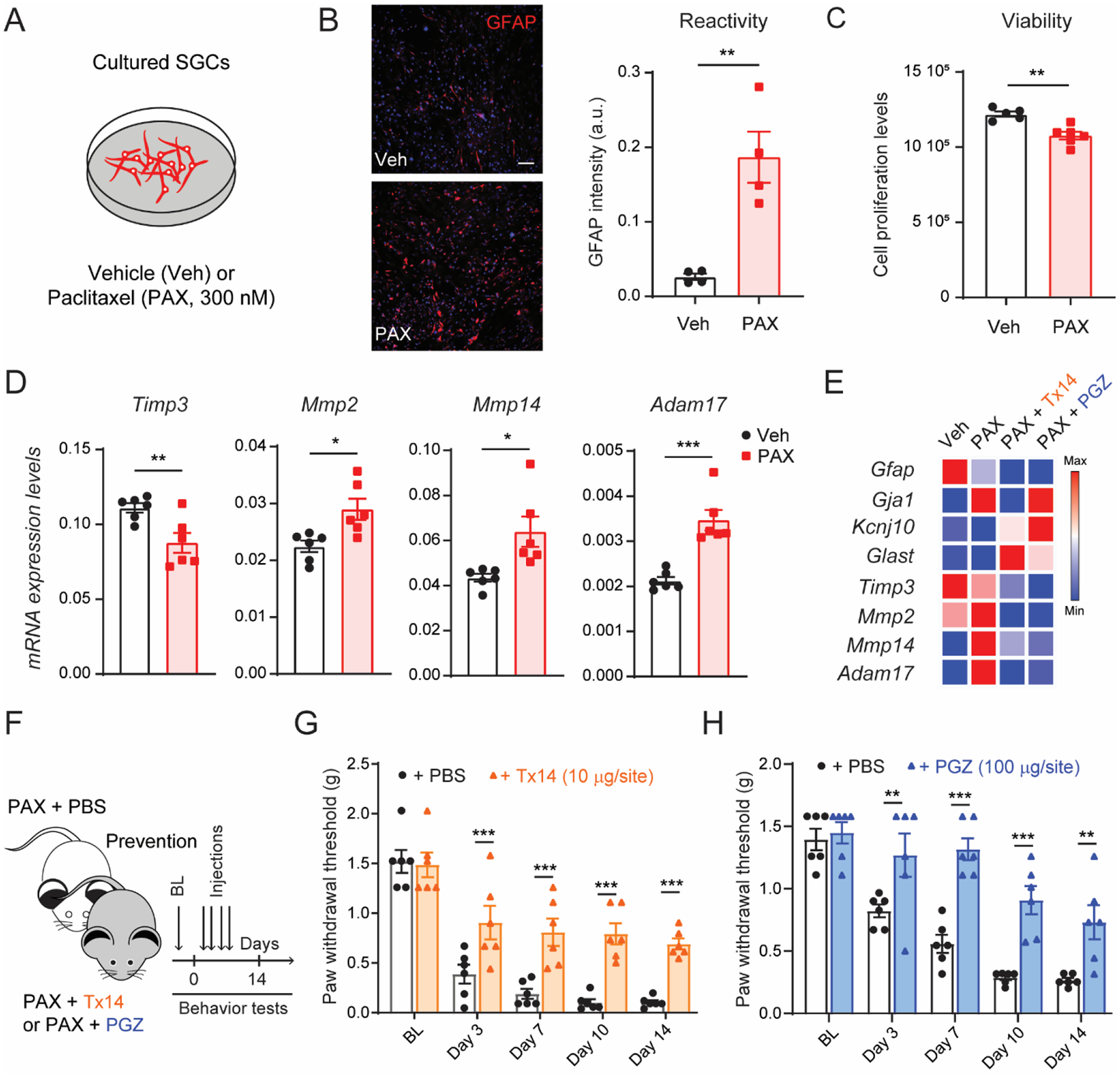
Satellite glial cells (SGCs) are among the most abundant non-neuronal cells in dorsal root ganglia (DRGs) and closely envelop sensory neurons that detect painful stimuli. However, little is still known about their homeostatic activities and their contribution to pain. Using single-cell RNA sequencing (scRNA-seq), we were able to obtain a unique transcriptional profile for SGCs. We found enriched expression of the tissue inhibitor metalloproteinase 3 (TIMP3) and other metalloproteinases in SGCs. Small interfering RNA and neutralizing antibody experiments revealed that TIMP3 modulates somatosensory stimuli. TIMP3 expression decreased after paclitaxel treatment, and its rescue by delivery of a recombinant TIMP3 protein reversed and prevented paclitaxel-induced pain. We also established that paclitaxel directly impacts metalloproteinase signaling in cultured SGCs, which may be used to identify potential new treatments for pain. Therefore, our results reveal a metalloproteinase signaling pathway in SGCs for proper processing of somatosensory stimuli and potential discovery of novel pain treatments.

Keywords: Metalloproteinases; Neuropathic pain; Satellite glial cells; Single-cell RNA sequencing.


Conflict of interest statement

Declaration of Competing Interest The authors declare that they have no known competing financial interests or personal relationships that could have appeared to influence the work reported in this paper.

Figures

Fig. 1.
Fig. 1.. Cellular heterogeneity of the DRG tissues.
(A) Overview of the experimental strategy is shown. Pooled DRG cell suspensions from three naïve mice were used for scRNA-seq, which was performed using the Chromium droplet encapsulating technology from 10X Genomics. Unsupervised clusters were generated by uniform manifold approximation and projection (UMAP). (B) UMAP plot of 10,172 DRG individual cells is shown. Each point represents an individual cell, and clusters are colored by cell type assignment. (C) Cluster names are followed by the number of cells per cluster, UMIs per cluster, and genes detected per cluster. (D) Heat map of expression of the top 50 most significantly enriched genes for each cluster is shown. (E) UMAP plots for the neuronal marker Tubb3, non-neuronal marker B2m, macrophage marker Aif1, and glial cell marker Sox10 are presented. The scale and color indicate log2 expression of each gene.
Fig. 2.
Fig. 2.. Identification of two distinct populations of satellite glial cells (SGCs).
(A) Illustration of SGCs wrapping around sensory neurons in DRGs. (B) Venn diagram of enriched genes of the two populations of SGCs and Schwann cells. (C) Expression levels of Plp1, Slc3a1, Gja1, and Kcnj10 in SGC clusters, as shown by violin plots with a log-normalized y-axis. (D) Violin plots for Entpd2 and representative RNAscope images of a DRG tissue for Gja1 and Entpd2. DAPI (blue) is used as nuclei counterstaining. (E) Gene ontology (GO) analysis of genes expressed in the SGCs I and SGCs II populations.
Fig. 3.
Fig. 3.. Transcriptional and protein expression of Timp3.
(A) Schematic illustration of TIMP3’s inhibitory activity on matrix metalloproteinases (MMPs) and on disintegrin and metalloprotease 17 (ADAM17), also called TACE (tumor necrosis factor-α-converting enzyme). (B, C) RNAscope localization of Timp3, Mmp14 and Adam17 mRNA expression in DRG of Plp1-Cre/tdTomato mice, wherein Cre recombinase is expressed in satellite glial cells (SGCs) (B), and in naïve CD1 mice (C). Arrowheads indicate mRNA colocalization of Mmp14 and Adam17 with Timp3 in SGCs, * indicates neurons. Scale bars = 25 μm in (B) and 5 μm in (C). (D) PCR in mouse and human DRG tissues. Samples with omitted RT (reverse transcriptase) show no bands, confirming the specificity of the amplification. (E) Immunofluorescence of TIMP3 in human DRG tissue. Scale bars = 50 μm. DAPI was used as counterstain. # indicates the fluorescent signal due to the presence of lipofuscins in human DRG neurons.
Fig. 4.
Fig. 4.. Timp3 controls mechanical and thermal sensitivities in naïve mice.
(A) Schematic illustration of the experiment showing the timeline of siRNA injections, pharmacological and biochemical studies. (B) Western blot representative image and quantification show that Timp3 siRNA significantly decreases Timp3 protein levels in DRGs tissues (n=4). (C) Timp3 siRNA injections do not cause locomotor dysfunction in the Rota-rod test (n=5). (D) Mechanical and thermal (von Frey, Hargreaves, and dry ice) allodynia induced by Timp3 siRNA compared to a control (Ctrl) non-targeting siRNA (2 μg of siRNA per delivery in the transfection agent PEI, n=7). (E) Anti-allodynic effect of exogenous recombinant TIMP3 (rTIMP3, 100 ng/site, i.t.), general endogenous tissue inhibitor of MMPs (TIMP-1, 4 pmol/site), MMP2 and MMP14 inhibitors (10 μg/site, i.t.), TACE/ADAM17 inhibitor (TAPI-2, 1 μg/site, i.t.), and a neutralizing antibody for TNF-α (5 μg/site, i.t.) on mechanical allodynia induced by Timp3 siRNA on day 2. (F) Anti-TIMP3 antibody (TIMP3 Ab, 10 μg/site, i.t.) induces mechanical allodynia compared to IgG control in male and female mice. BL = baseline. Data are expressed as mean ± SEM and statistically analyzed by two-tailed t-test (B, C, E) and Two-way ANOVA followed by Sidak’s post hoc test (D, F). *P < 0.05, **P < 0.01, ***P < 0.001, ****P < 0.0001.
Fig. 5.
Fig. 5.. Recombinant TIMP3 protein reverses and prevents mechanical and cold allodynia in a mouse model of chemotherapy-induced neuropathic pain.
(A) Schematic of the experiment showing the timeline of paclitaxel (PAX) or vehicle injections, pharmacological studies, and immunohistochemistry (IHC) analysis. (B) Representative image and (C) quantification of Timp3 protein in mouse DRG tissue 14 days after first injection of PAX or vehicle control (n = 5). (D, E) Paclitaxel-induced mechanical (von Frey) and (F) cold allodynia (dry ice) are significantly, dose-dependently reversed up to 6 h by single intrathecal administration of recombinant TIMP3 protein (rTIMP3, 3–100 ng/site) delivered at day 14 after chemotherapy injection (n = 4–5). (G) Schematic of experiment showing timeline of PAX injections concomitantly with rTIMP3 or PBS, behavioral tests, transcriptional, and histological analysis. (H, I) Repeated administrations of rTIMP3 (100 ng/site, i.t.) prevent paclitaxel-induced mechanical and cold allodynia (n = 5–6). BL = baseline. Data expressed as mean ± SEM, statistically analyzed by two-tailed t-test (C), two-way ANOVA followed by Sidak’s post hoc test (D, F, H, I), and one-way ANOVA followed by Turkey’s post hoc test (E). *P < 0.05, **P < 0.01, ***P < 0.001.
Fig. 6.
Fig. 6.. Transcriptional analyses of TIMP3 signaling in cultured SGCs after paclitaxel treatment.
(A) Schematic of the experimental design used in cultured SGCs. (B) Representative image and quantification of immunofluorescence intensity of GFAP protein in SGC culture after 24 h of incubation with paclitaxel (PAX, 300 nM) or vehicle control (Veh; n=4). (C) Quantification of SGC culture viability 24 h after PAX or Veh treatment (n=6). (D) Quantification of mRNA expression levels of Timp3, Mmp2, Mmp14 and Adam17 in SGC culture after PAX or Veh incubation (n=6). (E) Heat map of mRNA expression of SGC and metalloprotease signaling markers in SGC culture after incubation with prosaptide Tx14 (1 μM) or pioglitazone (PGZ, 10 μM) and PAX compared to vehicle (n = 3). (F) Schematic illustrating the timeline of Tx14, PGZ, or PBS concomitantly treated with PAX, and the behavioral assay. Repeated injections of (G) Tx14 (10μg/site, i.t.) or (H) PGZ (100μg/site, i.t.) prevent paclitaxel-induced mechanical allodynia (n=6). BL = baseline. Data are expressed as mean ± SEM and statistically analyzed by two-tailed t-test (B, C, D), and Two-way ANOVA followed by Sidak’s post hoc test (G, H): *P < 0.05, **P < 0.01, ***P < 0.001.

References

    1. Avraham O, Deng PY, Jones S, Kuruvilla R, Semenkovich CF, Klyachko VA, Cavalli V, 2020. Satellite glial cells promote regenerative growth in sensory neurons. Nat Commun 11, 4891. - PMC - PubMed
    1. Avraham O, Feng R, Ewan EE, Rustenhoven J, Zhao G, Cavalli V, 2021. Profiling sensory neuron microenvironment after peripheral and central axon injury reveals key pathways for neural repair. Elife 10. - PMC - PubMed
    1. Basbaum AI, Bautista DM, Scherrer G, Julius D, 2009. Cellular and molecular mechanisms of pain. Cell 139, 267–284. - PMC - PubMed
    1. Becht E, McInnes L, Healy J, Dutertre CA, Kwok IWH, Ng LG, Ginhoux F, Newell EW, 2018. Dimensionality reduction for visualizing single-cell data using UMAP. Nat Biotechnol. - PubMed
    1. Bennett GJ, Doyle T, Salvemini D, 2014. Mitotoxicity in distal symmetrical sensory peripheral neuropathies. Nat Rev Neurol 10, 326–336. - PMC - PubMed

Publication types