. 2023 Nov 27;60(12):1235-1244.
doi: 10.1136/jmg-2023-109362.

Use of genome sequencing to hunt for cryptic second-hit variants: analysis of 31 cases recruited to the 100 000 Genomes Project

Collaborators, Affiliations

Use of genome sequencing to hunt for cryptic second-hit variants: analysis of 31 cases recruited to the 100 000 Genomes Project

A Rachel Moore et al. J Med Genet. .

Abstract

Background: Current clinical testing methods used to uncover the genetic basis of rare disease have inherent limitations, which can lead to causative pathogenic variants being missed. Within the rare disease arm of the 100 000 Genomes Project (100kGP), families were recruited under the clinical indication 'single autosomal recessive mutation in rare disease'. These participants presented with strong clinical suspicion for a specific autosomal recessive disorder, but only one suspected pathogenic variant had been identified through standard-of-care testing. Whole genome sequencing (WGS) aimed to identify cryptic 'second-hit' variants.

Methods: To investigate the 31 families with available data that remained unsolved following formal review within the 100kGP, SVRare was used to aggregate structural variants present in <1% of 100kGP participants. Small variants were assessed using population allele frequency data and SpliceAI. Literature searches and publicly available online tools were used for further annotation of pathogenicity.

Results: Using these strategies, 8/31 cases were solved, increasing the overall diagnostic yield of this cohort from 10/41 (24.4%) to 18/41 (43.9%). Exemplar cases include a patient with cystic fibrosis harbouring a novel exonic LINE1 insertion in CFTR and a patient with generalised arterial calcification of infancy with complex interlinked duplications involving exons 2-6 of ENPP1. Although ambiguous by short-read WGS, the ENPP1 variant structure was resolved using optical genome mapping and RNA analysis.

Conclusion: Systematic examination of cryptic variants across a multi-disease cohort successfully identifies additional pathogenic variants. WGS data analysis in autosomal recessive rare disease should consider complex structural and small intronic variants as potentially pathogenic second hits.

Keywords: diagnosis; genetic diseases, inborn; genetics, medical; genomics; sequence analysis, DNA.


Conflict of interest statement

Competing interests: None declared.

Figures

Figure 1
Figure 1
Overall cohort structure. Summary of patients recruited to subcohort ‘single autosomal recessive mutation in rare disease’.
Figure 2
Figure 2
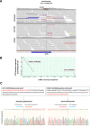
Validation of LINE1 insertion in CFTR by Sanger sequencing in P1. (A) Illumina short-read sequencing data supporting LINE1 insertion in P1. Approximate position of MLPA probes (MRC Holland) used during clinical testing is indicated by the grey bar above the coverage track. Left MLPA probe chr7:117,603,631-117,603,659; right MPLA probe chr7:117,603,660-117,603,706. Insertion sequences called by Manta are shown at the proximal (LEFT_INSSEQ) and distal (RIGHT_INSSEQ) breakpoints. Illumina short-read sequencing is unable to read through the full insertion sequence. (B) Dot plot showing comparison of insertion sequences called by Manta to the LINE1 consensus sequence using the BLAST tool at https://blast.ncbi.nlm.nih.gov/. (C) Sanger sequencing to confirm the breakpoint and LINE1 insertion sequence. Sanger sequences using reverse primer (not shown) also confirmed the breakpoint and insertion sequences. MLPA, multiplex ligation probe amplification.
Figure 3
Figure 3
Illumina short-read sequencing data supporting other SVs reported in this study. (A) P2: proband was recruited as part of a trio and data from both parents are also shown. Left panel: deletion of approximately 346.5 kb affecting exon 24 of RAB3GAP1 and exons 2–21 of ZRANB3. The deletion is also present in the father, but is mosaic rather than heterozygous (estimated to be present in 44% of cells (online supplemental figure S1)). Right panel: maternally inherited 4 bp deletion in exon 21 of RAB3GAP1. (B) P3: deletion of approximately 16.65 kb affecting exons 23–29 of ABCC6. SVs, structural variants.
Figure 4
Figure 4
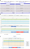
Illumina and Bionano optical genome mapping data supporting ENPP1 duplications in P4. DNA was extracted, labelled and stained according to Bionano Genomics protocols (Bionano Prep SP Frozen Human Blood DNA Isolation Protocol v2 (Document Number: 30395, Revision: B) and Bionano Prep Direct Label and Stain (DLS) Protocol (Document Number: 30206, Revision: F)). (A) Illumina short-read WGS showing duplications (gain of material) of ENPP1 exons 2–6 and region between LAMA2 and ARHGAP18. (B) Bionano data showing undisrupted intergenic locus (upper panel) and ENPP1 duplication (lower panel) with novel insertion (inversion of non-coding LOC102723409 sequence). Note the ENPP1 locus mapping to two genomic locations in the lower panel denoted by the vertical and diagonal grey lines spanning from the green reference genome to the blue proband sequence. The diagramatic schema in the lower panel shows the overall interpretation of the SV, with the duplicated region of ENPP1 sequence separated by the inverted insertion of LOC102723409 sequence. (C) Single molecule view at ENPP1 locus showing multiple molecules spanning the entire SV. SV, structural variant.
Figure 5
Figure 5
Radiological imaging supporting diagnosis of generalised arterial calcification of infancy in P4 (A–D) and Jeune thoracic dystrophy in P8 (E). (A) Anteroposterior radiograph of left knee aged 12 years demonstrating ‘wide epiphyseal plates of the knee’ with ‘frayed metaphyses of the femora’ and ‘frayed metaphyses of the tibiae’, the latter affecting the medial aspect of the proximal tibia. Appearances are consistent with hypophosphataemic rickets. (B) Axial image from dental CT examination performed aged 20 years. There is mural calcification of the facial arteries (black arrows) and calcification of the included portion of the pinnae of the ears (white arrows). (C) and (D) Axial images from non-contrast urinary tract CT examination aged 20 years. There is mural calcification of the superior mesenteric artery (black arrowhead in C) and both common femoral arteries (white arrowheads in D). Increased density is noted in the renal medullae (C), consistent with early medullary nephrocalcinosis. Calcification of hepatic artery branches was also noted (not shown). In addition, calcification of the popliteal arteries was noted on a CT examination of the lower limbs at age 14 years (not shown). (E) Anteroposterior chest radiograph (aged 6 months) shows a ‘narrow chest’ with ‘short ribs’ and ‘prominent ribs’'. Images annotated using terms from the dREAMS ontology (https://d-reams.org) in inverted commas.

References

    1. Carvill GL, Mefford HC. Microdeletion syndromes. Curr Opin Genet Dev 2013;23:232–9. 10.1016/j.gde.2013.03.004 - DOI - PubMed
    1. Sohn YB, Ko JM, Jang JY, et al. . Deletion of exons 16-17B of CFTR is frequently identified in Korean patients with cystic fibrosis. Eur J Med Genet 2019;62:103681. 10.1016/j.ejmg.2019.103681 - DOI - PubMed
    1. Pagnamenta AT, Yu J, Evans J, et al. . Conclusion of diagnostic odysseys due to inversions disrupting Gli3 and FBN1. J Med Genet 2023;60:505–10. 10.1136/jmg-2022-108753 - DOI - PMC - PubMed
    1. Schuy J, Grochowski CM, Carvalho CMB, et al. . Complex genomic rearrangements: an underestimated cause of rare diseases. Trends Genet 2022;38:1134–46. 10.1016/j.tig.2022.06.003 - DOI - PMC - PubMed
    1. Sawyer SL, Hartley T, Dyment DA, et al. . Utility of whole‐exome sequencing for those near the end of the diagnostic odyssey: time to address gaps in care. Clin Genet 2016;89:275–84. 10.1111/cge.12654 - DOI - PMC - PubMed

Publication types