

Ruptured Sinus of Valsalva Aneurysm in a Patient with a Ventricular Septal Defect Who Dropped Out of Lifelong Medical Follow-up
- PMID: 37558488
- PMCID: PMC11008991
- DOI: 10.2169/internalmedicine.1395-22
Ruptured Sinus of Valsalva Aneurysm in a Patient with a Ventricular Septal Defect Who Dropped Out of Lifelong Medical Follow-up
Abstract
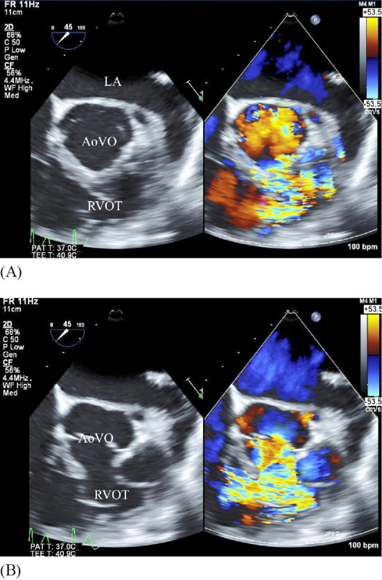
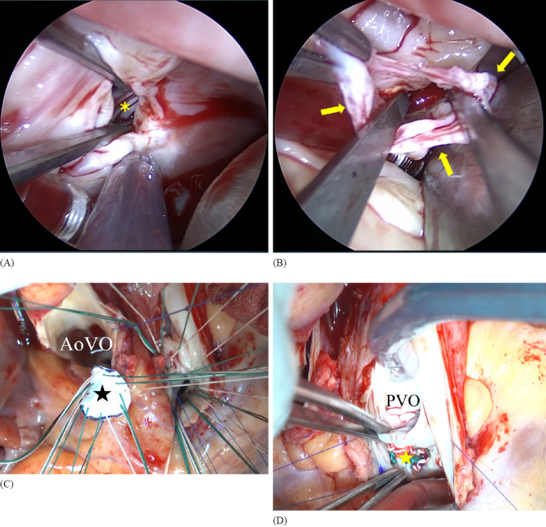
Ruptured sinus of Valsalva aneurysm (RSOVA) is a rare cardiac condition associated with high morbidity and mortality rates. We herein report a 35-year-old man with a history of ventricular septal defect (VSD). He had a history of interrupted hospital visits and presented to the emergency department with dyspnea, palpitations, and dizziness for a few days. Auscultation detected a continuous murmur. Transthoracic echocardiography followed by transesophageal echocardiography demonstrated RSOVA in the right ventricle with an aorto-right ventricular fistula. The fistula was resected, and the aneurysm was surgically repaired. The patient made a good recovery.
Keywords: continuous murmur; ruptured sinus of Valsalva aneurysm; ventricular septal defect.
Conflict of interest statement
Figures


References
-
- Tariq M, Andrew B, Bassent B, et al. Rupture of sinus of Valsalva aneurysm: Two case reports and a concise review of the literature. Heart Lung 47: 131-135, 2018. - PubMed
-
- Feldman DN, Roman MJ. Aneurysms of the sinuses of Valsalva. Cardiology 106: 73-81, 2006. - PubMed
-
- Smith WA. Aneurysm of the sinus of Valsalva, with report of 2 cases. JAMA 24: 1878-1880, 1914.
-
- Goldberg N, Krasnow N. Sinus of Valsalva aneurysms. Clin Cardiol 13: 831-836, 1990. - PubMed