. 2023 Aug 9;14(1):4816.
doi: 10.1038/s41467-023-40541-1.

A HIF independent oxygen-sensitive pathway for controlling cholesterol synthesis

Affiliations

A HIF independent oxygen-sensitive pathway for controlling cholesterol synthesis

Anna S Dickson et al. Nat Commun. .

Erratum in

Abstract

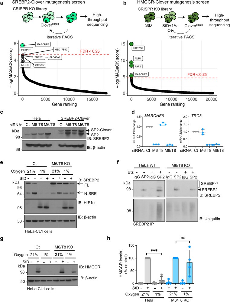
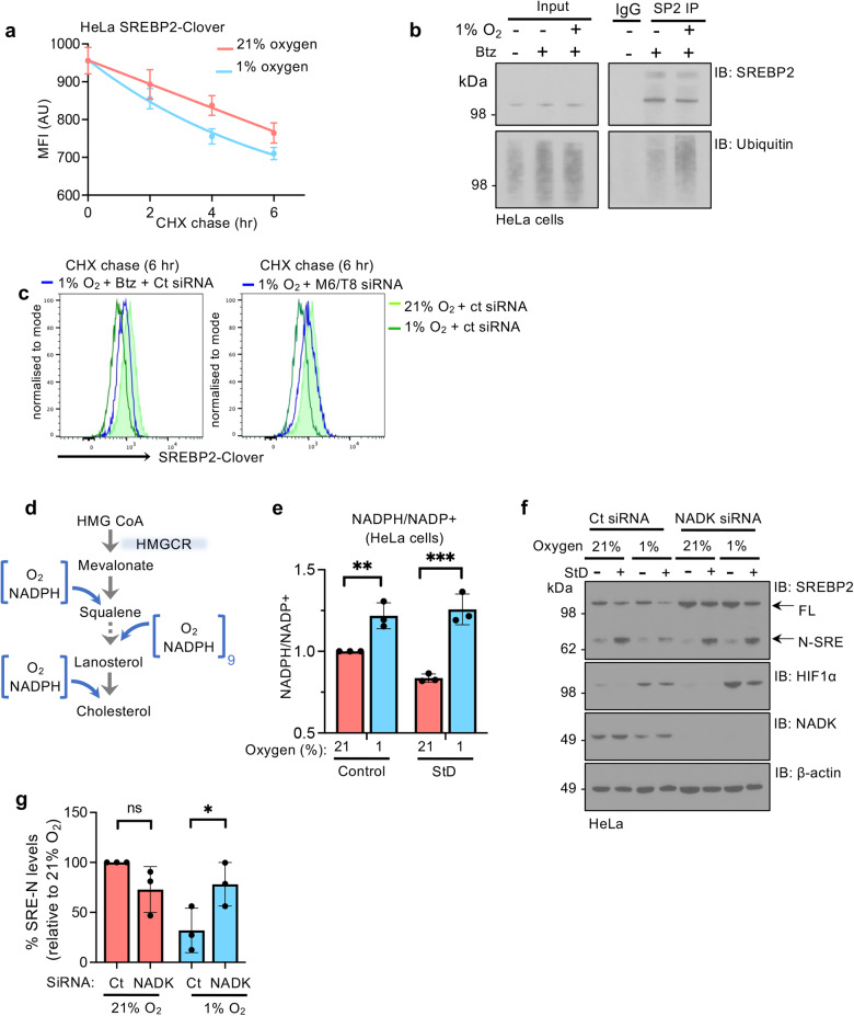
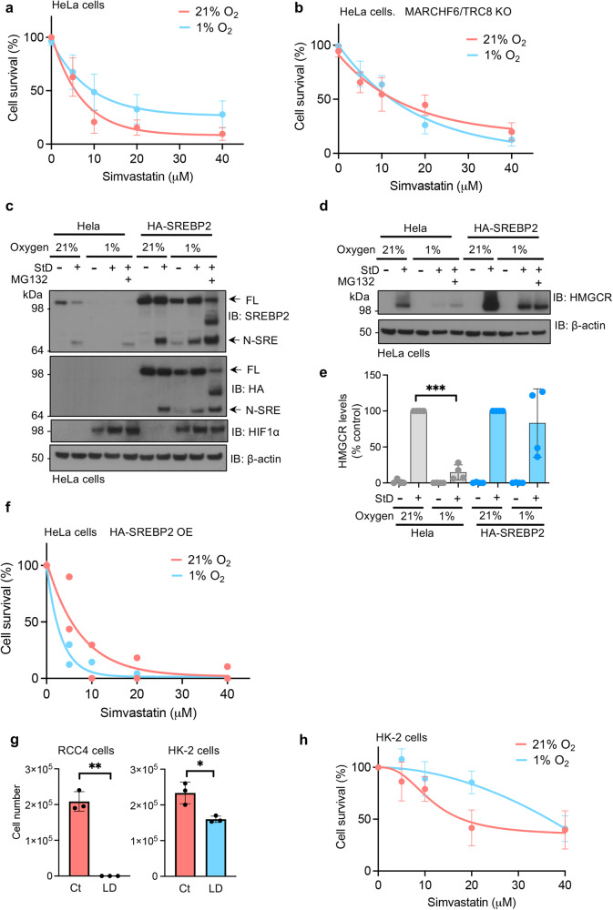
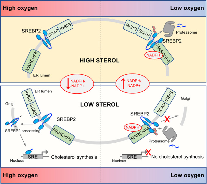
Cholesterol biosynthesis is a highly regulated, oxygen-dependent pathway, vital for cell membrane integrity and growth. In fungi, the dependency on oxygen for sterol production has resulted in a shared transcriptional response, resembling prolyl hydroxylation of Hypoxia Inducible Factors (HIFs) in metazoans. Whether an analogous metazoan pathway exists is unknown. Here, we identify Sterol Regulatory Element Binding Protein 2 (SREBP2), the key transcription factor driving sterol production in mammals, as an oxygen-sensitive regulator of cholesterol synthesis. SREBP2 degradation in hypoxia overrides the normal sterol-sensing response, and is HIF independent. We identify MARCHF6, through its NADPH-mediated activation in hypoxia, as the main ubiquitin ligase controlling SREBP2 stability. Hypoxia-mediated degradation of SREBP2 protects cells from statin-induced cell death by forcing cells to rely on exogenous cholesterol uptake, explaining why many solid organ tumours become auxotrophic for cholesterol. Our findings therefore uncover an oxygen-sensitive pathway for governing cholesterol synthesis through regulated SREBP2-dependent protein degradation.


Conflict of interest statement

J.A.N. receives a Pfizer ITEN discovery grant for unrelated work to this manuscript. Other authors declare no competing interests.

Figures

Fig. 1
Fig. 1. Hypoxia promotes SREBP2 degradation.
a Schematic of known sterol-sensing pathway for regulating cholesterol synthesis at the ER membrane. b, c SREBP2 processing in HeLa cells treated with or without sterol depletion (StD: DMEM supplemented with 10% lipid-depleted FCS and 10 μM mevastatin) in 21% and 1% oxygen for 40 h. SREBP2 processing to release N-terminal transcription factor (N-SRE) determined by immunoblot (b) and quantified using ImageJ. Full length SREBP2 21% versus 1%, P = 0.02, N-SRE 21% versus 1% P = 0.001 (c). n = 3 biological repeats, mean ± SD. *P ≤ 0.05 Two-way ANOVA. d SREBP2 mRNA levels with or without StD in 21% and 1% oxygen for 40 h. SREBP2 StD 21% versus 1%, P = 0.008. n = 5 biological repeats, mean ± SD. **P ≤ 0.01 Two-way ANOVA. e SREBP2 processing in normal media (N) or StD in 21% and 1% oxygen for 0 h, with or without addition of the proteasome inhibitor for 16 h prior to lysis (20 nM bortezomib, Btz). Immunoblot representative of 3 independent experiments. Flow cytometry analysis of HeLa SREBP2-Clover cells in 21% or 1% oxygen after 24 h (f), following incubation in 0–24 h 1% oxygen (g), and with proteasome inhibition (20 nM Btz 16 h) (h), and following StD and incubation in 21%, 5% and 1% oxygen for 24 h (i). Source data are provided as a Source Data file. n = 3 biological repeats. FL full length SREBP2, AU arbitrary units.
Fig. 2
Fig. 2. Hypoxia overrides the canonical sterol-sensing response.
a Flow cytometry analysis of endogenous HMGCR-Clover levels following incubation in 21% or 1% oxygen, with or without StD for 40 h. b Endogenous HMGCR levels in HeLa cells following incubation in 21% or 1% oxygen, with or without StD, as described. Immunoblots representative of 3 independent experiments. HMGCR-Clover knock-in (c) or wildtype HeLa (d) were treated with StD or hypoxia as described, but also with the proteasome inhibitor bortezomib (20 nM for final 18 h). Representative of 4 independent experiments. e Concurrent treatment of HMGCR-Clover cells with StD and either 21% or 1% oxygen for 24 h (left); or pre-treatment of HMGCR-Clover HeLa cells with StD for 24 h followed by incubation in 21% or 1% oxygen for 16 h (right). f Schematic of LC-MS analysis of [13C]glucose uptake to trace incorporation of 13C into newly synthesised cholesterol. g, h Cholesterol isotopomers in HeLa cells cultured in control or lipid-depleted media and incubated in 21% or 1% oxygen for 24 h. HeLa cells were then supplemented with [13C]glucose media for a further 24 h in the same conditions. Cells were also cultured in normal [13C]glucose DMEM media with or without a statin treatment to control for the detection of newly synthesised cholesterol isotopomers. Cholesterol (M + 0) levels (g) and the most abundant isotopomers are shown (h), relative to cell counts. Source data are provided as a Source Data file. P ≤ 0.0001. n = 2 biological repeats, mean ± SD. ***P ≤ 0.00.1 Two-way ANOVA. Arb. units arbitrary units.
Fig. 3
Fig. 3. Oxygen-mediated regulation of cholesterol synthesis is independent of HIFs.
a Flow cytometry analysis of HMGCR-Clover knock-in following StD and 1 mM DMOG or 100 μM Roxadustat (left panel), or following 1% oxygen (24 h) (right panel). b HMGCR levels in control or HIF1β null (mixed KO population) HMGCR-clover knock-in HeLa cells,with or without StD for 24 h, followed by incubation in 21% or 1% oxygen, or following 1 mM DMOG (16 h). c Flow cytometry analysis of SREBP2-Clover reporter levels in control HeLa or HIF1β null cells. Mixed KO populations of HIF1β were generated by sgRNA. Cells were cultured with or without StD for 40 h, and incubated in 21% or 1% oxygen for the final 16 h. Representative of 3 independent experiments. d Immunoblot of SREBP2 in HeLa HIF1β clonal KO cells compared to control HeLa (WT) following SD, with or without incubation in 1% oxygen. Representative of 3 independent experiments. e mRNA levels of the SREBP2 target genes HMGCR and HMGCS1 in HIF1β clonal KO or control HeLa cells following StD, with or without incubation in 1% oxygen. StD 21% oxygen versus 1% oxygen HMGCR, P = 0.011, HMGCS1, P = 0.05. n = 3 biological repeats, mean ± SD. *P ≤ 0.05, Two-way ANOVA. fh SREBP2 and HMGCR levels in HeLa control or Hela HIF1β KO cells cultured in 21%, 12%, 7%, 5% or 1% oxygen, with or without StD for 24 h. Immunoblot for proteins levels of SREBP2 and HMGCR for 21%, 5% and 1% (f). Protein levels in 12% and 7% oxygen shown in Supplementary Fig. 3j. Protein levels of N-SRE (g) and HMGCR (h) following StD were quantified by ImageJ following normalisation to β-actin. N-SRE Ct 21% versus 1% oxygen, P < 0.0001, N-SRE Ct 21% versus 1%, P < 0.0001, N-SRE HIF1β KO 21% versus 1% oxygen, P < 0.0001, N-SRE HIF1β KO 21% versus 1%, P < 0.0001, HMGCR Ct 21% versus 1% oxygen, P < 0.0001, HMGCR Ct 21% versus 1%, P < 0.0001, HMGCR HIF1β KO 21% versus 1% oxygen, P < 0.0001, HMGCR HIF1β KO 21% versus 1%, P < 0.0001. Source data are provided as a Source Data file. n = 3 biological repeats, mean ± SD. **P ≤ 0.01, ***P ≤ 0.001 Two-way ANOVA.
Fig. 4
Fig. 4. MARCHF6 degrades SREBP2 in hypoxia.
a, b Summary of parallel CRISPR/Cas9 mutagenesis screens using the SREBP2-Clover reporter (a) or HMGCR-Clover reporter cells (b). a Mutagenised SREBP2-Clover cells were sorted for a CloverHIGH population in 21% oxygen by FACS at day 9, and underwent a second sort at day 17 to enrich for this population. b Mutagenised HMGCR-Clover cells were sorted for a CloverHIGH population in StD conditions (42 h) and incubated in 1% oxygen for the last 18 h (day 9). The enriched population underwent a second sort at day 14 to enrich for this CloverHIGH population. SgRNA were identified by Illumina NovaSeq (a) or HiSeq (b), and compared to mutagenized population that had not undergone phenotypic selection. Comparative bubble plots are shown. Unadjusted P value calculated using MaGECK robust rank aggregation (RRA); red line false discovery rate (FDR) ≤ 0.25. Benjamini-Hocherg FDR. c, d Endogenous SREBP2 and SREBP2-Clover knock-in levels in HeLa cells following siRNA-mediated depletion of MARCHF6 (M6), TRC8 (T8), or combined M6/T8 depletion (c). mRNA levels of MARCHF6, TRC8 and SREBP2 confirm siRNA-mediated depletion of the E3 ligases (d). n = 3 biological repeats, mean ± SD. e HeLa mCherry-CL1 control (Ct) or MARCHF6/TRC8 double knockout clonal cells were treated StD for 42 h, with or without 1% oxygen for the final 18 h. SREBP2 processing was analysed by immunoblot. Representative of 3 biological repeats. f Ubiquitination of SREBP2. Immunoprecipitation of SREBP2 in HeLa control or combined MARCHF6/TRC8 siRNA-depleted cells. Prior to lysis, cells were treated with or without Btz (5 μM 6 h). Immunoprecipitated SREBP2 was analysed by SDS–PAGE and immunoblotted for ubiquitin. Representative of 3 biological repeats. g, h HeLa mCherry-CL1 control (Ct) or MARCHF6/TRC8 double knockout clonal cells were treated StD for 42 h, with or without 1% oxygen for the final 18 h. HMGCR levels (g) were analysed by immunoblot. HMGCR levels were quantified using ImageJ (h). HMGCR StD 21% versus 1% oxygen, P < 0.0001, M6/T8 KO StD 21% versus 1% oxygen, P = 0.89. Source data are provided as a Source Data file. n = 4 biological repeats, mean ± SD. ***P ≤ 0.001 Two-way ANOVA.
Fig. 5
Fig. 5. The hypoxia cholesterol response is mediated by MARCHF6 NADPH sensing.
a SREBP2-Clover levels by flow cytometry in HeLa SREBP2-Clover knock-in cells treated with 10 µg/ml of cycloheximide for 0–6 h with or without treatment with 1% oxygen. Quantification of mean fluorescence intensity (MFI) shown. n = 5 independent biological replicates, mean ± SD. b Ubiquitination of SREBP2 in hypoxia. Immunoprecipitation of SREBP2 in HeLa cells incubated in 21% or 1% oxygen for 6 h, with or without Btz (5 μM 6 h). Immunoprecipitated SREBP2 was analysed by SDS–PAGE and immunoblotted for ubiquitin. Representative of 3 biological replicates. c SREBP2-Clover levels by flow cytometry in reporter cells following treatment with 10 µg/ml of cycloheximide for 6 h and 1% oxygen, with or without Btz (5 μM 6 h) (left panel), or following combined siRNA-mediated depletion of MARCHF6 (M6) and TRC8 (T8) (right panel). The data depicted in the left and right panels originated from the same experiment and as such the control plots are the same in both. Representative of 3 biological replicates. d Schematic of oxygen consuming and NADPH oxidation steps within the cholesterol synthetic pathway. e Relative NADPH/NADP+ levels in HeLa cells following incubation in 21% or 1% oxygen, with or without StD for 24 h. Control 21% versus 1% oxygen, P = 0.006, StD 21% versus 1% oxygen, P < 0.0001. n = 3 biological repeats, mean ± SD. **P ≤ 0.01, ***P ≤ 0.001 Two-way ANOVA. f, g HeLa cells were depleted for NADK using siRNA or a mock control siRNA. After 48 h cells were incubated in 21% or 1% oxygen, with or without StD for 24 h. SREBP2 processing was visualised by immunoblot (f), and protein levels of N-SRE following sterol deplete conditions were quantified by ImageJ, following normalisation to β-actin (g). Control versus siRNA NADK in 21% oxygen, P = 0.232, Control versus siRNA NADK in 1% oxygen, P = 0.038. Source data are provided as a Source Data file. n = 3 biological repeats, mean ± SD. *P ≤ 0.05, Two-way ANOVA.
Fig. 6
Fig. 6. Hypoxia promotes cholesterol auxotrophy in tumour cells by suppressing cholesterol synthesis.
HeLa cells (a), or HeLa MARCHF6 and TRC8 KO cells (b) were incubated in 21% or 1% oxygen for 48 h with simvastatin as indicated. Cell viability was measured by Hoechst staining at 48 h. n = 7 (a) and n = 5 (b) biological repeats, mean ± SD. c HeLa cells were transduced with HA-SREBP2 and StD for 42 h, with or without incubation in 1% oxygen as described. Additionally, cells were treated with 10 μM MG132 4 h prior to harvest where appropriate. SREBP2 levels were measured by immunoblot. Immunoblot representative of 2 biological repeats. d, e HeLa cells were transduced with HA-SREBP2 and StD for 42 h, with or without incubation in 1% oxygen as described. HMGCR levels were measured by immunoblot (d) and quantified using ImageJ (e). HMGCR StD 21% versus 1% oxygen, P < 0.0001. n = 4 biological repeats, mean ± SD. ***p ≤ 0.001 Two-way ANOVA. f HeLa cells transduced with HA-SREBP2 were incubated in 21% or 1% oxygen for 48 h with simvastatin as indicated. Cell viability was measured by Hoechst staining at 48 h. n = 2 biological repeats. g 105 RCC4 renal cancer cells or HK-2 RTE cells were cultured in lipid-deplete media (LD) for 3 days and then the total number of viable cells was measured. RCC4 cells control versus LD media, P = 0.006, HK-2 cells control versus LD media 21%, P = 0.04. n = 3 biological repeats, mean ± SD. **P ≤ 0.05, **P ≤ 0.001 Unpaired two sample student T-test. h HK-2 cells were incubated in 21% or 1% oxygen for 48 h with simvastatin as indicated. Cell viability was measured by Hoechst staining at 48 h. Source data are provided as a Source Data file. n = 3 biological repeats, mean ± SD.
Fig. 7
Fig. 7. Model of oxygen-dependent regulation of cholesterol biosynthesis.
Schematic of the oxygen-dependent regulation of SREBP2. In 21% oxygen and sterol replete conditions, SREBP2 is held in the ER through its interaction with SCAP/INSIGs, and basal SREBP2 turnover by MARCHF6 is low (top left). Sterol depletion releases SREBP2 from SCAP/INSIGs, and SREBP2 undergoes processing in the Golgi to generate the N-SRE transcription factor (bottom left). In hypoxia, NAPDH accumulates, increasing MARCHF6 activity in a proportional manner to oxygen availability (top right). This promotes SREBP2 degradation in the ER, and this shuts down cholesterol synthesis in sterol deplete conditions (bottom right).

Similar articles

Cited by

References

    1. Simons K, Ikonen E. How cells handle cholesterol. Science. 2000;290:1721–1726. doi: 10.1126/science.290.5497.1721. - DOI - PubMed
    1. Libby P, Theroux P. Pathophysiology of coronary artery disease. Circulation. 2005;111:3481–3488. doi: 10.1161/CIRCULATIONAHA.105.537878. - DOI - PubMed
    1. Goldstein JL, Brown MS. The LDL receptor. Arterioscler. Thromb. Vasc. Biol. 2009;29:431–438. doi: 10.1161/ATVBAHA.108.179564. - DOI - PMC - PubMed
    1. Johnson BM, DeBose-Boyd RA. Underlying mechanisms for sterol-induced ubiquitination and ER-associated degradation of HMG CoA reductase. Semin. Cell Dev. Biol. 2018;81:121–128. doi: 10.1016/j.semcdb.2017.10.019. - DOI - PMC - PubMed
    1. Horton JD, Goldstein JL, Brown MS. SREBPs: activators of the complete program of cholesterol and fatty acid synthesis in the liver. J. Clin. Investig. 2002;109:1125–1131. doi: 10.1172/JCI0215593. - DOI - PMC - PubMed

Publication types

MeSH terms