. 2023 Aug 9;14(1):4798.
doi: 10.1038/s41467-023-40537-x.

Auranofin targets UBA1 and enhances UBA1 activity by facilitating ubiquitin trans-thioesterification to E2 ubiquitin-conjugating enzymes

Affiliations

Auranofin targets UBA1 and enhances UBA1 activity by facilitating ubiquitin trans-thioesterification to E2 ubiquitin-conjugating enzymes

Wenjing Yan et al. Nat Commun. .

Abstract

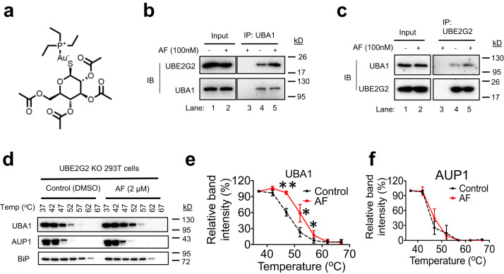
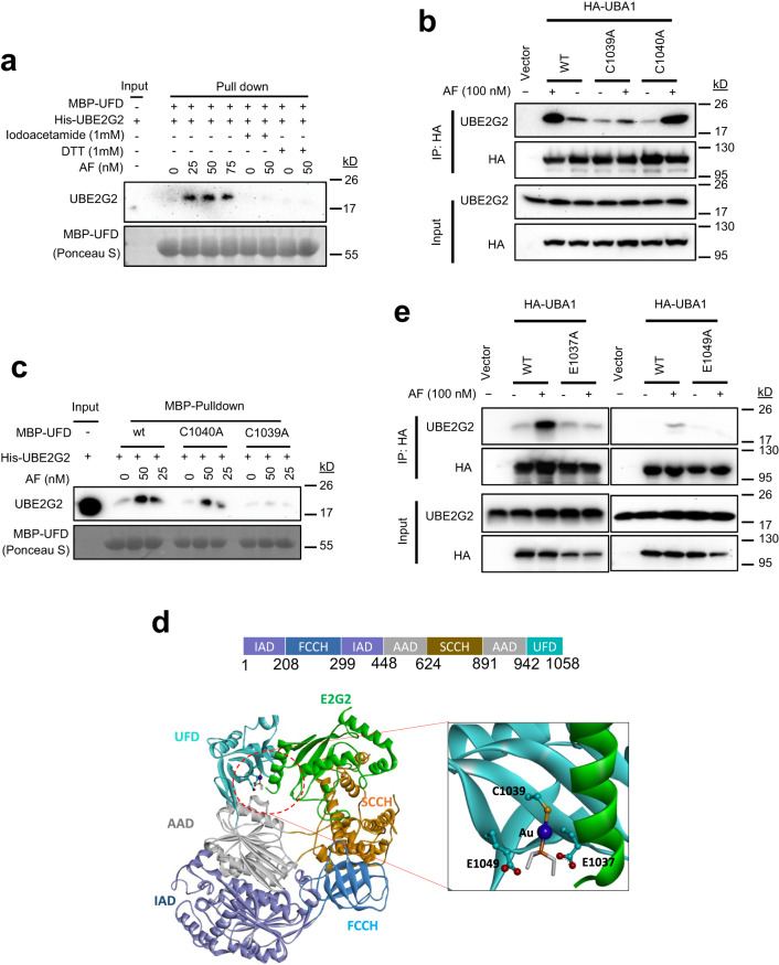
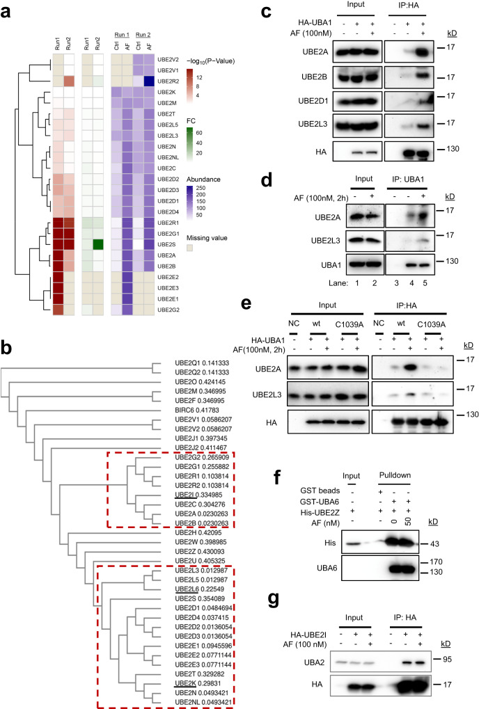
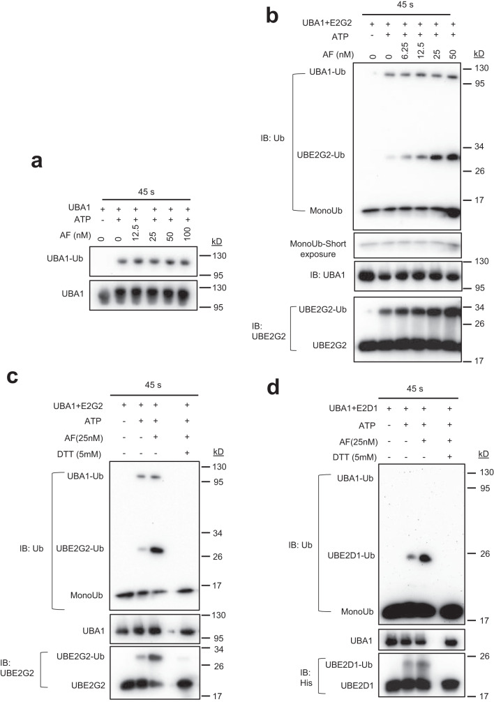
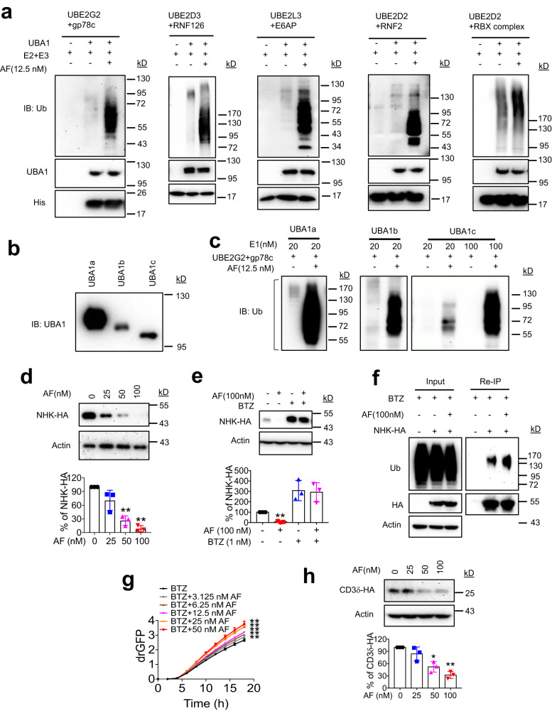
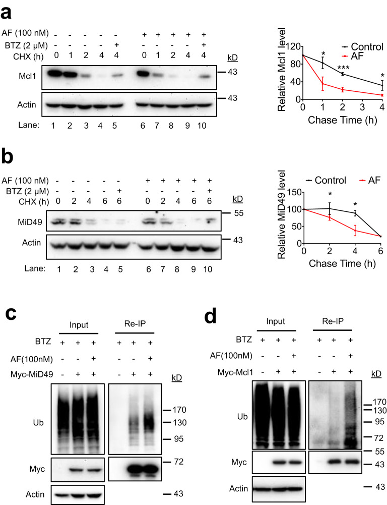
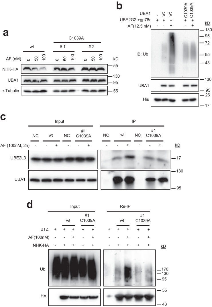
UBA1 is the primary E1 ubiquitin-activating enzyme responsible for generation of activated ubiquitin required for ubiquitination, a process that regulates stability and function of numerous proteins. Decreased or insufficient ubiquitination can cause or drive aging and many diseases. Therefore, a small-molecule enhancing UBA1 activity could have broad therapeutic potential. Here we report that auranofin, a drug approved for the treatment of rheumatoid arthritis, is a potent UBA1 activity enhancer. Auranofin binds to the UBA1's ubiquitin fold domain and conjugates to Cys1039 residue. The binding enhances UBA1 interactions with at least 20 different E2 ubiquitin-conjugating enzymes, facilitating ubiquitin charging to E2 and increasing the activities of seven representative E3s in vitro. Auranofin promotes ubiquitination and degradation of misfolded ER proteins during ER-associated degradation in cells at low nanomolar concentrations. It also facilitates outer mitochondrial membrane-associated degradation. These findings suggest that auranofin can serve as a much-needed tool for UBA1 research and therapeutic exploration.


Conflict of interest statement

The authors declare no competing interests.

Figures

Fig. 1
Fig. 1. Auranofin (AF) binds to UBA1 and enhances UBA1 interaction with UBE2G2.
a Chemical structure of AF. b, c AF enhances UBA1 interaction with UBE2G2 in cells. Total lysates from 293 T cells treated with 100 nM AF for 3 h were used for anti-UBA1 (a) or anti-UBE2G2 (b) co-immunoprecipitation (coIP) assays. Lane 3 is anti-IgG control. d Effects of AF on the thermal stability of UBA1 and AUP1 in UBE2G2 knockout 293 T cells as revealed by CETSA. The indicated proteins were blotted in soluble fractions. e, f The intensities of the bands for UBA1 and AUP1 in (d) were quantified. Data are presented as mean values ± S.D., n = 3 biologically independent experiments. p-value was calculated by two-tailed paired t-test. *p < 0.05 and **p < 0.01 for UBA1. p = 0.008, p = 0.0421 and p = 0.0285 at 47, 52 and 57 oC, respectively. No difference for AUP1. Source data are provided as a Source Data file.
Fig. 2
Fig. 2. AF binds to the UFD of UBA1 and conjugates to cysteine 1039.
a Effect of Iodoacetamide or DTT on the interaction of UBE2G2 with the UFD fragment of UBA1. b C1039A but not C1040A mutation diminishes AF-enhanced UBA1-UBE2G2 interaction in cells. HeLa cells transfected with the indicated plasmids were treated with 100 nM AF for 2 h, followed by processing for IP and IB. c C1039A but not C1040A mutation diminishes AF-enhanced MBP-UFD and UBE2G2 interaction in vitro. Recombinant MBP-UFD and its mutants were incubated with UBE2G2 along with different doses of AF. UBE2G2 bound to MBP-UFD, or its mutants was detected by IB. d Predicted binding complex of UBA1 with auranofin and E2G2. The binding interaction of the portion of Au-PEt3 of auranofin at the UFD domain is shown in a close-up panel. The protein structure of UBA1 (PDB 6DC6) and E2G2 (PDB 4LAD) are represented in ribbons. The gold Au (I) is coordinated to Cys1039 and rendered as balls in blue, the triethylphosphine ligand of auranofin in the pocket is shown in sticks. The domain structure of UBA1 is shown on top of the model. e Mutation of two predicted AF binding residues, E1037 and E1049, respectively, abrogates AF-enhanced UBA1-UBE2G2 interaction by coIP. Source data are provided as a Source Data file.
Fig. 3
Fig. 3. AF enhances UBA1 interaction with most of the ubiquitin-conjugating E2s.
a AF enhances UBA1 interactions with 20 E2s in cells. The heatmap was obtained by two independent ani-HA-UBA1 coIP followed by protein identification and quantification by mass spectrometry. The first four columns represent the summary p-value (columns 1,2) and fold change (columns 3,4) for control versus AF treatment groups. Columns 4–8 represent the UBA1-associated protein levels, calculated as an average from quadruplicate samples in each experimental run. b Guide tree of 36 human E2 ubiquitin-conjugating enzymes. Multiple sequence alignment and Guide tree of the 36 E2 proteins was performed by Clustal Omega. The E2s in two clusters in red dash-line rectangles are E2s whose interactions with UBA1 were enhanced by AF as shown in (a). The three E2s underlined are exceptions. The number following the E2s is indicative of the evolutionary distance between the sequences. c Validation of AF-enhanced UBA1-E2 interactions in cells by anti-HA-UBA1 coIP as in (a) followed by IB. d Validation of AF-enhanced UBA1-E2 interactions in cells by anti-UBA1 coIP as in (a) followed by IB. e C1039A mutation diminishes UBA1 interactions with E2s. f AF does not promote the UBA6 interaction with UBE2Z in a GST-UBA6 pull-down assay. g AF does not affect the UBA2 interaction with UBE2I as revealed by anti-HA-UBE2I coIP. Source data are provided as a Source Data file.
Fig. 4
Fig. 4. AF facilitates ubiquitin charging to E2s.
The effects of AF on ubiquitin charging in vitro to UBA1 (a), UBE2G2 (b, c), and UBE2D1 (d). The assays were performed using purified recombinant proteins. 250 nM UBA1 alone or in combination with 4 µM His-E2s (His-UBE2G2 or His-UBE2D1) were treated with increasing concentrations of AF in reaction buffer for 1 min. ATP (50 µM) was added to initiate the reaction. The reactions proceeded at 15 oC for 45 s and stopped by adding non-reducing loading buffer. 5 mM DTT was used to disrupt the thioester bond that links ubiquitin to the active cysteine of E2s. Source data are provided as a Source Data file.
Fig. 5
Fig. 5. AF potently enhances E3 activity in vitro and accelerates ERAD in cells.
a Effects of AF on E3 activity in in vitro E3 autoubiquitination assay. RBX complex: Elongin B/Elongin C/VHL/CUL2/RBX1. b IB of UBA1a (UBA1), UBA1b, and UBA1c used in the E3 activity assay in (c). UBA1a, UBA1b, and UBA1c translation start at methionine 1, 41, and 67, respectively. c In vitro gp78c autoubiquitination assay in the presence of UBA1a, UBA1b, and UBA1c, respectively, and the effects of AF. d AF decreased NHK protein levels in a dose-dependent manner. HeLa cells stably expressing NHK-HA were treated with AF for 24 h and then subject to IB. AF versus Control, p = 0.1482, p = 0.0065 and p = 0.0018 at 25, 50 and 100 µM, respectively. e AF-induced NHK downregulation is blocked by proteasome inhibitor, bortezomib (BTZ). AF versus Control, p = 0.0018. f AF increases NHK-HA ubiquitination in HeLa cells. Anti-HA reIP was performed to determine NHK-HA ubiquitination. g AF increases NHK dislocation in a dose-dependent manner. drGFP intensity was monitored and quantified in IncuCyte S3 Live-Cell Analysis System and expressed as mean ± S.D., n = 4 wells/treatment. **p < 0.01. AF versus Control, p = 0.0076, p = 0.0086, p = 0.008, p = 0.007 and p = 0.0079 at 3.125, 6.25, 12.5, 25 and 50 µM, respectively. p-values were calculated by two-tailed paired t-test. h AF decreased CD3δ protein levels in a dose-dependent manner. AF versus Control, p = 0.2442, p = 0.0238 and p = 0.0052 at 25, 50 and 100 µM, respectively The graphs in (d, e, and h) show % increase for each condition relative to DMSO-treated control. Data are presented as mean values ± S.D., n = 3 biologically independent experiments. *p < 0.05, **p < 0.01. p-values were calculated by two-tailed paired t-test. Source data are provided as a Source Data file.
Fig. 6
Fig. 6. AF accelerates ubiquitination-dependent OMMAD.
a, b Analyses of Mcl1 (a) and MiD49 (b) degradation using cycloheximide (CHX) chase. HeLa cells were incubated with 100 nM AF and CHX for the indicated durations. The relative band density of Mcl1 or MiD49 was normalized to that of Actin. Data are presented as mean values ± S.D., n = 3 biologically independent experiments. *p < 0.05, ***p < 0.001. p-values were calculated by two-tailed unpaired t-test. c, d AF promotes MiD49 (c) and Mcl1 (d) ubiquitination. HeLa cells transient expressing Myc-MiD49 or Myc-Mcl1 were treated with 100 nM AF in the presence or absence of 2 µM BTZ for 4 h. Anti-myc reIP was performed to determine Myc-MiD49 or Myc-Mcl1 ubiquitination. Three biological replicates were performed with one representative experiment shown. Source data are provided as a Source Data file.
Fig. 7
Fig. 7. AF does not enhance NHK ubiquitination and proteasomal degradation in HCT116 cells expressing C1039A mutant UBA1 (HCT116-UBA1(C1039A) cells).
a AF does not downregulate NHK in HCT116-UBA1(C1039A). HCT116-UBA1(C1039A) cells were generated by CRISPR/cas9. NHK-HA were transiently expressed in parental HCT116 and two different clones of HCT116-UBA1(C1039A) cells. Cells were treated with AF for 24 h and then processed for IB. b AF does not enhance gp78c-mediated ubiquitination in vitro in presence of UBA1(C1039A). c AF does not increase UBA1-UBE2L3 interaction in HCT116-UBA1(C1039A) cells as revealed by anti-UBA1 coIP. d AF does not promote NHK-HA ubiquitination in HCT116-UBA1(C1039A) cells as revealed by anti-HA reIP as described in Fig. 5f. Source data are provided as a Source Data file.

Similar articles

Cited by

References

    1. Rape M. Ubiquitylation at the crossroads of development and disease. Nat. Rev. Mol. Cell Biol. 2018;19:59–70. - PubMed
    1. Schulman BA, Harper Wade. J. Ubiquitin-like protein activation by E1 enzymes: the apex for downstream signalling pathways. Nat. Rev. Mol. Cell Biol. 2009;10:319–331. - PMC - PubMed
    1. Groettrup M, Pelzer C, Schmidtke G, Hofmann K. Activating the ubiquitin family: UBA6 challenges the field. Trends Biochem. Sci. 2008;33:230–237. - PubMed
    1. Hann ZS, et al. Structural basis for adenylation and thioester bond formation in the ubiquitin E1. Proc. Natl Acad. Sci. USA. 2019;116:15475–15484. - PMC - PubMed
    1. Weissman AM, Shabek N, Ciechanover A. The predator becomes the prey: regulating the ubiquitin system by ubiquitylation and degradation. Nat. Rev. Mol. Cell Biol. 2011;12:605–620. - PMC - PubMed

Publication types

MeSH terms