. 2023 Aug 9;14(1):4808.
doi: 10.1038/s41467-023-40482-9.

Molecular insights into intrinsic transducer-coupling bias in the CXCR4-CXCR7 system

Affiliations

Molecular insights into intrinsic transducer-coupling bias in the CXCR4-CXCR7 system

Parishmita Sarma et al. Nat Commun. .

Abstract

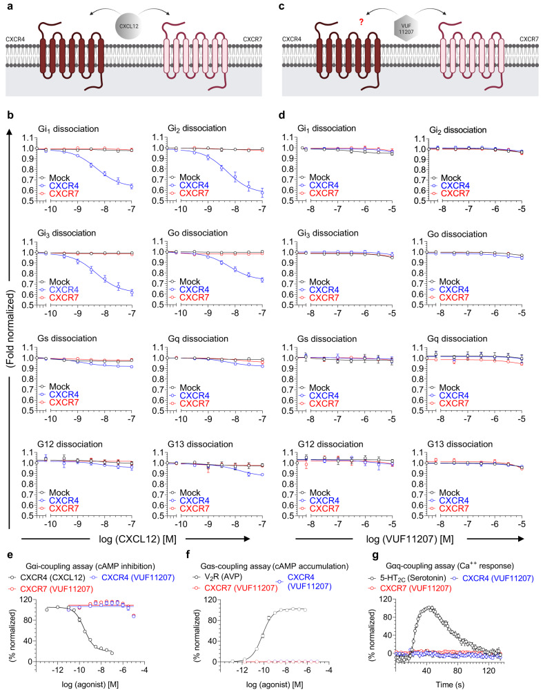
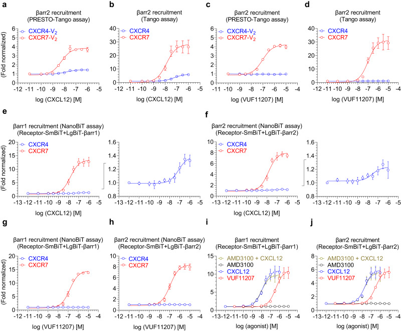
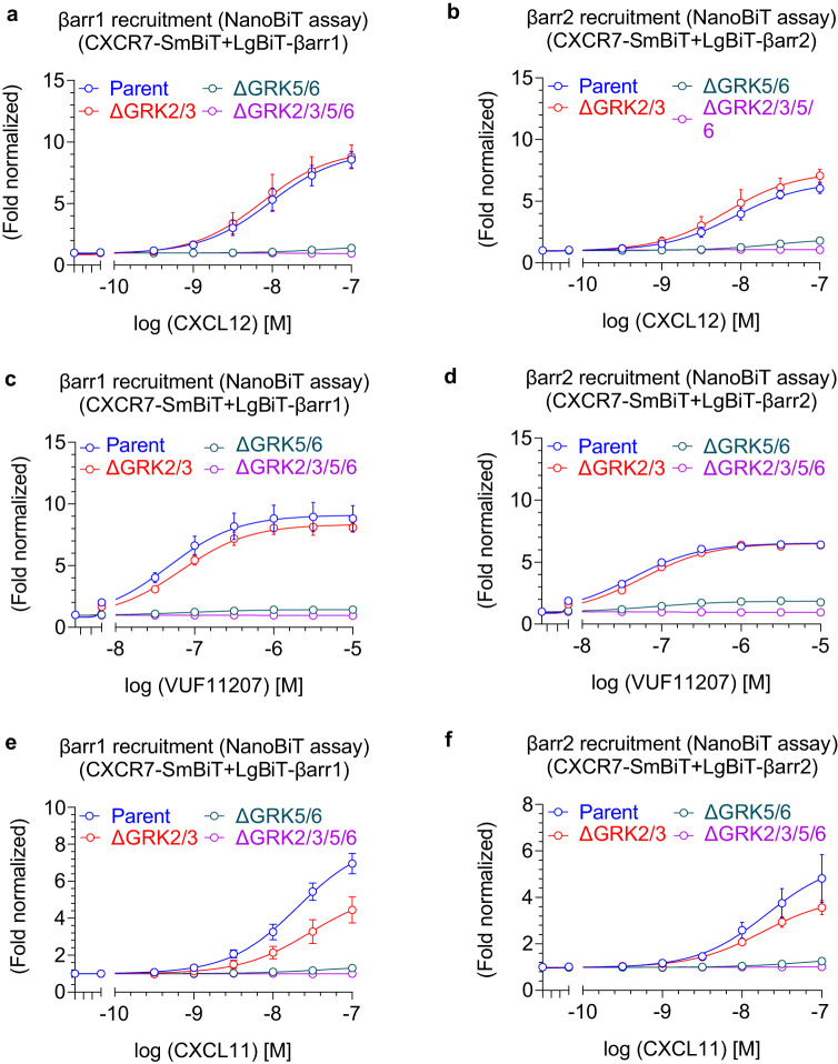
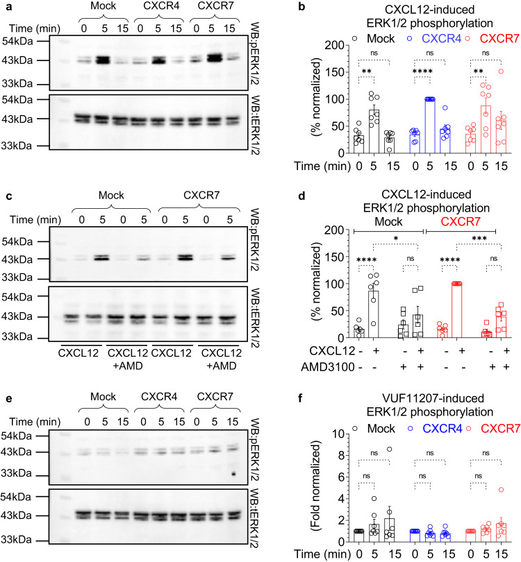
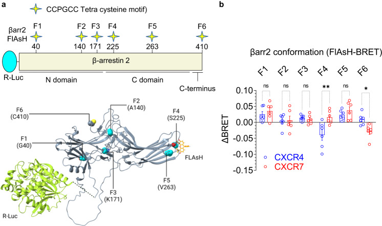
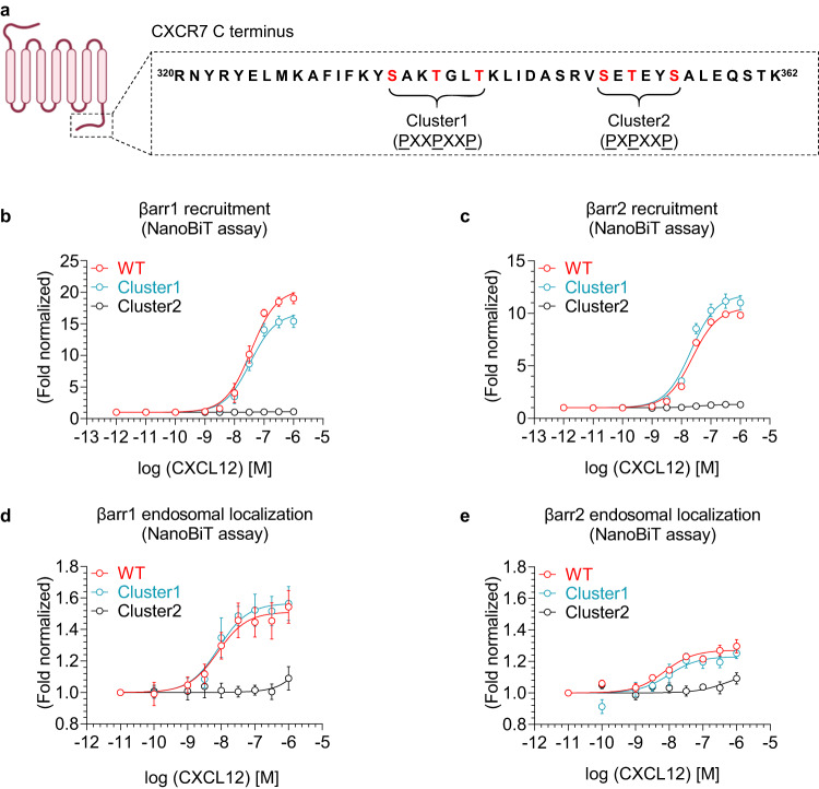
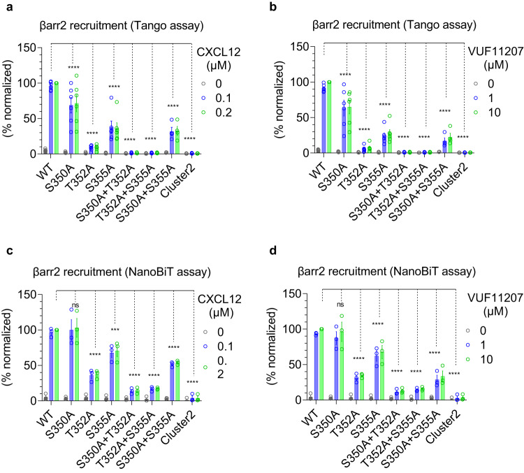
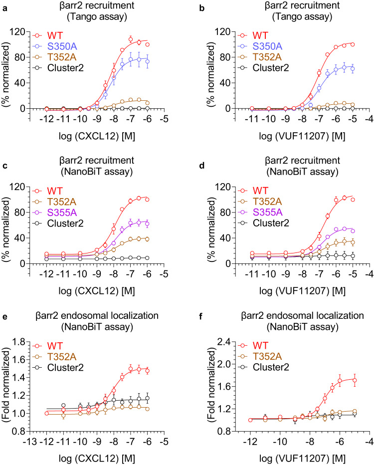
Chemokine receptors constitute an important subfamily of G protein-coupled receptors (GPCRs), and they are critically involved in a broad range of immune response mechanisms. Ligand promiscuity among these receptors makes them an interesting target to explore multiple aspects of biased agonism. Here, we comprehensively characterize two chemokine receptors namely, CXCR4 and CXCR7, in terms of their transducer-coupling and downstream signaling upon their stimulation by a common chemokine agonist, CXCL12, and a small molecule agonist, VUF11207. We observe that CXCR7 lacks G-protein-coupling while maintaining robust βarr recruitment with a major contribution of GRK5/6. On the other hand, CXCR4 displays robust G-protein activation as expected but exhibits significantly reduced βarr-coupling compared to CXCR7. These two receptors induce distinct βarr conformations even when activated by the same agonist, and CXCR7, unlike CXCR4, fails to activate ERK1/2 MAP kinase. We also identify a key contribution of a single phosphorylation site in CXCR7 for βarr recruitment and endosomal localization. Our study provides molecular insights into intrinsic-bias encoded in the CXCR4-CXCR7 system with broad implications for drug discovery.


Conflict of interest statement

G.D.L. and K.E.L. receive research funding from InterAx AG administered through the University of Michigan. All other authors declare no competing interests.

Figures

Fig. 1
Fig. 1. Lack of G-protein activation upon stimulation of CXCR7.
a CXCL12, a CXC type chemokine, is a common agonist for both, CXCR4 and CXCR7 (created with BioRender.com). b NanoBiT-based assay for CXCL12-induced dissociation of heterotrimeric G-proteins for CXCR4 and CXCR7 (mean ± SEM; n = 4-5 independent experiments; i.e., for Gi1 dissociation: Mock, n = 4; CXCR4, n = 5; CXCR7, n = 4; for Gi2 and Gi3 dissociation: n = 5; for Go dissociation: Mock, n = 4; CXCR4, n = 5; CXCR7, n = 5; for Gs, Gq, G12, and G13 dissociation: n = 4; normalized with luminescence signal under unstimulated condition taken as 1). Mock represents empty vector transfected cells as a negative control. c VUF11207 is a small molecule agonist for CXCR7 but its efficacy for CXCR4, if any, is not known (created with BioRender.com). d NanoBiT-based assay for VUF11207-induced dissociation of heterotrimeric G-proteins for CXCR4 and CXCR7 (mean ± SEM; n = 3 independent experiments; normalized with luminescence signal under unstimulated condition taken as 1). e Agonist-induced decrease in forskolin-induced cAMP level measured using the GloSensor assay for the indicated receptor-ligand combinations as a readout of Gαi-activation (mean ± SEM; n = 4; normalized with the signal at minimal ligand dose for CXCL12-CXCR4 combination as 100%). f Agonist-induced increase in cAMP level measured using the GloSensor assay for the indicated receptor-ligand combinations as a readout of Gαs-activation (mean ± SEM; n = 3; normalized with maximal signal for V2R as 100%). V2R (vasopressin receptor subtype 2) is used as a positive control. g Agonist-induced increase in Ca++ level measured using the GCaMP sensor for the indicated receptor-ligand combinations as a readout of Gαq-activation (mean ± SEM; n = 4; normalized with maximal signal for serotonin as 100%). 5-HT2C receptor is used as a positive control. Source data are provided as a source data file.
Fig. 2
Fig. 2. β-arrestin recruitment to CXCR7.
a, b CXCL12-induced βarr2 recruitment to CXCR4 and CXCR7 in PRESTO-Tango and Tango assays, respectively (mean ± SEM; n = 3 independent experiments; normalized with the luminescence signal at minimal ligand dose treated as 1). The PRESTO-Tango assay uses a chimeric receptor construct with the carboxyl-terminus of V2R while the Tango assay uses native receptors. c, d VUF11207-induced βarr2 recruitment to CXCR4 and CXCR7 in PRESTO-Tango and Tango assays, respectively (mean ± SEM; n = 3 independent experiments; normalized with the luminescence signal at minimal ligand dose treated as 1). e, f CXCL12-induced βarr1/2 recruitment to CXCR4 and CXCR7 in NanoBiT assay (mean ± SEM; n = 4 independent experiments; normalized with luminescence signal at minimal ligand dose treated as 1). Response for CXCR4 is also shown separately in the right panels. g, h VUF11207-induced βarr1/2 recruitment to CXCR4 and CXCR7 in NanoBiT assay (mean ± SEM; n = 4 independent experiments; normalized with luminescence signal at minimal ligand dose treated as 1). i, j A side-by-side comparison of CXCL12- vs. VUF11207-induced βarr1 and 2 recruitment to CXCR7, respectively (mean ± SEM; n = 4 independent experiments; normalized with luminescence signal at minimal ligand dose treated as 1). A CXCR4-specific antagonist AMD3100 is used either alone, or as pre-treatment to CXCL12, as a negative control and to rule out the possibility of any contribution from endogenous CXCR4. Source data are provided as a source data file.
Fig. 3
Fig. 3. Receptor sub-type selectivity of VUF11207 and its ability to promote cell migration.
a, b VUF11207-induced βarr2 recruitment for all the CXC chemokine receptors (CXCR1-7) in the PRESTO-Tango and Tango assay, respectively (mean ± SEM); n = 5 independent experiments; normalized with the luminescence signal at minimal ligand dose treated as 1. c VUF11207-induced Gαi-coupling for all the CXC chemokine receptors (CXCR1-7) in the GloSensor assay (mean ± SEM; n = 3 independent experiments; normalized with luminescence signal at minimal ligand dose treated as 100%). d Migration of MDA-MB-231 stably expressing CXCR7 in response to indicated ligands. The experiment was carried out using a 2D microfluidic device and each point on the graph represents an individual cell (n = 600 cells examined over 2 independent experiments, One-way ANOVA, Šídák’s multiple comparisons test). The exact p-values are as follows: Control vs. CXCL12 (p < 0.0001), Control vs. VUF11207 (p < 0.0001), Control vs. CXCL12 + VUF11207 (p < 0.0001). The lower and upper whiskers represent the 5th percentile and the 95th percentile respectively and the bounds of box correspond to 25th and 75th percentile. The cross inside the box indicates the mean (Control: 220.798, CXCL12: 390.201, VUF11207: 341.980, CXCL12 + VUF11207: 325.656). The solid line at the 50th percentile indicates the median (Control: 111.150, CXCL12: 325.000, VUF11207: 291.850, CXCL12 + VUF11207: 257.07)5. (****p < 0.0001). Source data are provided as a source data file.
Fig. 4
Fig. 4. Contribution of GRKs in β-arrestin1/2 recruitment to CXCR7.
a, b CXCL12-induced βarr1/2 recruitment to CXCR7 in GRK knock-out cells using the NanoBiT assay (mean ± SEM; n = 4–5 independent experiments; i.e., for βarr1 recruitment: Parent, n = 5; ∆GRK2/3, n = 4; ∆GRK5/6, n = 4; ∆GRK2/3/5/6, n = 5; for βarr2 recruitment: Parent, n = 5; ∆GRK2/3, n = 4; ∆GRK5/6, n = 4; ∆GRK2/3/5/6, n = 4; normalized with luminescence signal under unstimulated condition treated as 1). c, d VUF11207-induced βarr1/2 recruitment to CXCR7 in GRK knock-out cells using the NanoBiT assay (mean ± SEM; n = 4–5 independent experiments; i.e., for βarr1 recruitment: Parent, n = 5; ∆GRK2/3, n = 4; ∆GRK5/6, n = 4; ∆GRK2/3/5/6, n = 5; for βarr2 recruitment: Parent, n = 5; ∆GRK2/3, n = 4; ∆GRK5/6, n = 4; ∆GRK2/3/5/6, n = 4; normalized with luminescence signal under unstimulated condition treated as 1). e, f CXCL11-induced βarr1 and 2 recruitment to CXCR7 in GRK knock-out cells using the NanoBiT assay (mean ± SEM; n = 3–4 independent experiments; i.e., for βarr1 recruitment: Parent, n = 3; ∆GRK2/3, n = 4; ∆GRK5/6, n = 4; ∆GRK2/3/5/6, n = 4; for βarr2 recruitment: Parent, ∆GRK2/3, ∆GRK5/6, and ∆GRK2/3/5/6, n = 4; normalized with luminescence signal under unstimulated condition treated as 1). Source data are provided as a source data file.
Fig. 5
Fig. 5. Agonist-induced ERK1/2 phosphorylation for CXCR4 and CXCR7.
a, b CXCL12-induced ERK1/2 phosphorylation in Mock, CXCR4 or CXCR7 transfected HEK-293 cells measured by Western blotting. Densitometry-based quantification (mean ± SEM; n = 7 independent experiments, normalized with the 5 min signal for CXCR4 as 100%, Two-way ANOVA, Tukey’s multiple comparison test). The exact p-values are as follows: Mock: 0 min vs. Mock: 5 min (p = 0.0095), Mock: 0 min vs. Mock: 15 min (p > 0.9999), CXCR4: 0 min vs. CXCR4: 5 min (p < 0.0001), CXCR4: 0 min vs. CXCR4: 15 min (p = 0.9940), CXCR7: 0 min vs. CXCR7: 5 min (p = 0.0031), CXCR7: 0 min vs. CXCR7: 15 min (p = 0.5552). c, d CXCL12-induced ERK1/2 phosphorylation in CXCR7 expressing cells is blocked by pre-treatment with AMD3100 (10 μM, 30 min). Densitometry-based quantification (mean ± SEM; n = 6 independent experiments, normalized with CXCL12-induced signal for CXCR7 as 100%, Two-way ANOVA, Tukey’s multiple comparison test). The exact p-values are as follows: Mock(-AMD3100): 0 min vs. Mock(-AMD3100): 5 min (p < 0.0001), Mock(-AMD3100): 5 min vs. Mock(+AMD3100): 5 min (p = 0.0192), Mock(+AMD3100): 0 min vs. Mock(+AMD3100): 5 min (p = 0.8299), CXCR7(-AMD3100): 0 min vs. CXCR7(-AMD3100): 5 min (p < 0.0001), CXCR7(-AMD3100): 5 min vs. CXCR7(+AMD3100): 5 min (p = 0.0004), CXCR7(+AMD3100): 0 min vs. CXCR7(+AMD3100): 5 min (p = 0.3619). e, f VUF11207-induced ERK1/2 phosphorylation in Mock, CXCR4 or CXCR7 transfected HEK-293 cells as measured by Western blotting. Densitometry-based quantification (mean ± SEM; n = 7 independent experiments, normalized with respect to the 0 min signal for each condition treated as 1, Two-way ANOVA, Tukey’s multiple comparison test). The exact p-values are as follows: Mock: 0 min vs. Mock: 5 min (p = 0.9793), Mock: 0 min vs. Mock: 15 min (p > 0.6170), CXCR4: 0 min vs. CXCR4: 5 min (p > 0.9999), CXCR4: 0 min vs. CXCR4: 15 min (p > 0.9999), CXCR7: 0 min vs. CXCR7: 5 min (p > 0.9999), CXCR7: 0 min vs. CXCR7: 15 min (p = 0.9633) (*p < 0.05, **p < 0.01, ***p < 0.001, ****p < 0.0001, ns = non-significant). Source data are provided as a source data file.
Fig. 6
Fig. 6. Conformational changes in βarr2 upon interaction with CXCR4 vs. CXCR7.
a Schematic representation of intramolecular BRET-based sensors of βarr2 conformation where the N-terminus of βarr2 harbors R-Luc (Renilla luciferase) as the BRET donor, and the FlAsH motif are engineered at indicated positions in βarr2 as BRET acceptor. The structural representation in the lower panel is designed based on alpha fold generated model of βarr2. b CXCL12-induced BRET signal measured in HEK-293 cells expressing the indicated receptor and sensor constructs (mean ± SEM), n = 6 independent experiments; the difference of the net-BRET ratio between the stimulated and unstimulated condition were plotted, Two-way ANOVA, Sidak’s multiple comparison test. The exact p-values are as follows: F1:CXCR4 vs. F1:CXCR7 (p = 0.9746), F2:CXCR4 vs. F2:CXCR7 (p > 0.9999), F3:CXCR4 vs. F3:CXCR7 (p = 0.9991), F4:CXCR4 vs. F4:CXCR7 (0.0010), F5:CXCR4 vs. F5:CXCR7 (p = 0.9052), F6:CXCR4 vs. F6:CXCR7 (p = 0.0420) (*p < 0.05, **p < 0.01, ns non-significant). Source data are provided as a source data file.
Fig. 7
Fig. 7. Identification of the key phosphorylation site cluster in CXCR7.
a The carboxyl-terminus of CXCR7 harbors two potential phosphorylation site clusters indicated as cluster 1 and 2 with PXXPXXP and PXPXXP type phosphorylation codes (P is Ser/Thr; X is any other amino acid) (the schematic for the receptor is created with BioRender.com; same as in Fig. 1a/c). b, c Dose response curves for CXCL12-induced βarr1 and 2 recruitment to indicated CXCR7 constructs measured using the NanoBiT assay (Receptor-SmBiT+LgBiT-βarr1/2) (mean ± SEM; n = 4 independent experiments; normalized with luminescence signal at minimal ligand dose treated as 1). d, e Dose response curves of CXCL12-induced endosomal localization of βarr1/2 for the indicated CXCR7 constructs measured using the NanoBiT assay (Receptor+SmBiT-βarr1/2+LgBiT-FYVE) (mean ± SEM; n = 4 independent experiments; normalized with the luminescence signal at minimal ligand dose treated as 1). Source data are provided as a source data file.
Fig. 8
Fig. 8. Key phosphorylation sites in CXCR7 driving βarr1 recruitment and endosomal localization.
a, b CXCL12- and VUF11207-induced βarr1 recruitment, respectively, to the indicated phosphorylation site mutants of CXCR7 using the NanoBiT assay (mean ± SEM); n = 3–4 independent experiments; i.e., for CXCL12-induced βarr1 recruitment: WT, S350A, T352A, S355A, S350A + T352A, T352A + S355A, and Cluster2, n = 4; S350A + S355A, n = 3; for VUF11207-induced βarr1 recruitment: WT, S350A, T352A, S355A, S350A + T352A, T352A + S355A, and Cluster2, n = 4; S350A + S355A, n = 3; normalized with luminescence signal at maximal ligand dose for wild-type treated as 100%, Two-way ANOVA, Dunnett’s multiple comparisons test. The exact p-values are: for CXCL12-induced βarr1 recruitment; WT vs. S350A (p = 0.9972), WT vs. T352A (p < 0.0001), WT vs. S355A (p < 0.0001), WT vs. S350A + T352A (p < 0.0001), WT vs. T352A + S355A (p < 0.0001), WT vs. S350A + S355A (p < 0.0001), WT vs. Cluster2 (p < 0.0001). For VUF11207-induced βarr1 recruitment; WT vs. S350A (p = 0.4700), WT vs. T352A (p < 0.0001), WT vs. S355A (p < 0.0001), WT vs. S350A + T352A (p < 0.0001), WT vs. T352A + S355A (p < 0.0001), WT vs. S350A + S355A (p < 0.0001), WT vs. Cluster2 (p < 0.0001). c, d Dose response curves of CXCL12- and VUF11207-induced βarr1 recruitment to selected phosphorylation site mutants of CXCR7 in the NanoBiT assay (Receptor-SmBiT+LgBiT-βarr1) (mean ± SEM; n = 3 independent experiments; normalized with luminescence signal at maximal ligand dose for wild-type treated as 100%). e, f Dose response curves of CXCL12- and VUF11207-induced βarr1 endosomal localization for the selected phosphorylation site mutants of CXCR7 in the NanoBiT assay (receptor+SmBiT-βarr1 + LgBiT-FYVE) (mean ± SEM; n = 3–7 independent experiments; i.e., for CXCL12-induced βarr1 endosomal localization: WT, n = 6; T352A, n = 3; and Cluster2, n = 3; for VUF11207-induced βarr1 endosomal localization: WT, n = 7; T352A, n = 3; and Cluster2, n = 4; normalized with the luminescence signal at minimal ligand dose treated as 1). (****p < 0.0001, ns non-significant). Source data are provided as a source data file.
Fig. 9
Fig. 9. Contribution of different phosphorylation sites in CXCR7-mediated βarr1/2 recruitment.
a, b CXCL12- and VUF11207-induced βarr2 recruitment, respectively, to the indicated phosphorylation site mutants of CXCR7 using the Tango assay (mean ± SEM); n = 4–6 independent experiments; i.e., for CXCL12: WT, S350A, T352A, S355A, S350A + T352A, T352A + S355A, and Cluster2, n = 6; S350A + S355A, n = 4; for VUF11207-induced βarr2 recruitment: WT, S350A, T352A, S355A, S350A + T352A, T352A + S355A, and Cluster2, n = 6; S350A + S355A, n = 4; normalized with luminescence signal at maximal ligand dose for wild-type treated as 100%, Two-way ANOVA, Dunnett’s multiple comparisons test. The exact p-values are: for CXCL12-induced βarr2 recruitment; WT vs. S350A (p < 0.0001), WT vs. T352A (p < 0.0001), WT vs. S355A (p < 0.0001), WT vs. S350A + T352A (p < 0.0001), WT vs. T352A + S355A (p < 0.0001), WT vs. S350A + S355A (p < 0.0001), WT vs. Cluster2 (p < 0.0001). For VUF11207; WT vs. S350A (p < 0.0001), WT vs. T352A (p < 0.0001), WT vs. S355A (p < 0.0001), WT vs. S350A + T352A (p < 0.0001), WT vs. T352A + S355A (p < 0.0001), WT vs. S350A + S355A (p < 0.0001), WT vs. Cluster2 (p < 0.0001). c, d CXCL12- and VUF11207-induced βarr2 recruitment, respectively, to the indicated phosphorylation site mutants of CXCR7 using the NanoBiT assay (mean ± SEM); n = 3 independent experiments; normalized with luminescence signal at maximal ligand dose for wild-type treated as 100%, Two-way ANOVA, Dunnett’s multiple comparisons test. The exact p-values are: for CXCL12; WT vs. S350A (p = 0.9994), WT vs. T352A (p < 0.0001), WT vs. S355A (p = 0.0001), WT vs. S350A + T352A (p < 0.0001), WT vs. T352A + S355A (p < 0.0001), WT vs. S350A + S355A (p < 0.0001), WT vs. Cluster2 (p < 0.0001). For VUF11207; WT vs. S350A (p = 0.8957), WT vs. T352A (p < 0.0001), WT vs. S355A (p < 0.0001), WT vs. S350A + T352A (p < 0.0001), WT vs. T352A + S355A (p < 0.0001), WT vs. S350A + S355A (p < 0.0001), WT vs. Cluster2 (p < 0.0001) (***p < 0.001, **** p < 0.0001, ns non-significant). Source data are provided as a source data file.
Fig. 10
Fig. 10. Effect of Thr352Ala mutation in CXCR7 on βarr2 recruitment and endosomal localization.
ad Dose-response curves of CXCL12- and VUF11207-induced βarr2 recruitment to selected phosphorylation site mutants of CXCR7 in the Tango and NanoBiT assays (mean ± SEM; n = 6 independent experiments for a, b and n = 3 independent experiments for c, d; normalized with luminescence signal for WT at maximal ligand dose treated as 100%). e, f Dose-response curves of CXCL12- and VUF11207-induced βarr2 endosomal localization for the selected phosphorylation site mutants of CXCR7 in the NanoBiT assay (Receptor+SmBiT-βarr2 + LgBiT-FYVE) (mean ± SEM; n = 3–7 independent experiments; i.e., for CXCL12-induced βarr2 endosomal localization: WT, n = 6; T352A, n = 3; and Cluster2, n = 3; for VUF11207-induced βarr2 endosomal localization: WT, n = 7; T352A, n = 3; and Cluster2, n = 4; normalized with the luminescence signal at minimal ligand dose treated as 1). Source data are provided as a source data file.

References

    1. Hughes CE, Nibbs RJB. A guide to chemokines and their receptors. FEBS J. 2018;285:2944–2971. - PMC - PubMed
    1. Griffith JW, Sokol CL, Luster AD. Chemokines and chemokine receptors: positioning cells for host defense and immunity. Annu Rev. Immunol. 2014;32:659–702. - PubMed
    1. Proudfoot AE. Chemokine receptors: multifaceted therapeutic targets. Nat. Rev. Immunol. 2002;2:106–115. - PMC - PubMed
    1. Zlotnik A, Yoshie O, Nomiyama H. The chemokine and chemokine receptor superfamilies and their molecular evolution. Genome Biol. 2006;7:243. - PMC - PubMed
    1. Steen A, Larsen O, Thiele S, Rosenkilde MM. Biased and g protein-independent signaling of chemokine receptors. Front Immunol. 2014;5:277. - PMC - PubMed

Publication types

MeSH terms