Review
. 2023 Aug 10;7(1):41.
doi: 10.1186/s41747-023-00358-5.

Fetal MRI: what's new? A short review

Affiliations
Review

Fetal MRI: what's new? A short review

Lucia Manganaro et al. Eur Radiol Exp. .

Abstract

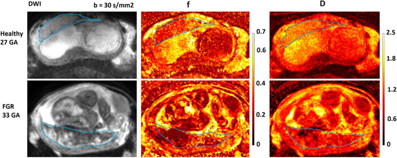
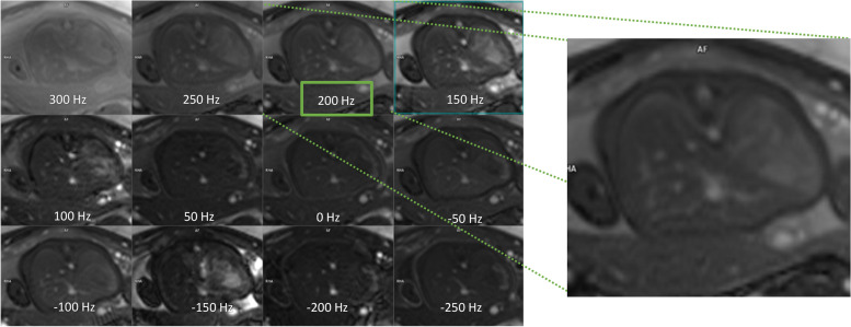
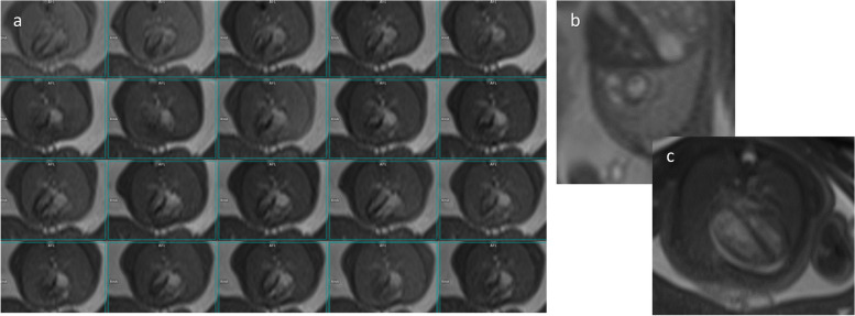
Fetal magnetic resonance imaging (fetal MRI) is usually performed as a second-level examination following routine ultrasound examination, generally exploiting morphological and diffusion MRI sequences. The objective of this review is to describe the novelties and new applications of fetal MRI, focusing on three main aspects: the new sequences with their applications, the transition from 1.5-T to 3-T magnetic field, and the new applications of artificial intelligence software. This review was carried out by consulting the MEDLINE references (PubMed) and including only peer-reviewed articles written in English. Among the most important novelties in fetal MRI, we find the intravoxel incoherent motion model which allow to discriminate the diffusion from the perfusion component in fetal and placenta tissues. The transition from 1.5-T to 3-T magnetic field allowed for higher quality images, thanks to the higher signal-to-noise ratio with a trade-off of more frequent artifacts. The application of motion-correction software makes it possible to overcome movement artifacts by obtaining higher quality images and to generate three-dimensional images useful in preoperative planning.Relevance statementThis review shows the latest developments offered by fetal MRI focusing on new sequences, transition from 1.5-T to 3-T magnetic field and the emerging role of AI software that are paving the way for new diagnostic strategies.Key points• Fetal magnetic resonance imaging (MRI) is a second-line imaging after ultrasound.• Diffusion-weighted imaging and intravoxel incoherent motion sequences provide quantitative biomarkers on fetal microstructure and perfusion.• 3-T MRI improves the detection of cerebral malformations.• 3-T MRI is useful for both body and nervous system indications.• Automatic MRI motion tracking overcomes fetal movement artifacts and improve fetal imaging.

Keywords: Artifacts; Artificial intelligence; Diffusion magnetic resonance imaging; Fetus; Prenatal diagnosis.


Conflict of interest statement

Ilaria Balba is an employee of Siemens Healthcare.

All remaining authors declare that they have no competing interests.

Figures

Fig. 1
Fig. 1
Diffusion-weighted imaging (DWI), obtained with b = 30 s/mm2, and intravoxel incoherent motion f and D maps of the placenta which is bordered by a thin blue line. Upper row: placenta from a woman with a normal 27-week healthy pregnancy. Bottom row: placenta from a female with fetal grow restriction fetus 33 weeks old. The colored bars indicate the percentage of perfusion fraction f and the value of the diffusion coefficient D in 10−3 mm2/s. The normal pregnancy placenta appears with homogeneous colors in the f and D maps. In contrast, the fetal grow restriction placenta appears heterogeneous characterized by lower f-values compared to the healthy placenta
Fig. 2
Fig. 2
Transverse view of two fetal brains in different gestational ages, acquired by varying flip angle (FA) and repetition time (TR) with a T1-weighted 3D GRE fat-saturated sequence. a Repetition time = 4.1 ms and flip angle = 9°. b Repetition time = 6 ms and flip angle = 9°. c Repetition time = 7 ms and flip angle = 14°. d Repetition time = 4 ms and flip angle = 4°. e Repetition time = 6 ms and flip angle = 9°. A 29-week fetus is shown in the first row (ac) and a 22-week fetus in the second row (de)
Fig. 3
Fig. 3
Sagittal and coronal views of a 34-week fetus acquired with a T2-weighted single-shot fast spin-echo sequence: sagittal plane of fetal brain corrupted by unpredicted fetal motion (arrow) (a). Coronal view of a fetal body corrupted by dielectric artifact (arrows) (b)
Fig. 4
Fig. 4
Coronal views of a fetal body before and after the setting of a prescan-B1 filter for T2-weighted single-shot fast spin-echo acquisitions, to describe dielectric artifacts. The signal intensity is spread to the image after the B1 filter was applied, showing up details around the mother’s organs (a, b). Images focused on the fetal brain, to describe how the body part of interest could be affected by the B1 filter: no B1-filter applied (c). B1-filter applied (d)
Fig. 5
Fig. 5
Transversal, coronal, and sagittal views of a T2-weighted single-shot fast spin-echo optimized sequence in fetuses of 22 weeks (a, b, c) and 28 weeks (d, e, f) 28 weeks
Fig. 6
Fig. 6
Single-slice transversal views of the “frequency scout” with twelve different offset frequencies. The frequency scout is planned in one or a few slices around the organ of interest. The “best” frequency is chosen by the operator and/or radiologist depending on the preferred contrast and/or the absence of the banding artifact around the point of interest, and the actual T2-weighted balanced steady-state-free precession is acquired
Fig. 7
Fig. 7
Single-slice cardiac views of a fetus' heart acquired with cine real-time bSSFP: 20-phases of 4-chamber view (a), short-axis view (b), selected 4-chamber view (c)

References

    1. Triulzi F, Manganaro L, Volpe P. Fetal magnetic resonance imaging: indications, study protocols and safety. Radiol Med. 2011;116:337–350. doi: 10.1007/s11547-011-0633-5. - DOI - PubMed
    1. Perrone A, Savelli S, Maggi C, et al. (2008) Magnetic resonance imaging versus ultrasonography in fetal pathology. Radiol Med 113:225–241. 10.1007/s11547-008-0242-0 - PubMed
    1. Powers AM, White C, Neuberger I, Maloney JA, Stence NV, Mirsky D. Fetal MRI neuroradiology: indications. Clin Perinatol. 2022;49:573–586. doi: 10.1016/j.clp.2022.05.001. - DOI - PubMed
    1. Masselli G, Cozzi D, Ceccanti S, Laghi F, Giancotti A, Brunelli R. Fetal body MRI for fetal and perinatal management. Clin Radiol. 2021;76:708.e1–708.e8. doi: 10.1016/j.crad.2021.05.006. - DOI - PubMed
    1. Dawood Y, Buijtendijk MFJ, Shah H, et al. Imaging fetal anatomy. Semin Cell Dev Biol. 2022;131:78–92. doi: 10.1016/j.semcdb.2022.02.023. - DOI - PubMed

LinkOut - more resources