. 2023 Aug 9;28(1):277.
doi: 10.1186/s40001-023-01182-8.

Human umbilical cord mesenchymal stem cell derived exosomes (HUCMSC-exos) recovery soluble fms-like tyrosine kinase-1 (sFlt-1)-induced endothelial dysfunction in preeclampsia

Affiliations

Human umbilical cord mesenchymal stem cell derived exosomes (HUCMSC-exos) recovery soluble fms-like tyrosine kinase-1 (sFlt-1)-induced endothelial dysfunction in preeclampsia

Xinwen Chang et al. Eur J Med Res. .

Abstract

Background: Preeclampsia is a unique multisystem disorder that affects 5-8% of pregnancies. A high level of soluble fms-like tyrosine kinase-1 (sFlt-1) is a hallmark of preeclampsia that causes endothelial dysfunction. Exosomes derived from mesenchymal stem cells (MSCs) have been indicated to improve endothelial performances by transporting signals to target cells. We hypothesized that exosomes derived from MSCs have potential effects against preeclampsia.

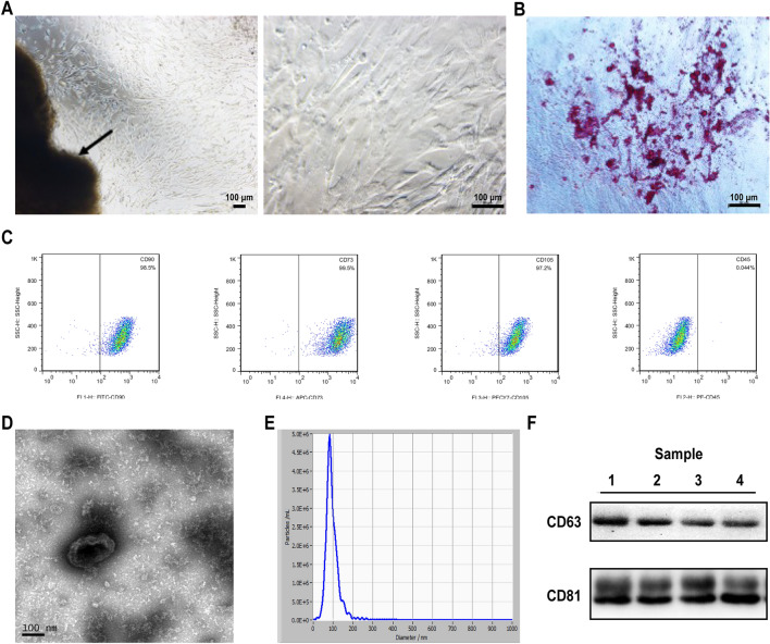
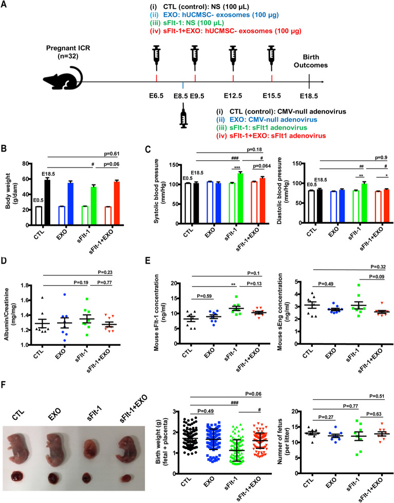
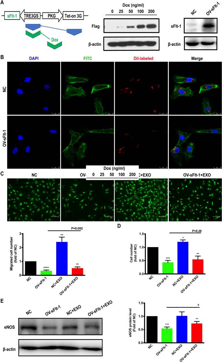
Methods: We collected human umbilical cord MSC-derived exosomes (HUCMSC-exos) by ultracentrifugation. The size and morphology of the exosomes were examined using a transmission electron microscope and nanoparticle tracking analysis. Pregnant mice were injected with murine sFlt-1 adenovirus to build the preeclampsia-like mouse model and then treated with HUCMSC-exos. Human umbilical vein endothelial cells (HUVECs) were infected with lentiviruses expressing tet-on-sFlt-1 to obtain cells overexpressing sFlt-1. Cell proliferation and migration assays were used to measure the endothelial functions. The exosomes enriched proteins underlying mechanisms were explored by proteomic analysis.

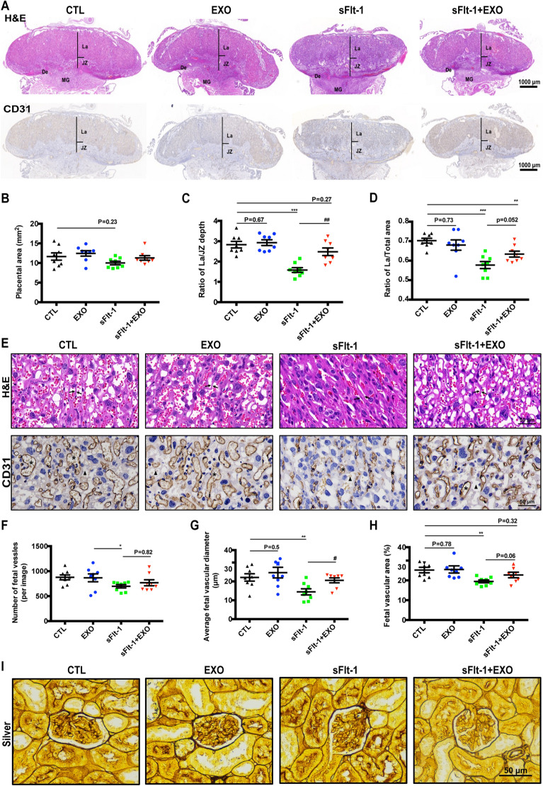
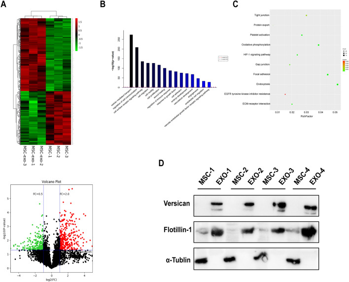
Results: In the current study, we successfully collected the cup-shaped HUCMSC-exos with diameters of 30-150 nm. In the sFlt-1-induced preeclampsia mouse model, HUCMSC-exos exhibited beneficial effects on adverse birth events by decreasing blood pressure and improving fetal birth weight. In addition, preeclamptic dams that were injected with HUCMSC-exos had rebuilt dense placental vascular networks. Furthermore, we observed that HUCMSC-exos partially rescued sFlt-1-induced HUVECs dysfunction in vitro. Proteomics analysis of HUCMSC-exos displayed functional enrichment in biological processes related to vesicle-mediated transport, cell communication, cell migration, and angiogenesis.

Conclusion: We propose that exosomes derived from HUCMSCs contain abundant Versican and play beneficial roles in the birth outcomes of sFlt-1-induced preeclamptic mice by promoting angiogenesis.

Keywords: Angiogenesis; Exosomes; Mesenchymal stem cell; Preeclampsia; Soluble fms-like tyrosine kinase-1/sFlt-1.


Conflict of interest statement

The authors declare that they have no competing interests.

Figures

Fig. 1
Fig. 1
Identification of HUCMSCs and their derived exosomes. a Cell morphology under an inverted microscope, scale bar, arrows indicated the tissue section. b Osteogenic differentiation of hUCMSCs stained positive with alizarin red. c Identification of HUCMSCs by flow cytometry for CD90, CD73, CD105 (positive markers), and CD45 (negative marker). df Exosomes collected from HUCMSCs supernatant by ultracentrifuge. Electron micrograph of exosomes. (d). Representative vesicle size (nm) distribution by NTA (e). Western blot analysis for the CD63 and CD81 exosomes enriched marker of HUCMSCs-exo from 4 individual pregnant women (f)
Fig. 2
Fig. 2
HUCMSC-exos improve the sFlt-1-induced preeclamptic mouse reproductive outcomes. a Pregnant mice were randomly divided into four groups and respectively treated with nature saline (i, CTL, n = 8, black), HUCMSC-exos (ii, EXO, 100 μg, n = 8, green), sFlt-1 adenovirus (iii, sFlt-1, n = 8, blue) or sFlt-1 adenovirus and HUCMSC-exos (iv, sFlt-1 + EXO, 100 μg, n = 8, red) injection via tail vein on E6.5, E9.5, E12.5, E15.5 and E15.5. (The syringes above indicated the injection of HUCMSC-exos or nature saline. The syringe below indicated the treatment of sFlt-1 adenovirus or CMV-null adenovirus). To establish PE-like mice model, pregnant mice were injected with CMV-null adenovirus (i, CTL; ii, EXO) and sFlt-1 adenovirus (iii, sFlt-1; iv, sFlt-1 + EXO) on E8.5. b Body weights of dams on E0.5 and E18.5 (one-way ANOVA and Dunett's post-hoc tests, sFlt-1 vs CTL on E18.5, #p < 0.05). c Blood pressure measurements on E0.5 and E18.5 (Paired students’ t-tests, E18.5 vs E0.5, *p < 0.05, **p < 0.01, ***p < 0.001; Systolic blood pressure on E18.5, one-way ANOVA and Dunett's post-hoc tests, sFlt-1 vs CTL, ###p < 0.001, sFlt-1 + EXO vs sFlt-1, #p < 0.05; Diastolic blood pressure on E18.5, sFlt-1 vs CTL, ##p < 0.01, sFlt-1 + EXO vs sFlt-1). d Ratio of albumin/creatinine in mice urine was measured by ELISA kit on E18.5. e Mouse sFlt-1 and sEng concentrations in serum were measured by ELISA kits on E18.5 (one-way ANOVA and Dunett’s post-hoc tests, sFlt-1 vs CTL, ##p < 0.01). f Effects of HUCMSC-exos-injection on morphology, birth weights and number of fetuses per litter. (n = 8 per group of dams, one-way ANOVA and Dunett's post-hoc tests, sFlt-1 vs CTL, ###p < 0.001, sFlt-1 + EXO vs sFlt-1, #p < 0.05)
Fig. 3
Fig. 3
Effects of HUCMSC-exos on histopathology of mice placenta and kidney. a H&E staining and IHC staining for CD31 of mice placentas (JZ, jountional zone; L, labyrinth), scale bar 2000 μm. b Placental area of pregnant mice. c Ratio of La/JZ depth of placenta. d Ratio of La/total area of placenta. e H&E and IHC staining for CD31 in mice placental tissues. Brown color indicates positive staining for CD31. Arrowheads: trophoblastic septa; asterisk: fetal vascular; triangle: intervillous space. Scale bar 50 μm. Numbers of fetal vessles (f), average fetal vascular diameter (g) and fetal vascular area (h) in JZ were measured (n = 8 per group of dams, one-way ANOVA and Dunett’s post-hoc tests, * sFlt-1 vs CTL, *p < 0.05, **p < 0.01, # sFlt-1-exo vs sFlt-1, #p < 0.05). i Staining of kidney sections with PAS, scale bar 50 μm
Fig. 4
Fig. 4
Proangiogenic effects of HUCMSC-exos. a Stably transfected HUVECs were treated with the indicated concentrations of Dox for 24 h. Western blot analysis of sFlt-1 in HUVECs was performed using anti-Flag and anti-sFlt-1 antibody. b NC-HUVECs and OV-sFlt-1-HUVECs were incubated with dil-labeled HUCMSC-exos (red) for 24 h before fluorescent and phase contrast images were captured, scale bar 20 μm. c Cell migration response to HUCMSC-exos (100 μg/mL) was determined by transwell assay, scale bar 100 μm. d After incubation with HUCMSC-exos (100 μg/mL), HUVEC proliferation was analyzed by CCK-8 assay. e Protein expression of eNOS in HUVECs were detected by western blot. All data were means ± S.E.M. of 3 pairs of independent experiments performed. One-way ANOVA, *p < 0.05, **p < 0.01, ***p < 0.001
Fig. 5
Fig. 5
Proteomic analysis of HUMSC and HUCMSC-exos. a Cluster analysis of differentially expressed genes. The colors from green to black and to red represent the expression values of the differentially expressed genes that have increased. Volcano plots for HUCMSCs vs HUCMSC-exos. Statistically significant proteins (P < 0.05) with a log2 fold change + 2 or − 0.5 are represented by red or green dots, respectively. Differentially expressed proteins that did not reach statistical significance are represented by black dots. Gene Ontology (GO) analysis of the top pathways (b) and bubble diagram of KEGG pathway enrichment (c) in both HUCMSC and HUCMSC-exos. (d) Protein expressions of Versican, Flotillin-1, and α-Tublin in HUCMSCs and HUCMSC-exos were detected by western blot

Similar articles

Cited by

References

    1. Redman CW, Sargent IL. Latest advances in understanding preeclampsia. Science. 2005;308(5728):1592–1594. doi: 10.1126/science.1111726. - DOI - PubMed
    1. Souza JP, Gülmezoglu AM, Vogel J, Carroli G, Lumbiganon P, Qureshi Z, et al. Moving beyond essential interventions for reduction of maternal mortality (the WHO Multicountry Survey on Maternal and Newborn Health): a cross-sectional study. Lancet. 2013;381(9879):1747–1755. doi: 10.1016/s0140-6736(13)60686-8. - DOI - PubMed
    1. Stepan H, Geide A, Faber R. Soluble fms-like tyrosine kinase 1. New England J Med. 2004;351(21):2241–2242. doi: 10.1056/nejm200411183512123. - DOI - PubMed
    1. Levine RJ, Maynard SE, Qian C, Lim KH, England LJ, Yu KF, et al. Circulating angiogenic factors and the risk of preeclampsia. New England J Med. 2004;350(7):672–683. doi: 10.1056/NEJMoa031884. - DOI - PubMed
    1. Yang W, Zhang J, Xu B, He Y, Liu W, Li J, et al. HucMSC-derived exosomes mitigate the age-related retardation of fertility in female mice. Mol Ther. 2020;28(4):1200–1213. doi: 10.1016/j.ymthe.2020.02.003. - DOI - PMC - PubMed

Substances

LinkOut - more resources