Review
. 2023 Jul;84(4):846-854.
doi: 10.3348/jksr.2023.0048. Epub 2023 Jul 26.

[Endovascular Treatment for Vascular Injuries of the Extremities]

[Article in Korean]
Review

[Endovascular Treatment for Vascular Injuries of the Extremities]

[Article in Korean]
Tae Won Choi et al. J Korean Soc Radiol. 2023 Jul.

Abstract

Vascular injuries of the extremities are associated with a high mortality rate. Conventionally, open surgery is the treatment of choice for peripheral vascular injuries. However, rapid development of devices and techniques in recent years has significantly increased the utilization and clinical application of endovascular treatment. Endovascular options for peripheral vascular injuries include stent-graft placement and embolization. The surgical approach is difficult in cases of axillo-subclavian or iliac artery injuries, and stent-graft placement is a widely accepted alternative to open surgery. Embolization can be considered for arterial injuries associated with active bleeding, pseudoaneurysms, and arteriovenous fistula and in patients in whom embolization can be safely performed without a risk of ischemic complications in the extremities. Endovascular treatment is a minimally invasive procedure and is useful as a simultaneous diagnostic and therapeutic approach, which serve as advantages of this technique that is widely utilized for vascular injuries of the extremities.

상하지 혈관 손상은 높은 사망률과 관계가 있다. 사지 혈관 손상에 대한 전통적인 치료법은 수술이었으나 최근 기술과 시술법의 비약적인 발달로 인해 혈관 내 치료법(endovascular treatment)의 효용 및 임상 적용이 증가하고 있다. 상하지 혈관 손상에서 시행할 수 있는 혈관 내 치료는 크게 스텐트 그래프트(stent graft) 설치술과 색전술로 나눠볼 수 있으며 일반적으로 손상 혈관의 위치와 크기, 혈관 손상의 성격에 따라 치료법이 달라진다. 겨드랑-쇄골하 동맥과 장골 동맥 손상의 경우 해부학적 위치상 수술적인 접근이 어려운 것으로 알려져 있으며 스텐트 그래프트 설치술이 수술을 대신할 수 있는 중요한 치료법으로 활용되고 있다. 활동성 출혈, 가성동맥류, 동정맥루 및 색전 시 허혈이 우려되지 않는 동맥의 손상에 대해서는 색전술을 고려해 볼 수 있다. 상하지의 혈관 손상에 대한 혈관 내 치료법은 최소침습적으로 진단과 치료를 동시에 할 수 있다는 장점이 있어 향후 그 적응증이 더 넓어질 것으로 기대된다.


Conflict of interest statement

Conflicts of Interest: The authors have no potential conflicts of interest to disclose.

Figures

Fig. 1
Fig. 1. A 74-year-old male patient involved in a motorcycle accident.
A. CT angiography shows active arterial bleeding (arrow) and hematoma involving the right axillary artery. B. Angiography of the right axillary artery shows arterial rupture and bleeding corresponding to CT findings. C. Final angiography shows disappearance of active bleeding after placement of a 9 mm × 6 cm stent-graft (Covera Plus, Bard, Tempe, AZ, USA) at the injured segment. D. Follow-up CT angiography (maximum intensity projection image) shows disappearance of the peripheral hematoma and preserved distal blood flow.
Fig. 2
Fig. 2. A 20-year-old male patient admitted to the trauma center following a motorcycle accident.
A, B. CT angiography (maximum intensity projection image) (A) and conventional angiography of the left subclavian artery (B) show transection of the left subclavian artery and compromised distal flow secondary to trauma. C. Attempt at passing a guidewire through the traumatic lesion via a n antergrade approach was unsuccessful. Therefore, retrograde approach via the brachial artery was tried to pass the lesion and it was successful. Fluoroscopy image shows the guidewire subsequently captured using a snare (arrow) via the femoral access route to establish through-and-through brachial-femoral guidewire access. D. Final angiography obtained after placement of a 7 mm × 10 cm bare-metal stent (SMART Flex, Cordis, Santa Clara, CA, USA) shows recovery of the distal blood flow.
Fig. 3
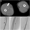
Fig. 3. A 80-year-old male patient involved in a pedestrian traffic accident.
A. CT angiography shows active bleeding (arrow) and hematoma involving the right thigh. B. Conventional angiography of the right common femoral artery shows definitive evidence of an active bleeding focus in a muscular branch (arrow). C. Image shows super-selective catheterization of the bleeding vessel using a microcatheter; embolization was performed using N-butyl cyanoacrylate. D. Final angiographic image shows disappearance of active bleeding.

References

    1. Doody O, Given MF, Lyon SM. Extremities--indications and techniques for treatment of extremity vascular injuries. Injury. 2008;39:1295–1303. - PubMed
    1. Mattox KL, Feliciano DV, Burch J, Beall AC, Jr, Jordan GL, Jr, De Bakey ME. Five thousand seven hundred sixty cardiovascular injuries in 4459 patients. Epidemiologic evolution 1958 to 1987. Ann Surg. 1989;209:698–705. - PMC - PubMed
    1. Cho H, Huh U, Lee CW, Song S, Kim SH, Chung SW. Traumatic peripheral arterial injury with open repair: a 10-year single-institutional analysis. Korean J Thorac Cardiovasc Surg. 2020;53:291–296. - PMC - PubMed
    1. Johnson CA. Endovascular management of peripheral vascular trauma. Semin Intervent Radiol. 2010;27:38–43. - PMC - PubMed
    1. Jeph S, Ahmed S, Bhatt RD, Nadal LL, Bhanushali A. Novel use of interventional radiology in trauma. J Emerg Crit Care Med. 2017;1:40