. 2023 Jul 15;13(7):3185-3202.
eCollection 2023.

Mettl3 synergistically regulates TGF-β/SMAD2/3 to promote proliferation and metastasis of gastric cancer

Affiliations

Mettl3 synergistically regulates TGF-β/SMAD2/3 to promote proliferation and metastasis of gastric cancer

Xiao-Ning Yuan et al. Am J Cancer Res. .

Abstract

Transforming Growth factor-β (TGF-β)/Smad signaling is a complex regulatory network that both inhibits and promotes tumorigenesis. However, the mechanisms underlying the function of TGF-β/Smad signaling pathway remain to be fully elucidated. As a methyltransferase, METTL3 is closely related to tumor development, but the role of METTL3 in the proliferation and metastasis of TGF-β/Smad-activated gastric cancer (GC) is unclear. In this study, we identified TGF-β/Smad2/3 axis as an important carcinogenic pathway in GC, which significantly promoted the proliferation and metastasis of GC. Furthermore, we found that Smad3 mRNA could be modified by m6A, which was subsequently recognized and stabilized by IGF2BP2, thereby enhancing Smad3 protein expression and promoting the activation of TGF-β/Smad pathway. Importantly, we also found that METTL3 could combine with p-Smad3 to regulate the transcription of downstream target genes. Therefore, this study revealed a novel mechanism by which METTL3 synergistically regulates TGF-β/Smad2/3 signaling and provide a new potential therapeutic target for the treatment of GC.

Keywords: Gastric cancer; METTL3; TGF-β/Smads; interaction.


Conflict of interest statement

The authors declare that the research was conducted in the absence of any commercial or financial relationships that could be construed as a potential conflict of interest.

Figures
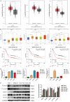
Figure 1
Figure 1
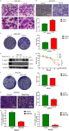
TGF-β/Smad pathway activation in GC. A: GEPIA website analyzes the mRNA expression of TGF-β/Smad signal pathway related genes in GC and corresponding normal tissues in TCGA and GTEx databases; B: UALCAN website analyzes the correlation of clinical stage with the expression of TGF-β, Smad2 and Smad3 in GC using GC data in TCGA database; C: Kaplan-Meier plotter displayed the correlation between the expression of TGF-β/Smad signal pathway related genes and the poor prognosis of patients; D: The mRNA levels of TGF-β/Smad signal pathway related genes in normal gastric epithelial and GC cells, as determined by qPCR; E: Western blot analysis of the protein levels of TGF-β/Smad signal pathway related genes in normal gastric epithelial and GC cells (*P<0.05, **P<0.01, ***P<0.001).
Figure 2
Figure 2
The activation of TGF-β/Smad2/3 pathway is crucial in the development of GC. A: Morphology changes in TGF-β-treated MGC803 cells; B: TGF-β promoted the migration of MGC803 cells, as determined by transwell assay; C: TGF-β promoted the colony formation of MGC803 cells; D: Inhibitor SB-431542 suppressed phosphor-Smad2/3 levels in MGC803 cells; E: The proliferation of MGC803 cells was detected by CCK-8; F: SB431542 inhibited the colony formation of MGC803 cells; G: The migration of MGC803 cells, as determined by transwell assay; H: The mRNA levels of Vimentin and FN was detected by qPCR (*P<0.05, **P<0.01, ***P<0.001).
Figure 3
Figure 3
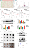
METTL3 expression was upregulated in GC cells. A, B: High expression of m6a related genes in TCGA database, as determined by “R” analysis; C: Kaplan-Meier plotter survival showed the correlation between the mRNA levels of METTL3, METTL14 and WTAP and the poor prognosis of GC patients; D: The protein levels of METTL3 and METTL14 were detected by Western blot; E: The mRNA levels of METTL3 and METTL14 in GC cells were detected by qPCR; F: The overall m6A modification level of cell mRNA was detected by Dot blot; G: The protein level of METTL3 was detected by Western blot; H: Tumor growth in nude mice (*P<0.05, **P<0.01, ***P<0.001).
Figure 4
Figure 4
METTL3 promoted the growth and migration of GC cells. A, B: METTL3 knockdown attenuated the migration of GC cells, as determined by transwell assay; C, D: METTL3 overexpression enhanced the colony formation of GC cells; E: The cell cycle of GC cells was measured by flow cytometry (*P<0.05, **P<0.01, ***P<0.001).
Figure 5
Figure 5
METTL3 knockdown reduced TGF-β-induced proliferation and metastasis of GC cells and improved the sensitivity to SB431542. A: Transwells assay of GC cells in the presence or absence of TGF-β1; B: Transwells assay of GC cells in the presence or absence of SB431542; C: The colony formation assay of GC cells in the presence or absence of TGF-β1; D: The colony formation assay of GC cells in the presence or absence of SB431542 (*P<0.05, **P<0.01, ***P<0.001).
Figure 6
Figure 6
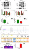
METTL3 positively modulated TGF-β/Smad2/3 signaling via the m6A modification of Smad3 mRNA in GC cells. A: Western blot analysis of the expression and phosphorylation of TGF-β/Smad protein after METTL3 knockdown by shMETTL3; B: Western blot analysis of the expression and phosphorylation of TGF-β/Smad protein in METTL3-expressing cells (OE); C: qPCR analysis of Smad3 mRNA level in shMETTL3 cells; D: Prediction of the m6A modification sites on Smad3 mRNA by SRAMP online database; E: Statistical analysis of m6A modification site on Smad3 mRNA; F: High confidence modification site in the 3’UTR region of Smad3; G: meRIP detected Smad3 mRNA modification by m6A (*P<0.05, **P<0.01, ***P<0.001).
Figure 7
Figure 7
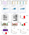
METTL3 regulated Smad3 expression via m6A reader IGF2BP2. A: Analysis of Smad3 mRNA binding proteins from CLIP-seq data in starBase; B: Correlation analysis between Smad3 and IGF2BP2 expression; C: Western blot analysis of the expression and phosphorylation of TGF-β/Smad protein in IGF2BP2 knockdown cells; D: Western blot analysis of the expression and phosphorylation of TGF-β/Smad protein inIGF2BP2-overexpressing cells; E: qPCR analysis of Smad3 mRNA expression in IGF2BP2-overexpressing cells; F: IGF2BP2 binding to Smad3 mRNA determined by RIP; G: Stability of Smad3 mRNA, as determined by the Act D assay (*P<0.05, **P<0.01 ***P<0.001).
Figure 8
Figure 8
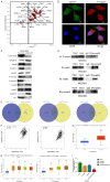
METTL3 combines with P-Smad3 to promote the transcription of downstream target genes. A: Correlation between METTL3 and p-Smad3 in a variety of tumors by Pearson analysis; B: Co-localization of METTL3 and p-Smad3 by immunofluorescence staining; C: Nucleoplasmic separation experiment; D: The interaction between proteins, as determined by Co-IP assay; E: Analyze the CHIP seq data of METTL3 and Smad3 using GTRD, Cistrome DB, and hTFarget databases; F: Correlation analysis of the correlation between HNRNPL expression and METTL3 and Smad3; G: UALCAN database analysis of the expression level of HNRNPL in gastric cancer; H: UALCAN database analysis of the relationship between HNRNPL and staging and grading of gastric cancer patients; I: qPCR analysis were used to investigate the relationship between HNRNPL and METTL3 and Smad3 expression (*P<0.05, **P<0.01, ***P<0.001).

Similar articles

Cited by

References

    1. Araújo D, Ribeiro E, Amorim I, Vale N. Repurposed drugs in gastric cancer. Molecules. 2022;28:319. - PMC - PubMed
    1. Hata A, Chen YG. TGF-β signaling from receptors to smads. Cold Spring Harb Perspect Biol. 2016;8:a022061. - PMC - PubMed
    1. Syed V. TGF-β signaling in cancer. J Cell Biochem. 2016;117:1279–1287. - PubMed
    1. Zhang J, Thorikay M, van der Zon G, van Dinther M, Ten Dijke P. Studying TGF-β signaling and TGF-β-induced epithelial-to-mesenchymal transition in breast cancer and normal cells. J Vis Exp. 2020 - PubMed
    1. Stojnev S, Krstić M, Čukuranović Kokoris J, Conić I, Petković I, Ilić S, Milosević-Stevanović J, Veličković LJ. Prognostic impact of canonical TGF-β signaling in urothelial bladder cancer. Medicina (Kaunas) 2019;55:302. - PMC - PubMed

LinkOut - more resources