Review
. 2023 Aug;28(8):080901.
doi: 10.1117/1.JBO.28.8.080901. Epub 2023 Aug 8.

Review of optical reporters of radiation effects in vivo: tools to quantify improvements in radiation delivery technique

Affiliations
Review

Review of optical reporters of radiation effects in vivo: tools to quantify improvements in radiation delivery technique

Jacob Sunnerberg et al. J Biomed Opt. 2023 Aug.

Abstract

Significance: Radiation damage studies are used to optimize radiotherapy treatment techniques. Although biological indicators of damage are the best assays of effect, they are highly variable due to biological heterogeneity. The free radical radiochemistry can be assayed with optical reporters, allowing for high precision titration of techniques.

Aim: We examine the optical reporters of radiochemistry to highlight those with the best potential for translational use in vivo, as surrogates for biological damage assays, to inform on mechanisms.

Approach: A survey of the radical chemistry effects from reactive oxygen species (ROS) and oxygen itself was completed to link to DNA or biological damage. Optical reporters of ROS include fluorescent, phosphorescent, and bioluminescent molecules that have a variety of activation pathways, and each was reviewed for its in vivo translation potential.

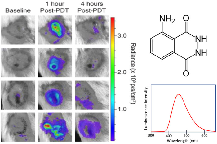
Results: There are molecular reporters of ROS having potential to report within living systems, including derivatives of luminol, 2'7'-dichlorofluorescein diacetate, Amplex Red, and fluorescein. None have unique specificity to singular ROS species. Macromolecular engineered reporters unique to specific ROS are emerging. The ability to directly measure oxygen via reporters, such as Oxyphor and protoporphyrin IX, is an opportunity to quantify the consumption of oxygen during ROS generation, and this translates from in vitro to in vivo use. Emerging techniques, such as ion particle beams, spatial fractionation, and ultra-high dose rate FLASH radiotherapy, provide the motivation for these studies.

Conclusions: In vivo optical reporters of radiochemistry are quantitatively useful for comparing radiotherapy techniques, although their use comes at the cost of the unknown connection to the mechanisms of radiobiological damage. Still their lower measurement uncertainty, compared with biological response assay, makes them an invaluable tool. Linkage to DNA damage and biological damage is needed, and measures such as oxygen consumption serve as useful surrogate measures that translate to in vivo use.

Keywords: fluorescence; phosphorescence; radiation; radiotherapy; reactive oxygen species.


Figures

Fig. 1
Fig. 1
Treatment dose schemes used and being examined and optimized for (a) maximizing the therapeutic ratio of tumor damage to normal tissue damage; (b) with the concept of conformal therapy, maximizing dose to tumor while minimizing it to normal tissue; (c) fractionated therapy, allowing for repair to increase the resistance of normal tissues; and (d) FLASH radiotherapy, providing a protective effect to normal tissues. (e) The problematic variance of biological assays is illustrated conceptually with large error bars and therefore overlapping confidence intervals for tumor and normal tissue assays. (f) The comparatively smaller variation leads to smaller error bars and more statistically separable data to allow for more definitive conclusions when varying irradiation parameters.
Fig. 2
Fig. 2
Radiation induced damage is illustrated as direct DNA damage (top) or indirect damage (bottom), each of which is largely proportional to the dose delivered. However, the microenvironment can alter the indirect damage more due to the highly complex interactions with lipids, proteins, and oxygen. The biochemical pathways of radiation-induced hydrolysis and damage are amplified by molecular oxygen, and the peroxyl radicals [ROO•] are a dominant factor in the biological effects.
Fig. 3
Fig. 3
Outline of the major types of optical reporter probes that have been or might be used as assay of radiation effects, including (1) DNA damage, (2) ROS reporter probes, (3) lipid peroxidation probes, and (4) sensors of oxygenation change.
Fig. 4
Fig. 4
Detection of x-ray radiation-induced changes in ROS content and cell apoptosis in eyes of live zebrafish embryos. (a)–(e) ROS content was measured using the fluorescent dye DCFH-DA. (f)–(j) Cell apoptosis was determined using AO staining. Figure adapted with permission from Ref. . The chemical pathway of DCFH-DA from extra cellular administration to hydrolysis with intracellular uptake and reaction with ROS to form the fluorescent DCF molecule is illustrated.
Fig. 5
Fig. 5
Murine mesothelioma flank tumors (AB12) treated with PDT and imaged for the inflammation response by the chemiluminescent reporter luminol, which reacts with an array of ROS and here shows a notable difference from baseline at both 1 and 4 h post-PDT. Figure adapted with permission from Ref. .
Fig. 6
Fig. 6
Photobleaching of 2  μM fluorescein measured in water and 5% bovine serum albumin solution, using conventional dose rate (0.03  Gy/s, denoted CONV) and UHDRs (100  Gy/s, denoted UHDR), illustrating the change in mechanisms with dose rate. The fluorescence intensity loss was measured as % decay in signal per Gy delivered to the solution.
Fig. 7
Fig. 7
Conceptual framework of caged luciferin (PCL-1) that is released by reaction with H2O2, HOCl, or ONOO for bioluminescent detection of ROS in vivo. In vivo images show the 30 min post-injection image of mice injected with PCL-1, which upon interacting with H2O2 show emission. The amount of H2O2 injected scales from 0 to 24 mM going from left to right.
Fig. 8
Fig. 8
(a) Oxygen measurements can be acquired in vivo with Oxyphor phosphorescence lifetime measurement and (b) imaged with phosphorescence lifetime imaging to show histograms of pO2 when excited by the radiation beam. (c) In separate experiments, the transient changes in pO2 were quantified during UHDR radiation treatment to quantify the change in oxygen, due to radiation chemistry-based consumption. (d) Differences in oxygen from UHDR irradiation of 20 Gy in 0.1 s could be seen as an abrupt decrease in both normal tissue and tumor tissues.
Fig. 9
Fig. 9
Oxygen measurements can be achieved in vivo through DF from protopoprphyrin IX molecules generated within cells. (a) This compound has a triplet state quenched by oxygen, and when not present, there is considerable reverse intersystem crossing to allow for (b) increased DF signal. In mice, (c) this DF can be seen from hypoxic tumors, and (d) the contrast relative to normal skin is high compared with the prompt fluorescence. The ability to measure oxygen with this molecule is evolving now.
Fig. 10
Fig. 10
Schematic illustrating the available reporters for in vivo radiation effects that could be used, for each step-in scaling from irradiation of protein solution to in vitro cells to in vivo experiments with eventual translations to clinical trials. Most optical reporters cannot translate to human use, but a few are emerging to translate to in vivo experimental use.
Fig. 11
Fig. 11
Visual illustrations of response measures. (a) Tumor regrowth assay from tumor volumes measured. (b) The zebrafish embryo undergoes the acute, quantifiable biological effect malformations observable from spine curvature.
Fig. 12
Fig. 12
Illustration of skin damage occurring in mice skin going from: (a) clinical appearance of shaved normal mouse skin; (b) histological appearance of normal mouse skin with 3–4 cell thick epidermis, normal hair follicles and sebaceous glands in dermis; (c) clinical epidermal desquamation in irradiated shaved skin; (d) histology of irradiated skin with loss of epithelial viability, sebaceous gland loss and excessive fibrosis; (e) clinical classic radiation induced skin most desquamation and ulceration; and (f) histologic radiation induced full thickness epidermal ulceration and necrosis, loss of epidermis and adnexal structures, with marked dermal inflammation and fibrosis. At right, a narrative illustration of one skin scoring system utilized for radiation response, often in combination with other clinical factors.

Similar articles

References

    1. Fowler J. F., “Review: total doses in fractionated radiotherapy—implications of new radiobiological data,” Int. J. Radiat. Biol. Relat. Stud. Phys. Chem. Med. 46(2), 103–120 (1984).IJRBA310.1080/09553008414551181 - DOI - PubMed
    1. Choi N., et al. , “Predictive factors in radiotherapy for non-small cell lung cancer: present status,” Lung Cancer 31(1), 43–56 (2001).10.1016/S0169-5002(00)00156-2 - DOI - PubMed
    1. Griffin R. J., et al. , “History and current perspectives on the biological effects of high-dose spatial fractionation and high dose-rate approaches: GRID, microbeam & FLASH radiotherapy,” Br. J. Radiol. 93(1113), 20200217 (2020).BJRAAP10.1259/bjr.20200217 - DOI - PMC - PubMed
    1. Favaudon V., Labarbe R., Limoli C. L., “Model studies of the role of oxygen in the FLASH effect,” Med. Phys. 49(3), 2068–2081 (2022).MPHYA610.1002/mp.15129 - DOI - PMC - PubMed
    1. Hirayama R., et al. , “OH radicals from the indirect actions of X-rays induce cell lethality and mediate the majority of the oxygen enhancement effect,” Radiat. Res. 180(5), 514–523 (2013).RAREAE10.1667/RR13368.1 - DOI - PubMed

Publication types