Review
. 2023 Jul 14;6(3):246-264.
doi: 10.31662/jmaj.2022-0220. Epub 2023 Jun 30.

Tohoku Medical Megabank Brain Magnetic Resonance Imaging Study: Rationale, Design, and Background

Affiliations
Review

Tohoku Medical Megabank Brain Magnetic Resonance Imaging Study: Rationale, Design, and Background

Makiko Taira et al. JMA J. .

Abstract

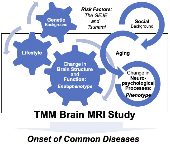
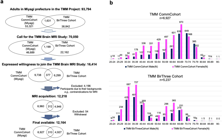
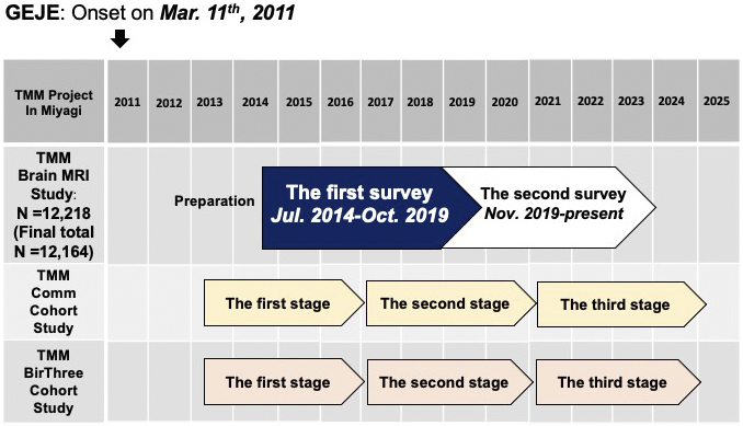
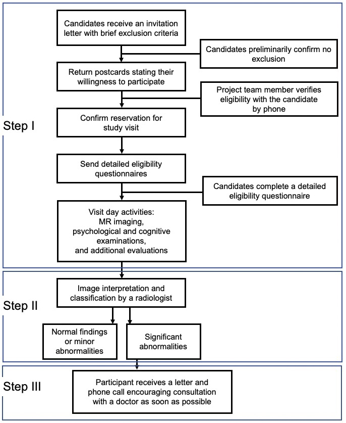
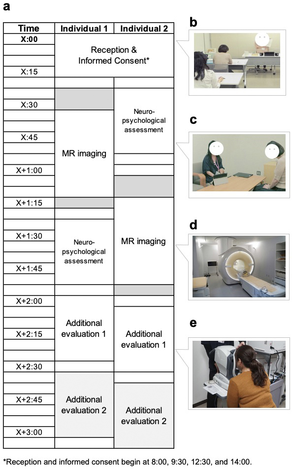
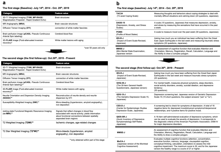
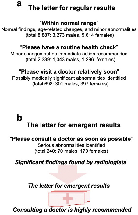
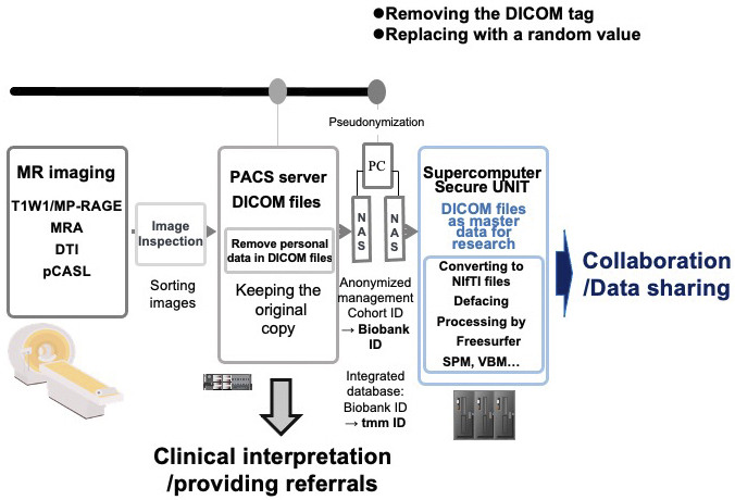
The Tohoku Medical Megabank Brain Magnetic Resonance Imaging Study (TMM Brain MRI Study) was established to collect multimodal information through neuroimaging and neuropsychological assessments to evaluate the cognitive function and mental health of residents who experienced the Great East Japan Earthquake (GEJE) and associated tsunami. The study also aimed to promote advances in personalized healthcare and medicine related to mental health and cognitive function among the general population. We recruited participants for the first (baseline) survey starting in July 2014, enrolling individuals who were participating in either the TMM Community-Based Cohort Study (TMM CommCohort Study) or the TMM Birth and Three-Generation Cohort Study (TMM BirThree Cohort Study). We collected multiple magnetic resonance imaging (MRI) sequences, including 3D T1-weighted sequences, magnetic resonance angiography (MRA), diffusion tensor imaging (DTI), pseudo-continuous arterial spin labeling (pCASL), and three-dimensional fluid-attenuated inversion recovery (FLAIR) sequences. To assess neuropsychological status, we used both questionnaire- and interview-based rating scales. The former assessments included the Tri-axial Coping Scale, Impact of Event Scale in Japanese, Profile of Mood States, and 15-item Depression, Anxiety, and Stress Scale, whereas the latter assessments included the Mini-Mental State Examination, Japanese version. A total of 12,164 individuals were recruited for the first (baseline) survey, including those unable to complete all assessments. In parallel, we returned the MRI results to the participants and subsequently shared the MRI data through the TMM Biobank. At present, the second (first follow-up) survey of the study started in October 2019 is underway. In this study, we established a large and comprehensive database that included robust neuroimaging data as well as psychological and cognitive assessment data. In combination with genomic and omics data already contained in the TMM Biobank database, these data could provide new insights into the relationships of pathological processes with neuropsychological disorders, including age-related cognitive impairment.

Keywords: TMM Birth and Three-Generation Cohort Study (TMM BirThree Cohort Study); TMM Community-Based Cohort Study (TMM CommCohort Study); The Tohoku Medical Megabank Brain Magnetic Resonance Imaging Study (TMM Brain MRI Study); cognitive impairment; neuroimaging; neuropsychological assessments.


Conflict of interest statement

None

Figures

Figure 1.
Figure 1.
Conceptual framework of the Tohoku Medical Megabank (TMM) Brain MRI Study. The TMM Brain MRI Study was designed to assess the risk factors for the onset of multifactorial neurological diseases as well as the possible influence of the Great East Japan Earthquake (GEJE) and tsunami. By examining the interaction of genetic, biological, social, and environmental factors in the development of manifestations, such as brain abnormalities (endophenotype) and clinical characteristics (phenotype) of common aging-associated diseases, the study could pave the way to the detection of prodromal features in older adults that precede age-related, systemic, or neurological deterioration.
Figure 2.
Figure 2.
Recruitment flow chart of the TMM Brain MRI Study. a. Participants in the TMM Brain MRI Study were recruited from those who had already participated in two cohort studies, the TMM Community-based Cohort Study (TMM CommCohort Study) and the TMM Birth and Three Generations Cohort Study (TMM BirThree Cohort Study), and have lived in Miyagi Prefecture. b. This figure presents the age and gender distribution of participants in the TMM CommCohort and TMM BirThree Cohort. Note that the age distributions are sharply different. Participants who participated in both the TMM CommCohort Study and TMM BirThree Cohort Study were counted as participants in the TMM BirThree Cohort Study.
Figure 3.
Figure 3.
Timeline of the TMM Brain MRI Study and its relation to the TMM CommCohort Study and TMM BirThree Cohort Study. The timeline of the TMM Brain MRI Study is presented in comparison with the timeline of the TMM CommCohort Study and the TMM BirThree Cohort Study. The first (baseline) survey of the TMM Brain MRI Study was conducted from July 2014 to Oct. 2019. It was conducted until late October 2019. The second (first follow-up) study began on the last business day of October 2019. Please note that the start and end dates for the TMM Brain MRI Study are later than the start and end dates for the two cohort studies.
Figure 4.
Figure 4.
Organization, leadership, and staffing of the TMM Brain MRI Study. Experts from a variety of disciplines bring together their academic knowledge and experience to manage the TMM Brain MRI Study through five special teams. Experts include clinical geneticists, radiologists, epidemiologists, bioinformaticians, and neurologists.
Figure 5.
Figure 5.
Steps from recruitment to return of the MRI results. The flowchart presents the implementation of this TMM Brain MRI Study in three phases. Phase I is from candidate recruitment to actual participation, and Phases II and III are from recruitment to return of the results. Note that Phase III indicates that a letter may be sent to the participant recommending immediate medical review and consultation at a medical institution based on findings incidentally obtained on the brain images that require medical attention.
Figure 6.
Figure 6.
Procedures experienced by participants on the day of the visit for each data acquisition. a. The procedure for the two participants who have their opening reception at the same time of day is divided into 15-min segments, and they alternate between each examination to avoid wasting time. b. Informed consent is provided at the same time, but participants are asked to sign individually to express their consent to participate in the testing. c. Neuropsychological evaluation is conducted by trained testers. d. The total image acquisition time per participant during MR imaging vary depending on the imaging conditions at each time point. e. Additional evaluations, such as retinal imaging, is performed on the same visit date.
Figure 7.
Figure 7.
MRI sequences and neuropsychological assessments during the first (baseline) and second (first follow-up) surveys. a. The figure presents MRI sequences in the first (baseline) and second (first follow-up) surveys. The first survey (baseline) included 3D T1-weighted imaging (T1W1; MP-RAGE), magnetic resonance angiography (MRA), diffusion tensor imaging (DTI), brain perfusion imaging (pCASL), and 3D-FLAIR sequences. The second (first follow-up) survey has added neurite orientation and dispersion density imaging (NODDI), susceptibility-weighted imaging (SWI), resting-state functional magnetic resonance imaging (rs-fMRI), T2-weighted imaging (T2WI), and T2*-weighted imaging (T2*WI). b. The figure presents questionnaires and on-site (interview) assessments in the first (baseline) and second (first follow-up) surveys at the top and bottom tables, respectively. Questionnaires are shown above the double line, whereas on-site assessments are shown below the line. In both surveys, the participants completed questionnaires assessing stress, psychiatric, personality, and neuropsychological factors at home and brought the completed forms with them on the day of the visit. On-site neuropsychological testing using the MMSE-J has been administered by trained testers. In the second (first follow-up) survey, the Montreal Cognitive Assessment, Japanese version (MoCA-J), is now administered to all participants.
Figure 8.
Figure 8.
Return of the MRI results. TMM radiologists reviewed MRI scans and categorized abnormalities. Thereafter, participants are sent one of two types of letters communicating the scan findings, depending on the significance of the abnormalities. For both letters, the total number and sex distribution of participants are presented, along with the recommended follow-up. a. This is the content for the letter about regular results (including descriptions for each subcategory: normal, mild abnormalities, or noteworthy but nonemergent findings). b. This is the content for the letter about emergent findings.
Figure 9.
Figure 9.
Pipeline for sharing MRI data. The processing scheme/flow from MRI data acquisition to distribution and sharing is shown: raw images in the DICOM format are transferred to the PACS server through a closed network. To share these data for research purposes, they are transferred to a standalone PC for pseudonymization via a USB hard disk drive (HDD) after removing personal information. Furthermore, to transfer the data to the supercomputer, the data after the pseudonymization process is exported to another USB HDD for further processing and research use.
Figure 10.
Figure 10.
Preliminary analyses of brain morphometry and cognitive function. a-f. These figures present the distribution of anatomical volumes and the correlation between anatomical volume and age for 12,164 participants in the Brain MRI Study. a, d. Each figure presents the relationship between total gray matter volume and age. b e. Each figure presents the relationship between left hippocampal volume and age. c, f. Each figure presents the relationship between right hippocampal volume and age. Pearson’s correlation analysis was conducted to evaluate the correlation between anatomical volume and age. g. The figure presents the distribution of MMSE-J (Mini-Mental State Examination, Japanese version) scores (total and by sex), as well as the number and percentage of individuals scoring below the normal range.

References

    1. Fuse N, Sakurai-Yageta M, Katsuoka F, et al. Establishment of integrated biobank for precision medicine and personalized healthcare: the Tohoku Medical Megabank project. JMA J. 2019;2(2):113-22. - PMC - PubMed
    1. Hozawa A, Tanno K, Nakaya N, et al. Study profile of the Tohoku Medical Megabank community-based cohort study. J Epidemiol. 2021;31(1):65-76. - PMC - PubMed
    1. Kuriyama S, Yaegashi N, Nagami F, et al. The Tohoku Medical Megabank project: design and mission. J Epidemiol. 2016;26(9):493-511. - PMC - PubMed
    1. Kuriyama S, Metoki H, Kikuya M, et al. Cohort profile: Tohoku Medical Megabank project birth and three-generation cohort study (TMM BirThree cohort study): rationale, progress and perspective. Int J Epidemiol. 2020;49(1):18-19m. - PMC - PubMed
    1. The reconstruction design council in response to the great East Japan earthquake. Cabinet Secretariat [Internet]. 2011 Jun [cited 2022 Dec 14]. Available from: https://www.cas.go.jp/jp/fukkou/english/index.html

LinkOut - more resources