. 2023 Jul 31:42:15-30.
doi: 10.1016/j.jot.2023.07.002. eCollection 2023 Sep.

Kartogenin-enhanced dynamic hydrogel ameliorates intervertebral disc degeneration via restoration of local redox homeostasis

Affiliations

Kartogenin-enhanced dynamic hydrogel ameliorates intervertebral disc degeneration via restoration of local redox homeostasis

Xin Tian et al. J Orthop Translat. .

Abstract

Introduction: Over-activation of oxidative stress due to impaired antioxidant functions in nucleus pulpous (NP) has been identified as a key factor contributing to intervertebral disc degeneration (IVDD). While Kartogenin (KGN) has previously demonstrated antioxidant properties on articular cartilage against osteoarthritis, its effects on NP degeneration have yet to be fully understood.

Objectives: This study aimed to investigate the protective effects of KGN on nucleus pulpous cells (NPCs) against an inflammatory environment induced by interleukin (IL)-1β, as well as to explore the therapeutic potential of KGN-enhanced dynamic hydrogel in preventing IVDD.

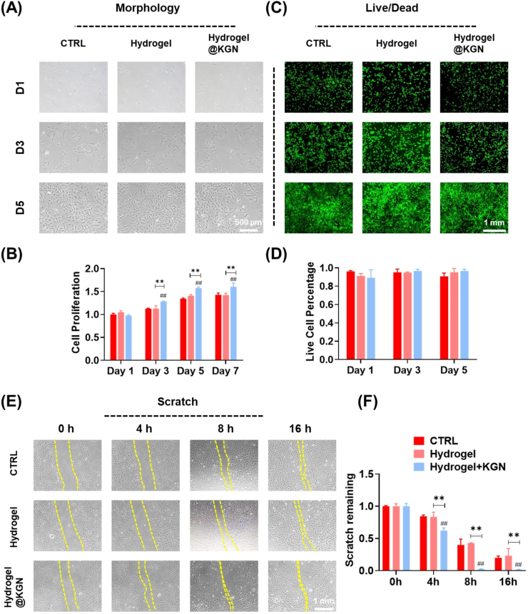
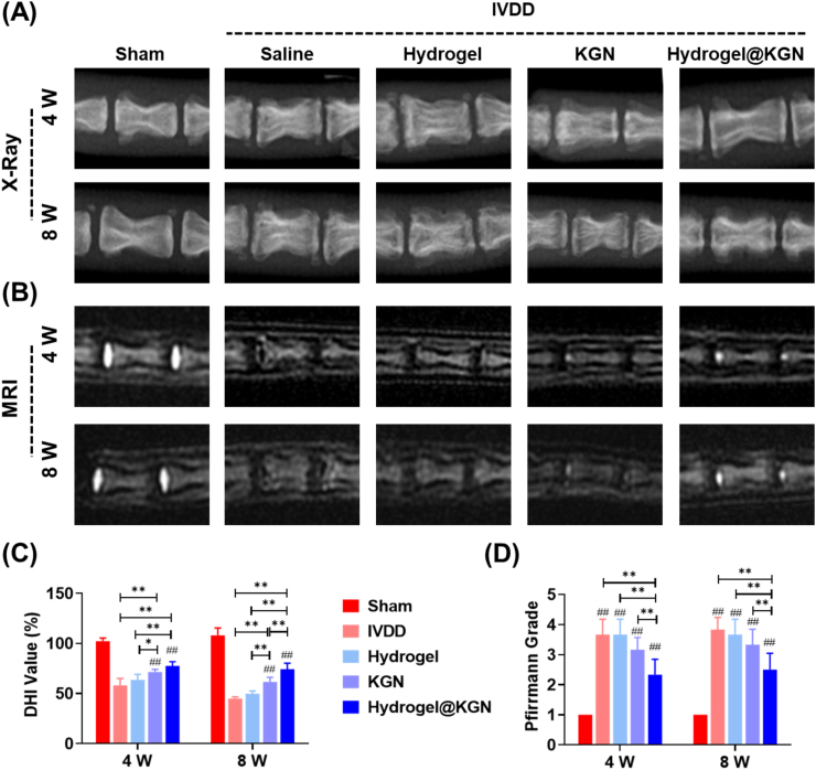
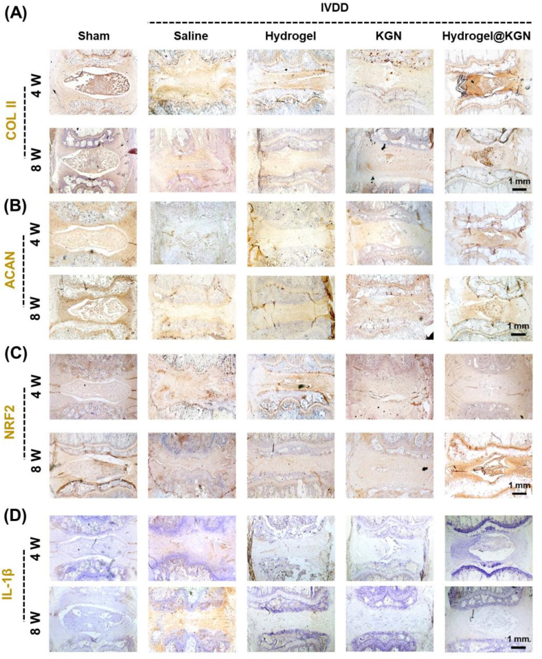
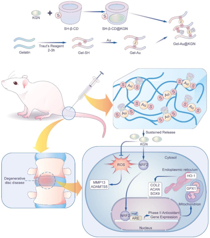
Methods: NPCs were isolated from rat caudal IVDs and subjected to treatment with KGN at varying concentrations (ranging from 0.01 to 1 ​μM) in the presence of IL-1β. The expression of extracellular matrix (ECM) anabolism markers was quantitatively assessed at both the mRNA and protein levels. Additionally, intracellular reactive oxygen species and antioxidant enzyme expression were evaluated, along with the role of nuclear factor erythroid 2-related factor 2 (NRF2). Based on these findings, a dynamic self-healing hydrogel loaded with KGN was developed through interconnecting networks. Subsequently, KGN-enhanced dynamic hydrogel was administered into rat caudal IVDs that had undergone puncture injury, followed by radiographic analysis and immunohistochemical staining to evaluate the therapeutic efficacy.

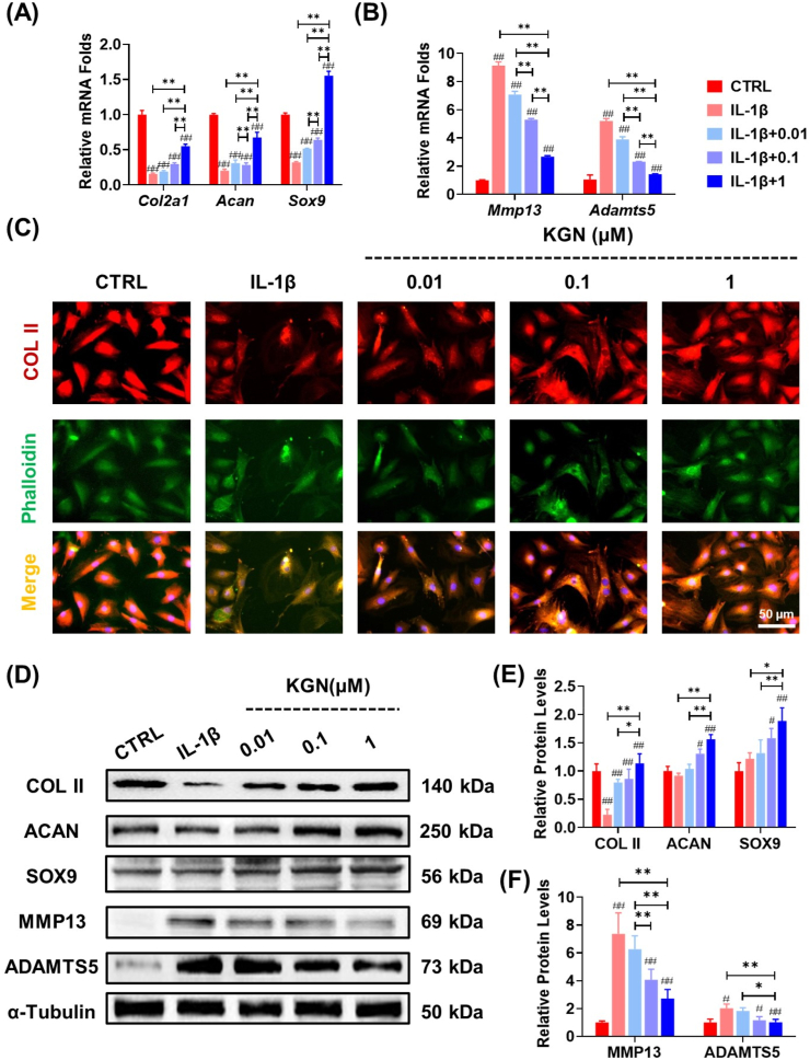
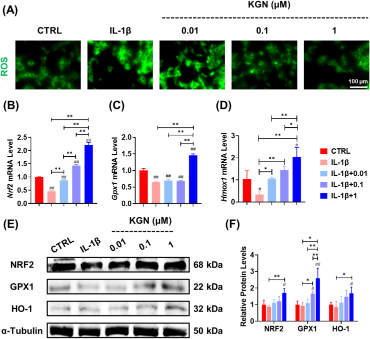
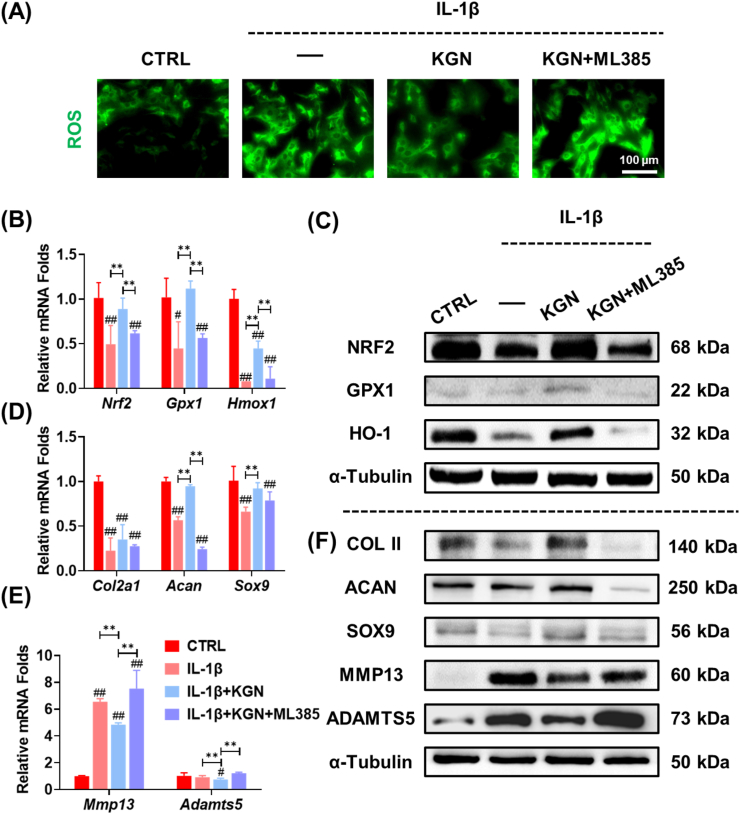
Results: In vitro treatments utilizing KGN were observed to maintain ECM synthesis and inhibit catabolic activities in IL-1β-stimulated NPCs. The mechanism behind this protective effect of KGN on NPCs was found to involve the asctivation of NRF2 and downstream antioxidant enzymes, including glutathione peroxidase 1 and heme oxygenase 1. This was further supported by the loss of both antioxidant and anabolic effects upon pharmacological inhibition of NRF2. Furthermore, a self-healing hydrogel was developed and loaded with KGN to achieve localized and sustained release of the compound. The injection of KGN-enhanced hydrogel effectively ameliorated the degradation of NP ECM and mitigated inflammation in a rat model of puncture-induced IVDD.

Conclusions: Our results indicate that KGN exhibits potential as a therapeutic agent for NP degeneration, and that KGN-enhanced dynamic hydrogel represents a novel approach for treating IVDD by restoring redox homeostasis in NP.The translational potential of this article: The dysregulation of oxidant and antioxidant balance has been shown to impede the repair and regeneration of NP, thereby hastening the progression of IVDD following injury. The present investigation has demonstrated that the sustained release of KGN promotes the synthesis of ECM in vitro and mitigates the progression of IVDD in vivo by restoring redox equilibrium, thereby presenting a novel therapeutic candidate based on the antioxidant properties of KGN for the treatment of IVDD.

Keywords: Dynamic hydrogel; Intervertebral disc degeneration; Kartogenin; NRF2; Nucleus pulposus.


Conflict of interest statement

A conflict of interest occurs when an individual's objectivity is potentially compromised by a desire for financial gain, prominence, professional advancement or a successful outcome. The Editors of the Journal of Orthopaedic Translation strive to ensure that what is published in the Journal is as balanced, objective and evidence-based as possible. Since it can be difficult to distinguish between an actual conflict of interest and a perceived conflict of interest, the Journal requires authors to disclose all and any potential conflicts of interest.

Figures

Image 1
Graphical abstract
Fig. 1
Fig. 1
The effects of KGN on the matrix synthesis of nucleus pulposus cells (NPCs) under normal culture conditions. NPCs were treated with KGN at concentrations of 0.01, 0.1, and 1 ​μM. (A) Cell proliferation was examined at 1, 3, 5 and 7 days using CCK-8 assay, n ​= ​6. (B) The mRNA expression levels of Col2a1, Acan, and Sox9 as determined by RT-PCR, n ​= ​4. (C) Immunofluorescence staining images showed the expression of COL II proteins in KGN-treated cells. (D–E) The effects of KGN on the protein levels of NP matrix components in NPCs were determined using Western blot, n ​= ​3. Data are shown as the mean ​± ​standard deviation. Statistically significant differences are indicated by # where P ​< ​0.05 or ## where P ​< ​0.01 compared to the control (CTRL) group; ∗ where P ​< ​0.05 or ∗∗ where P ​< ​0.01 between the indicated groups.
Fig. 2
Fig. 2
The protective effects of KGN on ECM metabolism of NPCs in the presence of IL-1β. NPCs were exposed to 5 ​ng/mL IL-1β and treated with KGN at concentrations of 0.01, 0.1, and 1 ​μM. (A–B) The mRNA expression levels of Col2a1, Acan, Sox9, Mmp13, and Adamts5 in IL-1β-treated NPCs were evaluated to assess the effect of KGN, n ​= ​4. (C) Immunofluorescence staining was performed to assess the expression of COL II in NPCs, n ​= ​3. (D–F) The effects of KGN on the protein levels of anabolic or catabolic markers in IL-1β-stimulated NPCs were determined using Western blot, n ​= ​3. Data are presented as the mean ​± ​standard deviation. Statistically significant differences are indicated by # where P ​< ​0.05 or ## where P ​< ​0.01 compared to the control (CTRL) group; ∗ where P ​< ​0.05 or ∗∗ where P ​< ​0.01 between the indicated groups.
Fig. 3
Fig. 3
Impact of KGN on oxidative stress of IL-1β-stimulated NPCs. (A) Immunofluorescence staining indicated the effect of KGN on intracellular ROS in IL-1β-stimulated cells. (B–D) The gene expression of Nrf2, Gpx1, and Hmox1 was quantified using RT-PCR, n ​= ​4. (E–F) The effects of KGN on the protein levels of antioxidant enzymes in IL-1β-stimulated NPCs, n ​= ​3. Data are presented as the mean ​± ​standard deviation. Statistically significant differences are indicated by # where P ​< ​0.05 or ## where P ​< ​0.01 compared to the control (CTRL) group; ∗ where P ​< ​0.05 or ∗∗ where P ​< ​0.01 between the indicated groups.
Fig. 4
Fig. 4
The role of NRF2 in KGN-modulated antioxidant functions in IL-1β-stimulated NPCs. Prior to exposure to 5 ​ng/mL IL-1β and 1 ​μM KGN, NPCs were pre-treated with an NRF2-specific inhibitor, ML385. (A) Immunofluorescence staining revealed intracellular ROS in NPCs. (B) The gene expression of Nrf2, Gpx1, and Hmox1 was quantified using RT-PCR, n ​= ​4. (C) The protein levels of NRF2, GPX1, and HO-1 were measured using Western blot, n ​= ​3. (D–E) The gene expression of Col2a1, Acan, Sox9, Mmp13, and Adamts5 was quantified using RT-PCR, n ​= ​4. (C) The protein levels of COL II, ACAN, SOX9, MMP13, and ADAMTS5 were measured using Western blot. Data are presented as the mean ​± ​standard deviation. Statistically significant differences are indicated by # where P ​< ​0.05 or ## where P ​< ​0.01 compared to the control (CTRL) group; ∗ where P ​< ​0.05 or ∗∗ where P ​< ​0.01 between the indicated groups.
Fig. 5
Fig. 5
Characterization of Au-Gel dynamic hydrogel loaded with KGN. (A) Representative SEM images of Au-Gel, Au-Gel@β-CD, and Au-Gel@KGN hydrogels. (B) The gelling process of hydrogel was determined by the inverting experiment. (C–D) The self-healing hydrogel exhibited injectability that can form letter patterns (Soochow university, SU). (E) The self-healing capacity of hydrogel after fragmentation. (F–I) The dynamic oscillatory frequency sweeps (strain ​= ​1%), the strain amplitude sweeps (frequency ​= ​1 ​rad/s), and the step-strain sweeps (strain ​= ​1 or 300%, frequency ​= ​1 ​rad/s), the shear viscosity (shear rates ranging from 0.1 to 50 ​rad/s) of the hydrogel (G′, storage modulus; G″, loss modulus).
Fig. 6
Fig. 6
The biocompatibility of the self-healing Hydrogel@KGN. (A) The cell morphology of NPCs cultured with hydrogel leachate. (B) Cell proliferation of NPCs treated with hydrogel leachate was determined using CCK-8 assays, n ​= ​6. (C) Representative images of Live/dead staining of NPCs treated with hydrogel leachate. (D) Quantification of live and dead cells, n ​= ​3. (E–F) Cell migration at 0, 4, 8, 16 ​h after cultured with hydrogel leachate, n ​= ​3. Data are presented as the mean ​± ​standard deviation. Statistically significant differences are indicated by # where P ​< ​0.05 or ## where P ​< ​0.01 compared to the control (CTRL) group; ∗ where P ​< ​0.05 or ∗∗ where P ​< ​0.01 between the indicated groups.
Fig. 7
Fig. 7
Diagnostic imaging analyses were conducted to evaluate the impact of Hydrogel@KGN on puncture-induced NP degeneration. A) Representative X-ray images of treated sites in rats' caudal IVDs at 4 and 8 weeks post-surgery. (B) Representative MRI images of treated sites in rats' caudal IVDs 4 and 8 weeks after surgery. (C) The disc height index (DHI) was calculated from the radiograph images, n ​= ​6. (D) The optical density values of IVD based on MRI signals were utilized to indicate the degeneration of rats' caudal IVDs, n ​= ​6. Statistically significant differences are indicated by # where P ​< ​0.05 or ## where P ​< ​0.01 compared to the Sham group; ∗ where P ​< ​0.05 or ∗∗ where P ​< ​0.01 between the indicated groups.
Fig. 8
Fig. 8
Histological analysis was conducted to investigate the effect of Hydrogel@KGN on protecting NP matrix in rats' caudal intervertebral discs (IVDs) with puncture injury. (A) Representative images of hematoxylin and eosin (H&E) of IVDs at 4 and 8 weeks post-surgery. Scale bar: 1 ​mm.(B) Representative images of Safranin O (S.O.) staining of IVDs at 4 and 8 weeks post-surgery. Scale bar: 1 ​mm. (C–D) Histological scores and the modified Thompson score of the untreated and treated IVDs at 4 and 8 weeks post-surgery, n ​= ​6. Statistically significant differences are indicated by # where P ​< ​0.05 or ## where P ​< ​0.01 compared to the Sham group; ∗ where P ​< ​0.05 or ∗∗ where P ​< ​0.01 between the indicated groups.
Fig. 9
Fig. 9
Immunohistochemical (IHC) analysis was conducted to investigate the NP matrix preservation in rats' caudal intervertebral discs (IVDs) with puncture injury. (A–D) Representative images of IHC staining for collagen II (COL II), aggrecan (ACAN), NRF2, and IL-1β in the NP site at 4 and 8 weeks post-surgery. Scale bar: 1 ​mm.
Fig. 10
Fig. 10
Kartogenin-enhanced dynamic hydrogel ameliorates IVDD progression. Mechanistically, NRF2 plays an important role in reducing oxidative stress and preventing ECM degradation in NPCs. To achieve localized and sustained release, a self-healing hydrogel loaded with Kartogenin was developed. In situ injection of KGN-enhanced hydrogel mitigates NP degeneration in a rat IVDD model induced by puncture.

Similar articles

Cited by

References

    1. Mohd Isa I.L., Mokhtar S.A., Abbah S.A., Fauzi M.B., Devitt A., Pandit A. Intervertebral disc degeneration: biomaterials and tissue engineering strategies toward precision medicine. Adv. Healthc. Mater. 2022;11 - PMC - PubMed
    1. Armbrecht G., Felsenberg D., Ganswindt M., Lunt M., Kaptoge S.K., Abendroth K., et al. Degenerative inter-vertebral disc disease osteochondrosis intervertebralis in Europe: prevalence, geographic variation and radiological correlates in men and women aged 50 and over. Rheumatology. 2017;56:1189–1199. - PMC - PubMed
    1. Berman B.M., Langevin H.M., Witt C.M., Dubner R. Acupuncture for chronic low back pain. N Engl J Med. 2010;363:454–461. - PubMed
    1. Chen S., Wu X., Lai Y., Chen D., Bai X., Liu S., et al. Kindlin-2 inhibits Nlrp3 inflammasome activation in nucleus pulposus to maintain homeostasis of the intervertebral disc. Bone Res. 2022;10:5. - PMC - PubMed
    1. Zhou Z., Cui S., Du J., Richards R.G., Alini M., Grad S., et al. One strike loading organ culture model to investigate the post-traumatic disc degenerative condition. J. Orthop. Transl. 2020;26:141–150. - PMC - PubMed

LinkOut - more resources