

Visual-evoked potential predicts the efficacy of the optical canal decompression for traumatic optic nerve neuropathy showing blindness: A case report
- PMID: 37560571
- PMCID: PMC10408612
- DOI: 10.25259/SNI_450_2023
Visual-evoked potential predicts the efficacy of the optical canal decompression for traumatic optic nerve neuropathy showing blindness: A case report
Abstract
Background: The indication for surgical optic canal decompression (OCD) for traumatic optic neuropathy (TON) remains controversial because there is no reliable predictor of a good outcome. We report the case of a blind patient with TON whose remaining visual-evoked potential (VEP) suggested recovery potential of the injured optic nerve after OCD.
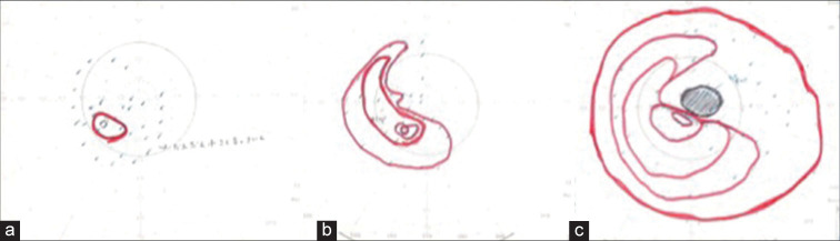
Case description: A 48-year-old man had fallen from a height of 7 m, striking his head. He immediately complained of right-eye blindness. He had no light perception and the direct light reflex disappeared from the right pupil, although there was no fracture or traumatic lesion on computed tomography and magnetic resonance imaging. Because the amplitude of the VEP with the right eye stimulation remained unchanged, we performed the right OCD. During surgical OCD, the amplitude and latency of VEP began to improve. Finally, the visual field improved in almost all directions, and eyesight improved to 0.2.
Conclusion: The retained VEP activity in TON may suggest the recovery potential of the injured optic nerve, even in cases of blindness. It is possible that VEP is an indicator of aggressive treatment for TON such as OCD.
Keywords: Optic canal decompression; Traumatic optic neuropathy; Visual-evoked potential.
Copyright: © 2023 Surgical Neurology International.
Conflict of interest statement
There are no conflicts of interest.
Figures


References
-
- Agarwal A, Mahapatra AK. Visual outcome in optic nerve injury patients without initial light perception. Indian J Ophthalmol. 1999;47:233–6. - PubMed
-
- Chen B, Zhang H, Zhai Q, Li H, Wang C, Wang Y. Traumatic optic neuropathy: A review of current studies. Neurosurg Rev. 2022;45:1895–913. - PubMed
-
- Chen HH, Lee MC, Tsai CH, Pan CH, Lin YT, Chen CT. Surgical decompression or corticosteroid treatment of indirect traumatic optic neuropathy: A randomized controlled trial. Ann Plast Surg. 2020;84(Suppl 1):S80–3. - PubMed
-
- Entezari M, Rajavi Z, Sedighi N, Daftarian N, Sanagoo M. High-dose intravenous methylprednisolone in recent traumatic optic neuropathy; a randomized double-masked placebo-controled clinical trial. Graefes Arch Clin Exp Ophthalmol. 2007;245:1267–71. - PubMed
-
- Fujitani T, Inoue K, Takahishi T, Ikushima K, Asai T. Indirect traumatic optic neuropathy--visual outcome of operative and nonoperative cases. Jpn J Opthalmol. 1986;30:125–34. - PubMed
Publication types
LinkOut - more resources
Full Text Sources