. 2023 Oct 12;66(19):13665-13683.
doi: 10.1021/acs.jmedchem.3c01030. Epub 2023 Aug 10.

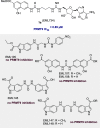
Identification of a Protein Arginine Methyltransferase 7 (PRMT7)/Protein Arginine Methyltransferase 9 (PRMT9) Inhibitor

Affiliations

Identification of a Protein Arginine Methyltransferase 7 (PRMT7)/Protein Arginine Methyltransferase 9 (PRMT9) Inhibitor

Alessandra Feoli et al. J Med Chem. .

Abstract

Less studied than the other protein arginine methyltransferase isoforms, PRMT7 and PRMT9 have recently been identified as important therapeutic targets. Yet, most of their biological roles and functions are still to be defined, as well as the structural requirements that could drive the identification of selective modulators of their activity. We recently described the structural requirements that led to the identification of potent and selective PRMT4 inhibitors spanning both the substrate and the cosubstrate pockets. The reanalysis of the data suggested a PRMT7 preferential binding for shorter derivatives and prompted us to extend these structural studies to PRMT9. Here, we report the identification of the first potent PRMT7/9 inhibitor and its binding mode to the two PRMT enzymes. Label-free quantification mass spectrometry confirmed significant inhibition of PRMT activity in cells. We also report the setup of an effective AlphaLISA assay to screen small molecule inhibitors of PRMT9.


Conflict of interest statement

The authors declare no competing financial interest.

Figures

Figure 1
Figure 1
Architecture of PRMT7 and PRMT9 (prepared using Illustrator for Biological Sequences, IBS) and phylogenetic tree of SAM-dependent class I methyltransferases (obtained with the Structural Genomic Consortium ChromoHub and modified with Adobe Illustrator CC 2023). PRMTs are highlighted in the orange area, whereas non-SET domain-containing KMTs are in the gray area.
Figure 2
Figure 2
Inhibitory activities of compounds 1ah against PRMT7: the heatmaps depict the IC50 values (nM) for compounds 1a1h against PRMT7 (left panel, in shades of green) and the selectivity index (fold) for PRMT7 over the specified PRMT (right, shades of blue and orange).
Figure 3
Figure 3
Inhibitory activities of selected PRMT modulators (from in-house libraries) against PRMT9.
Figure 4
Figure 4
Inhibition of GST-tagged HsPRMT9 and HsPRMT7 (panels a and b, respectively) or CePRMT7 and CePRMT9 (panels c and d, respectively) by compounds 1ac as detected by a radioisotope-based assay. The experiments were performed as reported in the Experimental procedures section. GST-HsPRMT9 (a) and GST-HsPRMT7 (b) were incubated with human GST-SF3B2 (401–550) peptide or recombinant HsH2B, respectively (1 μg of enzyme, 5 μg of substrate), 0.14 μM [3H]SAM, and the indicated concentrations of tested compounds at the corresponding optimal reaction temperature (37 °C for HsPRMT9, 15 °C for HsPRMT7). C. elegans GST-tagged enzymes PRMT9 (c) and PRMT7 (d) were incubated with GST-CeSFTB-2 (99–248) fragment or recombinant HsH2B, respectively (1 μg of enzyme, 5 μg of substrate), 0.14 μM [3H]SAM, and the indicated concentrations of tested compounds at the corresponding optimal reaction temperature (25 °C for CePRMT9, 15 °C for CePRMT7). After SDS-PAGE, the gels were treated as previously described and densitometry analysis was done using ImageJ software, and data was plotted as normalized activity to the no inhibitor controls.
Figure 5
Figure 5
Binding mode of 1a in complex with the PRMT9 3D structure (PDB entry 7RBQ) as predicted by docking calculations (a) and representative frame of the 500 ns long MD simulation (b). The ligand and enzyme are represented as orange and cyan stick and ribbons, respectively. L-RMSD (c) and L-RMSF (d) plots obtained from the analysis of MD simulations.
Figure 6
Figure 6
Design of compound 1j to strengthen π-stacking interaction with the PRMT9 W152 residue (in blue).
Figure 7
Figure 7
Sensorgrams obtained from the SPR interaction analysis of compound 1j binding to immobilized PRMT9. The compound was injected at different concentrations (from 25 to 0.05 mM) with an association and a dissociation time of 90 and 180 s, respectively, and with a flow rate of 30 μL/min. The equilibrium dissociation constant (KD) was derived from the ratio between kinetic dissociation (koff) and association (kon) constants.
Figure 8
Figure 8
Testing the effects of compounds 1a (EML734), 1b (EML736), 1e (EML979), and 1f (EML980) on PRMT9 activity in MCF7 (top) and MDA-MB-436 breast cancer cell lines. MCF7 and MDA-MB-436 cells were treated with 4 candidate inhibitors at indicated concentrations for 72 h. The total cell lysates were harvested in RIPA buffer and the levels of SF3B2 R508me2s, SF3B2, and PRMT9 were detected by using Western blot assays. Anti-Tubulin antibody was used as a loading control.
Figure 9
Figure 9
Schematic description of the MS proteomic experiment. Created with BioRender.com.
Figure 10
Figure 10
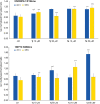
Compounds 1a and 1j inhibit PRMT7 in cells. The bar graphs plot the ratio between the abundance of nonmethylated over methylated peptides in HEK392T cells treated with compounds 1a or 1j or untreated for 24 h (blue) or 48 h (yellow). The top panel shows the ratio between HNRNPA1 unmethylated over R194me peptides, and the bottom panel shows the ratio between HSP70 unmethylated over R469me peptides.
Scheme 1
Scheme 1. Synthesis of Compounds 1i and 1j
Reagents and conditions: (a) zinc dust, acetic acid, 1 h (97–98%); (b) phenyl chloroformate, TEA, AcOEt, r. t., 12 h (65–70%); (c) TEA, dry DMF, r. t., 2 h (68–70%); (d) DCM/TFA 9:1, r. t., 2 h (80–92%); (e) EDC hydrochloride, TEA, dry DCM, r. t., 18 h (60–74%); (f) DCM/TFA 1:1, r. t., 2 h (60–76%).
Scheme 2
Scheme 2. Synthesis of Derivative 2b
Reagents and conditions: (a) NaBH4, I2, dry THF, 0 °C to reflux, 18 h (88%); (b) Dess-Martin periodinane, dry DCM, 2 h, r.t. (74%); (c) toluene, r. t., 48 h (63%); (d) sodium acetate, acetic anhydride, 120 °C (MW), 25 min (67%).

References

    1. Wu Q.; Schapira M.; Arrowsmith C. H.; Barsyte-Lovejoy D. Protein arginine methylation: from enigmatic functions to therapeutic targeting. Nat. Rev. Drug Discovery 2021, 20 (7), 509–530. 10.1038/s41573-021-00159-8. - DOI - PubMed
    1. Turner B. M. Reading signals on the nucleosome with a new nomenclature for modified histones. Nat. Struct. Mol. Biol. 2005, 12 (2), 110–112. 10.1038/nsmb0205-110. - DOI - PubMed
    1. Bedford M. T.; Clarke S. G. Protein Arginine Methylation in Mammals: Who, What, and Why. Mol. Cell 2009, 33 (1), 1–13. 10.1016/j.molcel.2008.12.013. - DOI - PMC - PubMed
    1. Yang Y.; Bedford M. T. Protein arginine methyltransferases and cancer. Nat. Rev. Cancer 2013, 13 (1), 37–50. 10.1038/nrc3409. - DOI - PubMed
    1. Schubert H. L.; Blumenthal R. M.; Cheng X. Many paths to methyltransfer: a chronicle of convergence. Trends Biochem. Sci. 2003, 28 (6), 329–335. 10.1016/S0968-0004(03)00090-2. - DOI - PMC - PubMed

Publication types

MeSH terms

LinkOut - more resources