Clinical Trial
. 2023 Oct 2;133(19):e164918.
doi: 10.1172/JCI164918.

A phase III randomized crossover trial of plerixafor versus G-CSF for treatment of WHIM syndrome

Affiliations
Clinical Trial

A phase III randomized crossover trial of plerixafor versus G-CSF for treatment of WHIM syndrome

David H McDermott et al. J Clin Invest. .

Abstract

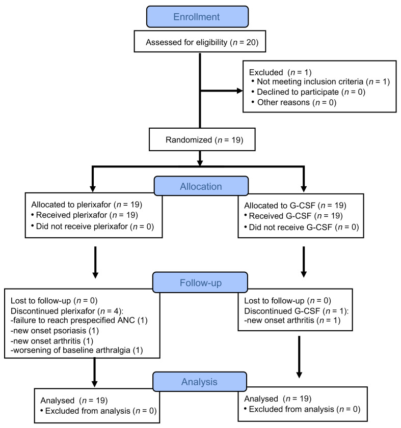
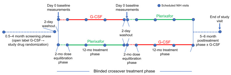
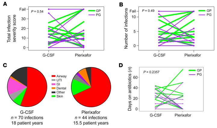
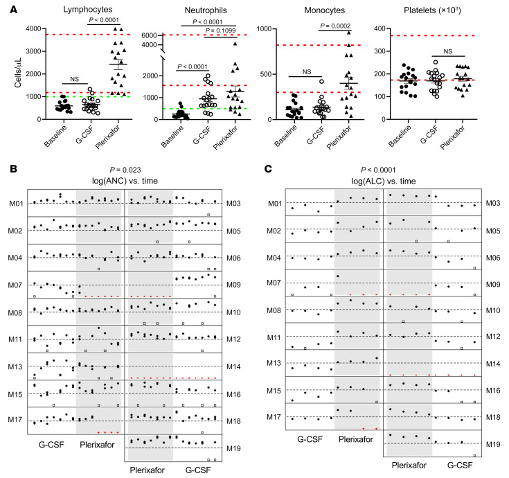
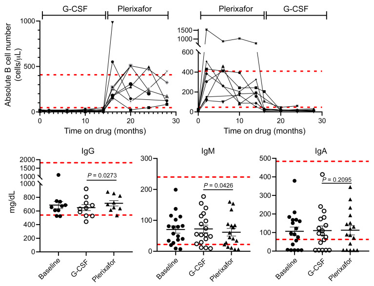
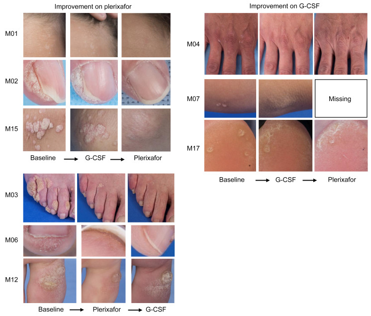
BACKGROUNDWarts, hypogammaglobulinemia, infections, and myelokathexis (WHIM) syndrome is a primary immunodeficiency disorder caused by heterozygous gain-of-function CXCR4 mutations. Myelokathexis is a kind of neutropenia caused by neutrophil retention in bone marrow and in WHIM syndrome is associated with lymphopenia and monocytopenia. The CXCR4 antagonist plerixafor mobilizes leukocytes to the blood; however, its safety and efficacy in WHIM syndrome are undefined.METHODSIn this investigator-initiated, single-center, quadruple-masked phase III crossover trial, we compared the total infection severity score (TISS) as the primary endpoint in an intent-to-treat manner in 19 patients with WHIM who each received 12 months treatment with plerixafor and 12 months treatment with granulocyte CSF (G-CSF, the standard of care for severe congenital neutropenia). The treatment order was randomized for each patient.RESULTSPlerixafor was nonsuperior to G-CSF for TISS (P = 0.54). In exploratory endpoints, plerixafor was noninferior to G-CSF for maintaining neutrophil counts of more than 500 cells/μL (P = 0.023) and was superior to G-CSF for maintaining lymphocyte counts above 1,000 cells/μL (P < 0.0001). Complete regression of a subset of large wart areas occurred on plerixafor in 5 of 7 patients with major wart burdens at baseline. Transient rash occurred on plerixafor, and bone pain was more common on G-CSF. There were no significant differences in drug preference or quality of life or the incidence of drug failure or serious adverse events.CONCLUSIONPlerixafor was not superior to G-CSF in patients with WHIM for TISS, the primary endpoint. Together with wart regression and hematologic improvement, the infection severity results support continued study of plerixafor as a potential treatment for WHIM syndrome.TRIAL REGISTRATIONClinicaltrials.gov NCT02231879.FUNDINGThis study was funded by the Division of Intramural Research, National Institute of Allergy and Infectious Diseases.

Keywords: Clinical Trials; Genetic diseases; Immunology; Innate immunity; Neutrophils.


Figures

Figure 1
Figure 1. Flow diagram of the progress of the participants through the phases of the study.
The diagram was created using CONSORT (http://www.consort-statement.org/consort-statement/flow-diagram).
Figure 2
Figure 2. Trial design.
Study phase durations, treatment, and interval NIH visits (designated by blue dots) are indicated.
Figure 3
Figure 3. Low dose G-CSF versus plerixafor for 19 patients with WHIM.
Drug doses for the 3 phases of the study are shown, stratifying patients by randomization order. P, plerixafor; G, G-CSF. Horizontal dashed red lines indicate the package insert-recommended total daily dosage of G-CSF for severe congenital neutropenia or the single injection daily FDA-approved dose of plerixafor for HSC mobilization. Vertical dashed green lines indicate day 56, the final day of the equilibration phase (Equil. phase). Horizontal dashed black lines indicate target total daily dose ranges for the study. Children are indicated by asterisks. Changes in drug dose were to stay within the target ANC range or to mitigate side effects. (fail), patient dropouts due to side effects or drug failure (see main text for details).
Figure 4
Figure 4. Infection severity and distribution for patients with WHIM during the plerixafor and G-CSF treatment phases.
(A, B, and D) Drug order is color-coded in the top right corner. GP, G-CSF first followed by plerixafor; PG, plerixafor first followed by G-CSF. Each line represents a single patient, connecting results for each treatment phase. “Fail” in A and B refers to patients who dropped out because of drug intolerance or failure to meet the prespecified ANC threshold during the equilibration phase. (C) The infection distribution by site and the total number of patient-years of drug exposure for each treatment phase. UTI, urinary tract infection; GI, gastrointestinal infection. P values at the top left of each graph were determined using a Wilcoxon’s rank-sum analysis, as specified in the Supplemental Statistical Analysis Plan.
Figure 5
Figure 5. Effects of plerixafor versus G-CSF on circulating blood cell counts in patients with WHIM.
(A) Plerixafor, but not G-CSF, reversed panleukopenia without affecting circulating platelet concentration. Baseline refers to the day 0 value of the equilibration phase after 2-day washout of G-CSF or plerixafor from the preceding phase. G-CSF and plerixafor values are the final values obtained for the approximately 3-hour postmorning dose at the end of each treatment phase. Dashed green horizontal lines indicate the prespecified thresholds for judging success for improving neutropenia and lymphopenia. Each symbol represents a different patient. Dashed red horizontal lines demarcate the normal range for adults for each parameter established by the NIH-CC Department of Laboratory Medicine. P values were determined by a 2-sided Wilcoxon’s matched pairs rank test. (B and C) Plerixafor is noninferior to G-CSF for durably reversing neutropenia and is superior to G-CSF for durably reversing lymphopenia for 1 year. Filled black circles indicate individual values at the indicated times; open black squares indicate missing values due to scheduling conflicts; and red triangles indicate missing data due to drug failure. The y axes are on a log scale from 50 to 5,000 for ANC and from 100 to 10,000 for ALC. The thresholds for judging success are indicated by dashed horizontal lines at 500 and 1,000 cells/μL for ANC and ALC, respectively. P values (at the top of each panel) were determined by a Wilcoxon’s matched pairs rank test, as specified in the Supplemental Statistical Analysis Plan and the Supplemental Statistical Analysis Plan Supplement.
Figure 6
Figure 6. Normalization of circulating B cell levels by plerixafor did not affect immunoglobulin levels.
Dashed horizontal lines demarcate the normal range for each parameter established by the NIH-CC Department of Laboratory Medicine. Top: B cells. The time on each drug includes both equilibration and treatment phases and is indicated at the top by brackets. Each graphed line represents data for a single patient. Bottom: Immunoglobulin levels. G-CSF and plerixafor data designate the final value obtained at the end of each treatment phase. IgG data are only shown for patients not receiving supplemental immunoglobulin. P values are only shown for the drug comparisons and were determined by a 2-sided Wilcoxon’s matched pairs rank test. Comparisons of day –0 baseline data to data on each drug were not significant.
Figure 7
Figure 7. Plerixafor and G-CSF effects on wart burden in patients with WHIM.
Representative images of warts at baseline and at the end of the indicated drug treatment period are shown for patients demonstrating improvement in wart areas during the study. Images are for the patient indicated to the left of the corresponding row. Comprehensive assessments of wart changes on drug treatment are detailed in Table 2, Supplemental Tables 7–13, and Supplemental Figure 7.

References

    1. Beaussant Cohen S, et al. Description and outcome of a cohort of 8 patients with WHIM syndrome from the French Severe Chronic Neutropenia Registry. Orphanet J Rare Dis. 2012;7:71. doi: 10.1186/1750-1172-7-71. - DOI - PMC - PubMed
    1. Dale DC, et al. Family studies of warts, hypogammaglobulinemia, immunodeficiency, myelokathexis syndrome. Curr Opin Hematol. 2020;27(1):11–17. doi: 10.1097/MOH.0000000000000554. - DOI - PMC - PubMed
    1. Geier CB, et al. Disease progression of WHIM syndrome in an international cohort of 66 pediatric and adult patients. J Clin Immunol. 2022;42(8):1748–1765. doi: 10.1007/s10875-022-01312-7. - DOI - PMC - PubMed
    1. Heusinkveld LE, et al. WHIM syndrome: from pathogenesis towards personalized medicine and cure. J Clin Immunol. 2019;39(6):532–556. doi: 10.1007/s10875-019-00665-w. - DOI - PMC - PubMed
    1. Krill CE, Jr, et al. Chronic idiopathic granulocytopenia. N Engl J Med. 1964;270:973–979. doi: 10.1056/NEJM196405072701902. - DOI - PubMed

Publication types

MeSH terms

Substances

Supplementary concepts

Associated data