. 2023 Oct 2;133(19):e171356.
doi: 10.1172/JCI171356.

Nonviral base editing of KCNJ13 mutation preserves vision in a model of inherited retinal channelopathy

Affiliations

Nonviral base editing of KCNJ13 mutation preserves vision in a model of inherited retinal channelopathy

Meha Kabra et al. J Clin Invest. .

Abstract

Clinical genome editing is emerging for rare disease treatment, but one of the major limitations is the targeting of CRISPR editors' delivery. We delivered base editors to the retinal pigmented epithelium (RPE) in the mouse eye using silica nanocapsules (SNCs) as a treatment for retinal degeneration. Leber congenital amaurosis type 16 (LCA16) is a rare pediatric blindness caused by point mutations in the KCNJ13 gene, a loss of function inwardly rectifying potassium channel (Kir7.1) in the RPE. SNCs carrying adenine base editor 8e (ABE8e) mRNA and sgRNA precisely and efficiently corrected the KCNJ13W53X/W53X mutation. Editing in both patient fibroblasts (47%) and human induced pluripotent stem cell-derived RPE (LCA16-iPSC-RPE) (17%) showed minimal off-target editing. We detected functional Kir7.1 channels in the edited LCA16-iPSC-RPE. In the LCA16 mouse model (Kcnj13W53X/+ΔR), RPE cells targeted SNC delivery of ABE8e mRNA preserved normal vision, measured by full-field electroretinogram (ERG). Moreover, multifocal ERG confirmed the topographic measure of electrical activity primarily originating from the edited retinal area at the injection site. Preserved retina structure after treatment was established by optical coherence tomography (OCT). This preclinical validation of targeted ion channel functional rescue, a challenge for pharmacological and genomic interventions, reinforced the effectiveness of nonviral genome-editing therapy for rare inherited disorders.

Keywords: Gene therapy; Ion channels; Nanotechnology; Ophthalmology.


Figures

Figure 1
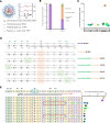
Figure 1. Evaluation of ABE8e RNP and ABE8e mRNA to correct hKCNJ13W53X/W53X allele in HEK293 FRT stable cells.
(A) Construct design to generate HEK293 FRT stable cells harboring the KCNJ13 W53X allele. (B) Chromatogram generated from HEK293 FRT stable cells showing the W53X codon marked in the red box and the downward black arrow indicating the specific nucleotide change (G>A).(C) Schematic of the hKCNJ13 locus highlighting the mutation c.158G>A (blue box marked with asterisk) and position of the W53X targeting sgRNA (black line) with TGG PAM (red line). (D) Base-editing efficiencies are shown as the percentages of sequencing reads with the corrected WT allele (and no other silent changes, bystander edits, or indels) in HEK293W53X cells following electroporation of ABE8e protein+sgRNA (RNP) or ABE8e mRNA+sgRNA (n = 3). Markers (diamonds) represent the individual biological replicates (n = 3), and error bars represent SEM by 2-tailed Student’s t test. (E) Percentages of sequencing reads with indels in ABE8e RNP– and ABE8e mRNA–treated stable cells (n = 3). Markers (diamonds) represent the individual biological replicates (n = 3), and error bars represent SEM by 2-tailed Student’s t test. (F) Kir7.1 expression in ABE8e mRNA–treated cells assessed by immunocytochemistry. GFP primary antibody was used to enhance the endogenous signal. DAPI was used to stain the nucleus. Scale bars: 50 μm. White arrows mark membrane localization in cells.
Figure 2
Figure 2. Kir7.1 current recording in WT, W53X, and base-edited W53X HEK293 FRT stable cells.
(A) Left: snapshots of Kir7.1 current profile in WT stable cells. Center: current-sweep plot represents the experimental timeline and is shown for 1 representative cell. Right: Rb+- and Cs+-sensitive current in HEKWT stable cells (B) Left: snapshots of Kir7.1 current profile in HEKW53X stable cells. Center: current-sweep plot is shown for 1 representative cell. Right: Rb+- and Cs+-sensitive current in HEKW53X stable cells. (C) Left: snapshots of Kir7.1 current profile in HEKW53X base-edited cells using ABE8e mRNA. Cells marked with asterisks showed recovery of K+ channel functions after base editing. Center: current-sweep plot is shown for 1 representative cell. Right: Rb+- and Cs+-sensitive current in HEKW53 base-edited cells.
Figure 3
Figure 3. Evaluation of ABE8e mRNA+sgRNA combinations to correct the W53X allele in LCA16 patient fibroblasts.
(A) Design of SNCs used to encapsulate ABE8e mRNA and sgRNA. (B) Base-editing efficiencies are shown as the percentages of total DNA sequencing reads, classified as unedited, correctly edited, or incorrectly edited due to bystander A edits, and with indels in treated and untreated (UT) cells. (C) Percentage editing of the target (A6) and bystander (A–9, A–8, A–4, A–2, A14, A17) A to G by ABE8e mRNA as observed in 3 independent experiments. (D) Amino acid conversion at the respective location was generated due to target and bystander edits. The protospacer sequence is underlined, the pathogenic early stop codon is in a purple box, the target A>G edit is marked in orange, and bystander A edits are in green. (E) The sgRNA location is marked by a black line, PAM is marked by a red line, and mutation is in the blue box. All the A bases within the protospacer are numbered 1–20 based on location. The A bases downstream of the protospacer are numbered from –1 to –9, considering +1 as the first base of the protospacer. The top 10 most frequent alleles generated by ABE8e mRNA treatment show the nucleotide distribution around the cleavage site for sgRNA. Substitutions are highlighted in bold, insertions are shown in the red box, and dashes show deletions. The scatterplot shows the frequency of reads observed in treated cells (n = 3 biological replicates). Data from replicates are represented as means ± SEM.
Figure 4
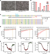
Figure 4. Evaluation of ABE8e to correct W53X alleles in iPSC-RPEW53X/W53X.
(A) Representative bright-field images of base-editor treated and untreated iPSC-RPEW53X/W53X. Scale bars: 100 μm. (B) Base-editing efficiencies following treatment (BE) with ABE8e mRNA and sgRNA encapsulated in SNC-PEG in iPSC-RPEW53X/W53X as compared with untreated cells. Reads from the untreated and treated cells (n = 3) were categorized into 4 subtypes based on their sequences, unedited, W53*>WT, indels, and substitutions. (C) Reads generated by ABE8e mRNA treatment showing the nucleotide distribution around the cleavage site for sgRNA. Substitutions are highlighted in bold. The scatterplot shows the frequency of alleles observed in treated cells (n = 3). Data are represented as means ± SEM. (D) Manual single-cell patch-clamp assays on iPSC-RPEW53X/W53X cells after treatment with ABE8e. Of the 13 cells assessed for Kir7.1 activity, each could be binned into 1 of 3 classes: low-responding single cells, which appeared to be unedited mutant cells; medium-responding single cells, which showed a low level of Rb+ response; and high-responding single cells, which showed Rb+ response like WT iPSC-RPE cells. The number (n) of cells binned into each class is shown at the top of each graph. (E) Current-sweep plot from a representative cell of each bin across a time course of being exposed to physiological HR solution (gray), Rb+ stimulation (red), and subsequent wash with HR solution (green).
Figure 5
Figure 5. Visual function of Kcnj13W53X/+ mice is similar to that of Kcnj13+/+ mice.
(A and B) Two different sgRNAs targeting the Kcnj13 gene at exon 2 and a ssODN sequence with the desired nucleotide change to generate the Kcnj13W53X/W53X mouse model by CRISPR/Cas9 and HDR genome-editing technique by microinjecting them into the pronuclei of the zygote. Double asterisks indicate postnatal day 1 lethal. (C) RFLP analysis of the Kcnj13 gene from the generated mice digested with Nhe1 enzyme on 2% agarose gel. (D) Chromatograph confirming the mouse genotype. (E) OCT images showing comparison between Kcnj13+/+, Kcnj13W53X/+, and WT allele–disrupted Kcnj13W53X/+ΔR mice. (F) Averaged c wave response confirming WT allele disruption in the RPE of Kcnj13W53X/+ΔR using the targeted guide (T). One-way ANOVA with post hoc Tukey’s HSD test was used for comparisons between the groups. NT, nontargeting sgRNA.
Figure 6
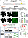
Figure 6. Phenotypic reversal of RPEW53X mice following in vivo ABE8e treatment.
(A) Kcnj13W53X allele–specific sgRNA. Black arrow represents the sgRNA spacer sequence, the desired base editing site is indicated by an asterisk, and the PAM is shown in yellow. (B) Workflow of in vivo base-editing strategy. (C) RPE florets after SNC-PEG-ATRA packaged ABE8e mRNA, W53X sgRNA, and GFP mRNA or empty SNC-PEG-ATRA/PBS as a mock treatment subretinal delivery. (D) W53X>WT corrected cell percentages observed in Kcnj13W53X/– mice treated with 2 μg or 3 μg of ABE8e. (E) Indel percentages observed in Kcnj13W53X/– mice treated with 2 μg or 3 μg of ABE8e. (F) In vivo experiment time line. Baseline ERG prior to the disruption of the WT allele and after 6 weeks follow-up. ERG prior to injection of the base editor. Recovery monitored for 10 weeks. (G) Representation of the c wave amplitude in Kcnj13W53X/+ mice with retina OCT image. (H) Reduced c wave amplitude in the Kcnj13W53X/+ mice at 6 weeks after disrupting the WT allele with Cas9 protein and WT-specific sgRNA. (I) The c wave and mfERG traces following the injection of base editor with a nontargeting guide (red) and base editor with a targeting guide (green). The faded traces represent comparisons before the disruption of the WT allele (gray) and injection of the base editor (orange). (J) Average c wave amplitude 6 weeks after the disruption of the WT allele (blue) or after the injection of base editor with a nontargeting guide (red) and targeting guide (green). (K) Normalized c wave amplitude in the eyes injected with nontargeting and targeting guides at weeks 2, 6, and 10. One-way ANOVA with post hoc Tukey’s HSD test was used for comparisons between the groups.

References

    1. Kumar M, Pattnaik BR. Focus on Kir7.1: physiology and channelopathy. Channels (Austin) 2014;8(6):488–495. doi: 10.4161/19336950.2014.959809. - DOI - PMC - PubMed
    1. Kabra M, Pattnaik BR. Sensing through non-sensing ocular ion channels. Int J Mol Sci. 2020;21(18):6925. doi: 10.3390/ijms21186925. - DOI - PMC - PubMed
    1. Zhang W, et al. Characterization of the R162W Kir7.1 mutation associated with snowflake vitreoretinopathy. Am J Physiol Cell Physiol. 2013;304(5):C440–C449. doi: 10.1152/ajpcell.00363.2012. - DOI - PMC - PubMed
    1. Toms M, et al. Missense variants in the conserved transmembrane M2 protein domain of KCNJ13 associated with retinovascular changes in humans and zebrafish. Exp Eye Res. 2019;189:107852. doi: 10.1016/j.exer.2019.107852. - DOI - PMC - PubMed
    1. Sergouniotis PI, et al. Recessive mutations in KCNJ13, encoding an inwardly rectifying potassium channel subunit, cause leber congenital amaurosis. Am J Hum Genet. 2011;89(1):183–190. doi: 10.1016/j.ajhg.2011.06.002. - DOI - PMC - PubMed

Publication types

Substances