. 2023 Aug 29;42(8):112942.
doi: 10.1016/j.celrep.2023.112942. Epub 2023 Aug 9.

Zika-specific neutralizing antibodies targeting inter-dimer envelope epitopes

Affiliations

Zika-specific neutralizing antibodies targeting inter-dimer envelope epitopes

Rajeshwer S Sankhala et al. Cell Rep. .

Abstract

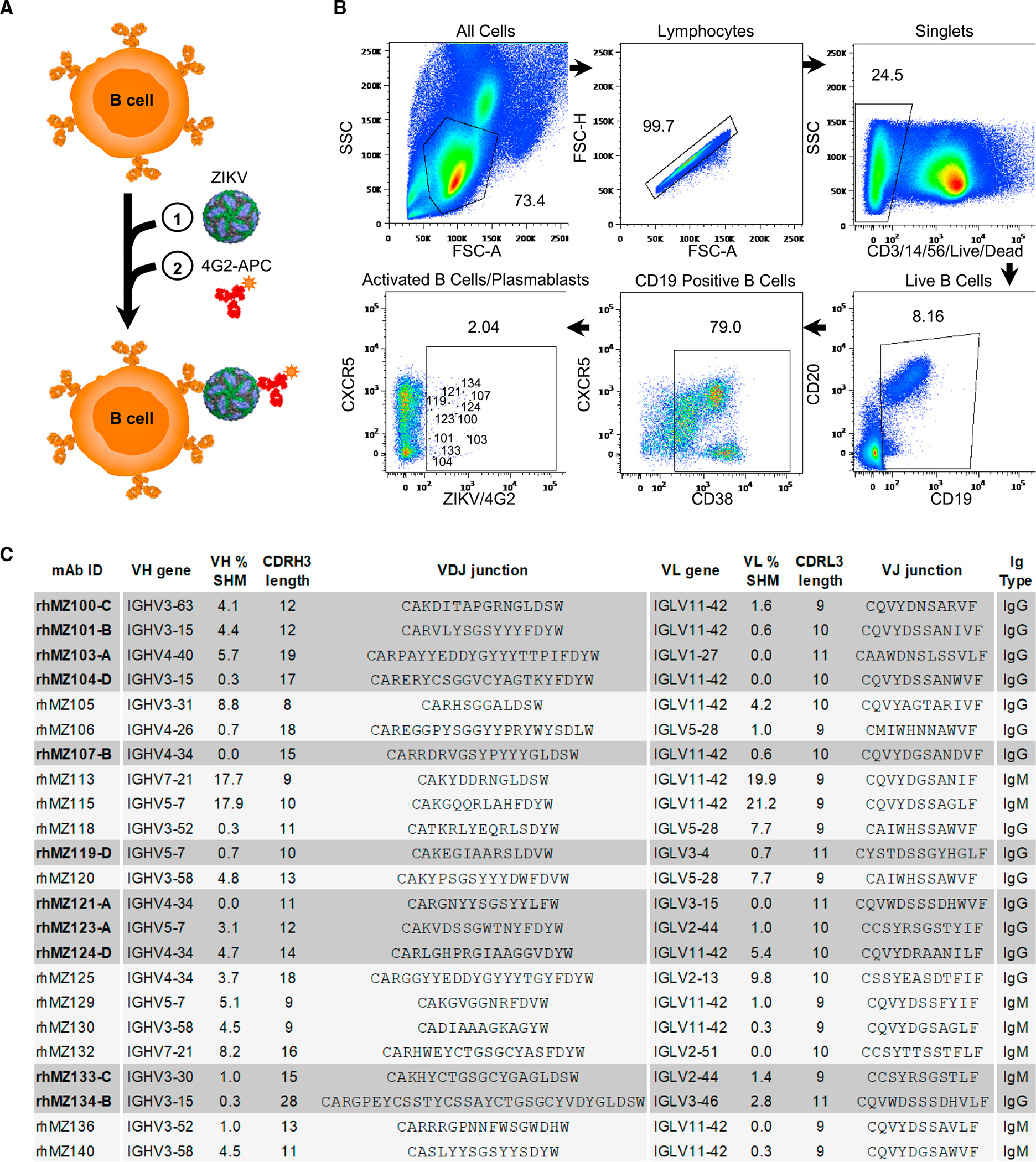
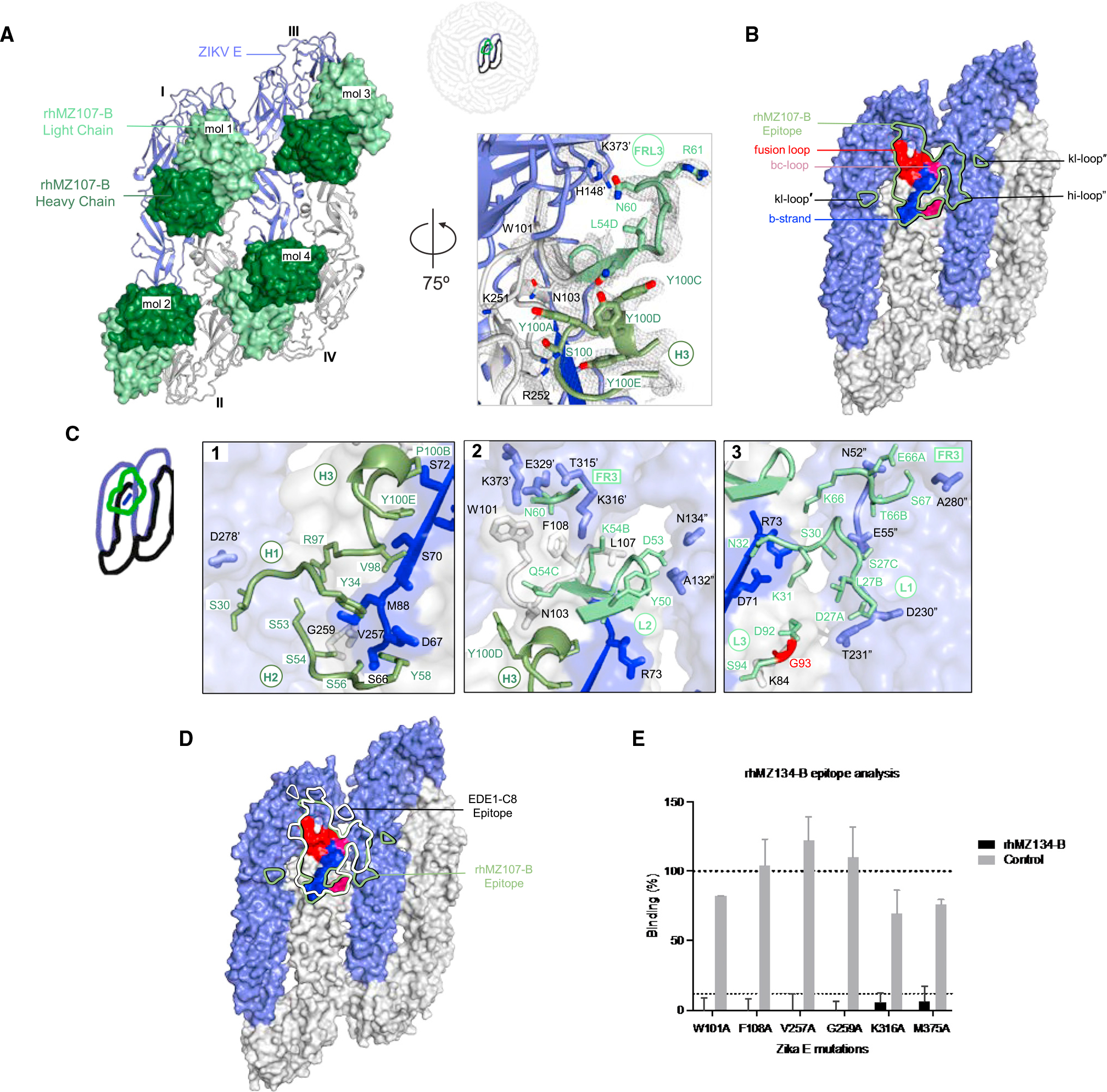
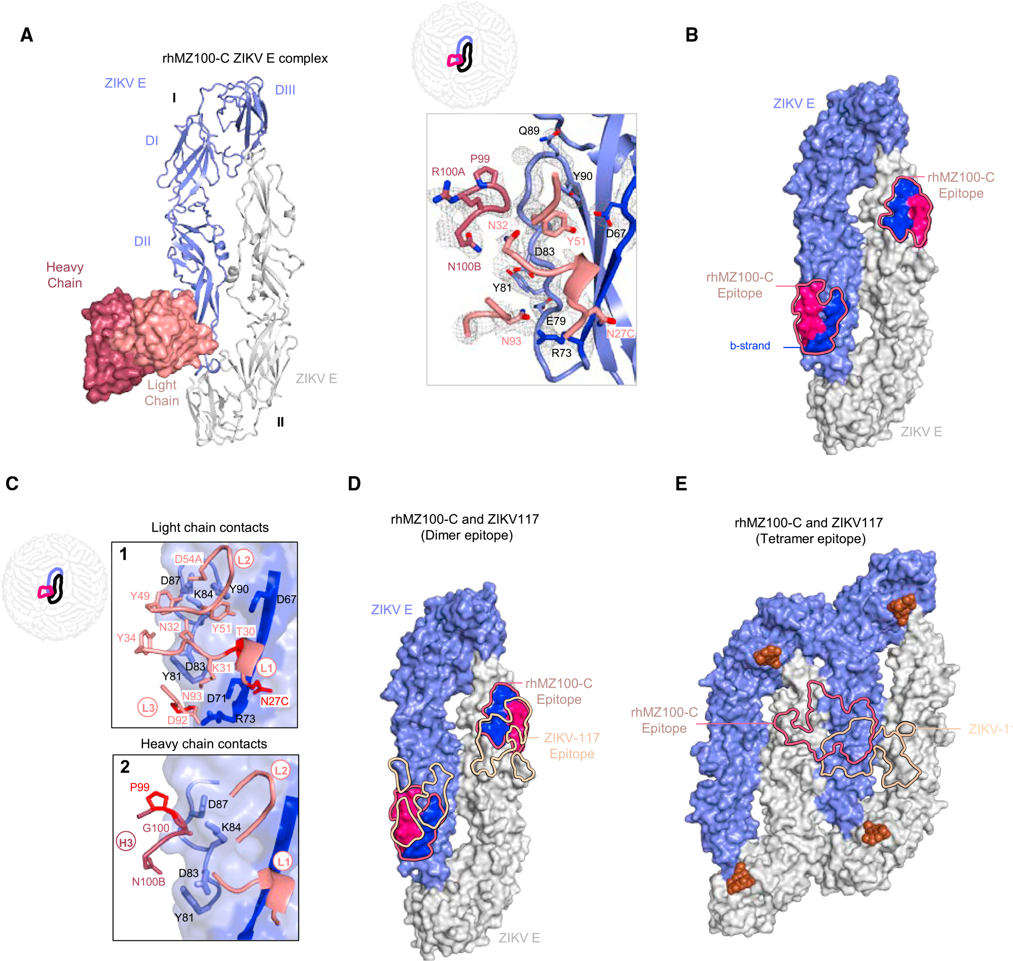
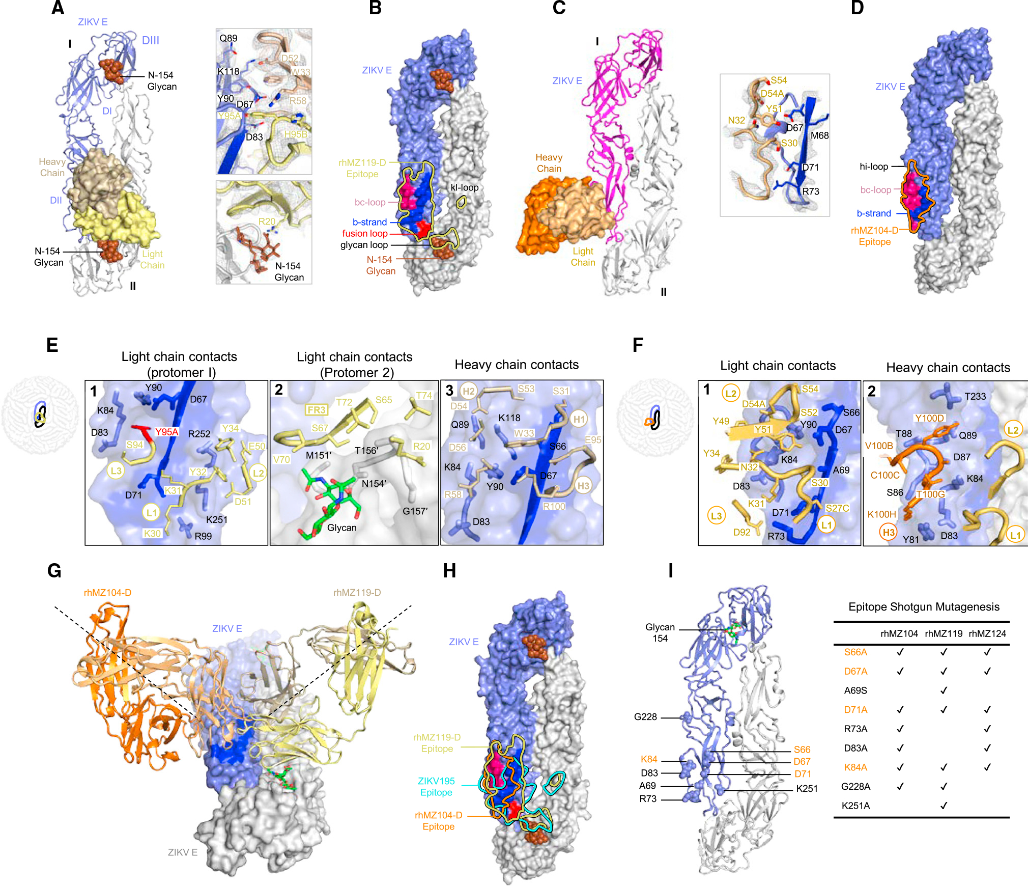
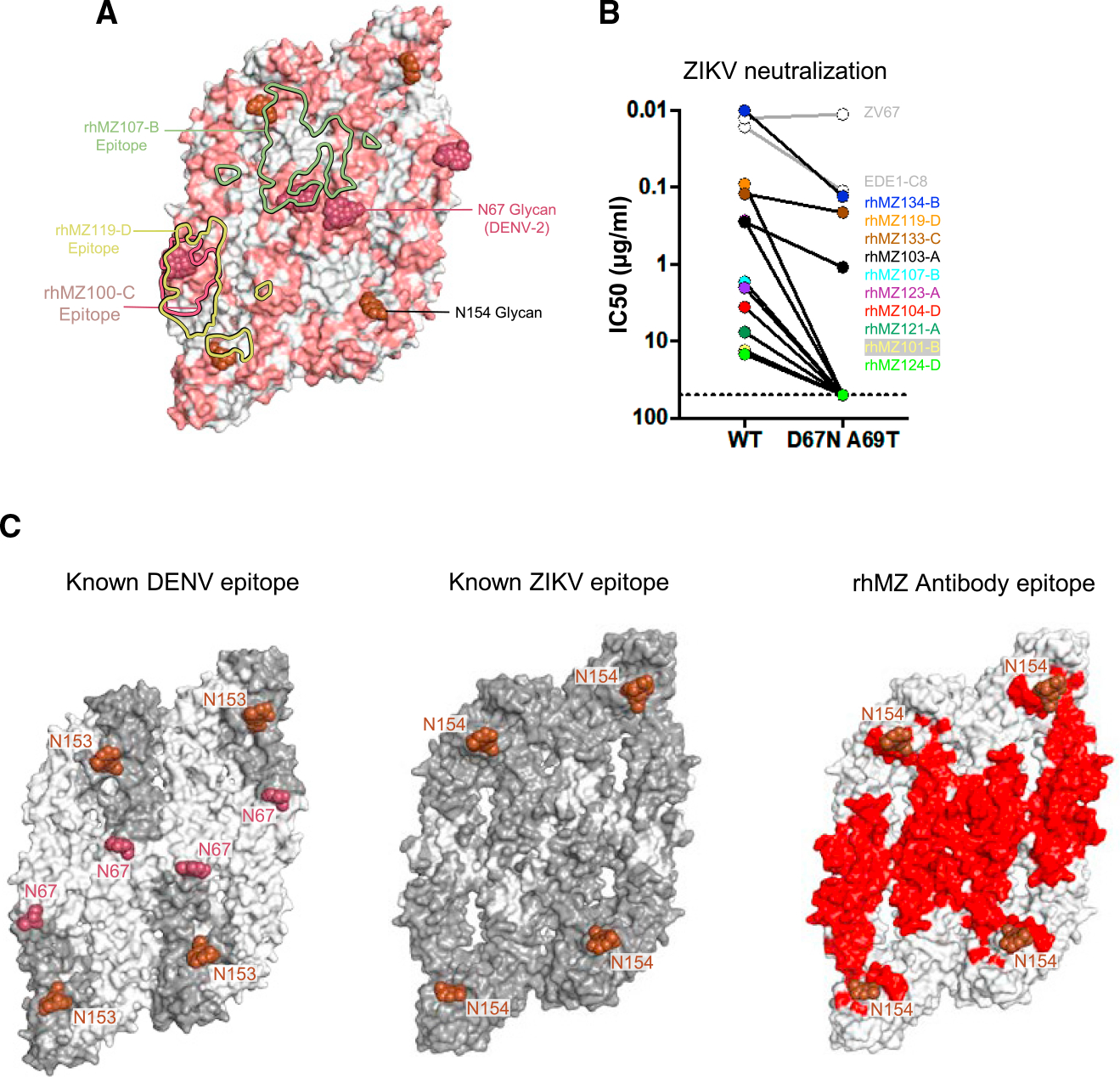
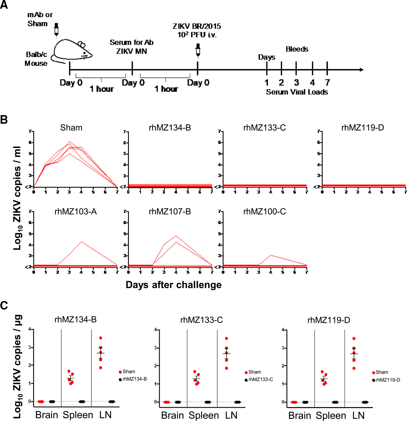
Zika virus (ZIKV) is an emerging pathogen that causes devastating congenital defects. The overlapping epidemiology and immunologic cross-reactivity between ZIKV and dengue virus (DENV) pose complex challenges to vaccine design, given the potential for antibody-dependent enhancement of disease. Therefore, classification of ZIKV-specific antibody targets is of notable value. From a ZIKV-infected rhesus macaque, we identify ZIKV-reactive B cells and isolate potent neutralizing monoclonal antibodies (mAbs) with no cross-reactivity to DENV. We group these mAbs into four distinct antigenic groups targeting ZIKV-specific cross-protomer epitopes on the envelope glycoprotein. Co-crystal structures of representative mAbs in complex with ZIKV envelope glycoprotein reveal envelope-dimer epitope and unique dimer-dimer epitope targeting. All four specificities are serologically identified in convalescent humans following ZIKV infection, and representative mAbs from all four groups protect against ZIKV replication in mice. These results provide key insights into ZIKV-specific antigenicity and have implications for ZIKV vaccine, diagnostic, and therapeutic development.

Keywords: CP: Immunology; X-ray crystallography; Zika virus; dengue virus; flavivirus; monoclonal antibodies; neutralizing antibodies; rhesus macaque; structural biology.


Conflict of interest statement

Declaration of interests S.J.K., V.D., G.D., K.M., N.M., D.H.B., M.G.J., R.S.S., and R.G.J. are named inventors on a PCT patent application WO 2019/209974 describing ZIKV neutralizing antibodies and their use. D.H.B. has received grants from Novavax and personal fees from IGM Biosciences. M.H., A.A., E.D., and B.J.D. are employees of Integral Molecular. B.J.D. is also a shareholder of the company.

Figures

Figure 1.
Figure 1.. Isolation of ZIKV-reactive antibodies from a flavivirus-naive ZIKV-infected macaque
(A) Schematic of the sequential staining strategy of ZIKV-specific B cells. (1) Incubation of PBMCs with unlabeled whole ZIKV virions followed by (2) incubation with fusion-loop-targeting antibody 4G2-APC conjugate. (B) Isolation of ZIKV-reactive activated B cells and plasmablasts from rhesus macaque 10U032 peripheral blood at 14 days post infection. Flow cytometry gates show the percentage of cells identified for each phenotypic population. CD19+CD38+CXCR5hi/lo4G2+ ZIKV-specific B cells sorted and sequenced are indicated in the last gate, with cells encoding neutralizing antibodies labeled with the matching rhMZ antibody number. (C) Genetic, binding, and neutralization characteristics of isolated ZIKV-reactive mAbs. Antibody nomenclature is in an abbreviated form that indicates species, source, viral probe, clone, and antigenic group. V(D)J assignments were performed using IgBLAST. Antibodies positive in the ZIKV (PRVABC59) MN screen are highlighted in gray. See also Figures S1, S3, and Tables S1 and S2.
Figure 2.
Figure 2.. Neutralizing and binding characteristics define four ZIKV-specific epitopes targeted on the E glycoprotein
(A) Neutralization against ZIKV PRVABC59 was assessed by MN assay in Vero cells. Shown are neutralization curves compared to the EDE1-C8 and Z004 controls. (B) Summary of mAb binding and neutralization activities. Antibodies were screened for binding to recombinant ZIKV E by BLI. Values indicate mean binding responses calculated from two independent experiments. Neutralization IC50 values (ng/mL) are shown for MN, and flow-based assay (FlowNT) in Vero, and U937-DC-SIGN cells, respectively. Cross-neutralization screen of a panel of seven flaviviruses (DENV1–4, JEV, WNV, and YFV). Shading represents binding or neutralization strength ranging from strong (dark) to weak (light); NT, not tested. (C and D) mAb binding of monomeric ZIKV sE (C) and ZIKV (D) by ELISA. Antibodies were titrated using 4-fold dilution series starting from 20 μg/mL. Values indicate mean binding responses calculated from two independent experiments. (E) Relative ratio of binding to Zika virions compared to E indicating quaternary targeting (calculated from C and D). To directly compare binding activities between the isolated macaque mAbs and human mAb controls, we calculated whole-virus/E binding ratios by using A450 values obtained at 20 μg/mL with their respective secondary antibodies. Binding ratio of 2A10G6, an FLE-directed antibody, was arbitrarily set at 1 (dotted line) as this antibody binds equally well to both monomeric E and ZIKV virion. Antibodies with ratio values >1 indicated preferential targeting of quaternary epitopes (such as EDE), whereas ratios at or below 1 were characteristic of monomeric recognition similar to FLE antibodies. (F) Binding competition with control antibodies defined four targeting epitope groups. Left: control antibody epitopes mapped on the ZIKV E dimer structure. Right: four distinct antibody competition profiles were identified in a BLI-based competition assay. Values represent the percentage of residual binding of the indicated second antibody after prior saturation of ZIKV E with the indicated first antibody. Shading from dark to light indicates competition strength ranging from strong (0%–30%), to intermediate (31%–69%), to weak/none (70%–100%). The negative control mAb was an HIV-specific mAb VRC01 that has no reactivity to ZIKV E. See also Figures S1 and S2 and Tables S2 and S3.
Figure 3.
Figure 3.. Crystal structure of ZIKV-specific EDE antibody rhMZ107-B in complex with ZIKV E glycoprotein
(A) Left: top view of the co-crystal structure of rhMZ107-B in complex with ZIKV E (PRABC59). rhMZ107-B Fv heavy and light chains are shown in surface representation and are colored dark and light green, respectively, while four ZIKV E protomers are shown in ribbon representation and colored blue and gray. Four ZIKV E protomers, left to right, are labeled as I–IV. Right: 2Fo-Fc electron density for the rhMZ107-B (mol 1) and ZIKV E interface residues is shown as gray mesh (contoured at 1.5σ). (B) Epitope footprint of rhMZ107-B antibody (mol 1) is shown as solid green line, displayed on four ZIKV E protomers shown in surface representation. TherhMZ107-B epitope extends across protomers I–III. Relevant antigenic ZIKV E regions within the epitope are labeled (fusion loop is in red, b strand in blue, bc-loop in magenta, and other loop regions are labeled). Antigenic regions of protomers I and III are marked with prime (′) and double prime (″), respectively. (C) rhMZ107-B contact residues are shown as sticks based on (1) CDRs H1, H2, and H3; (2) CDR H3 and L2; (3) CDR L1, L3, and FR L3 antibody-contacting regions. ZIKV E contact residues of protomers I and III are marked with prime (′) and double prime (″), respectively. The b strand residues 63–73 on protomer II are colored dark blue. mAb somatic hypermutation (SHM) residues are colored bright red. (D) Epitopes for rhMZ107-B and EDE1-C8 are represented with green and white lines, respectively. EDE1-C8 (PDB: 5LBS) antibody was overlaid onto therhMZ107-B ZIKV E structure to map the epitopes. (E) Shotgun mutagenesis ZIKV E epitope analysis for rhMZ134-B antibody. Relative binding to ZIKV prM/E for individual mutations is plotted. Residues from which substitution to alanine causes >90% loss in binding were considered important for binding; limit of detection: 10% (gray dotted line). Error bars indicate mean ± SEM from two independent experiments. See also Figures S3–S5 and Tables S4 and S5.
Figure 4.
Figure 4.. Crystal structure of inter-dimer-epitope antibody rhMZ100-C Fab in complex with ZIKV E glycoprotein
(A) Left: top view of the co-crystal structure of rhMZ100-C in complex with ZIKV E (PRABC59). rhMZ100-C Fv heavy and light chains are colored dark and light raspberry, respectively, and are shown in surface representation, while two ZIKV E protomers are shown in ribbon representation colored blue and gray. ZIKV E protomers, left to right, are labeled as I and II. Right: 2Fo-Fc electron density for the rhMZ100-C and ZIKV E interface residues is shown as gray mesh (contoured at 1.5σ). (B) Epitope footprint of rhMZ100-C antibody is indicated with a solid raspberry-colored line displayed on two ZIKV E protomers (surface representation). Relevant antigenic ZIKV E regions within the epitope are labeled (b strand is shown in blue and rest of the rhMZ100 epitope is shown in raspberry color). (C) rhMZ100-C contact residues are shown as sticks based on (1) CDRs L1, L2, and L3; (2) CDR H3 antibody-contacting regions; b strand residues 63–73 on protomer I are highlighted in dark blue color. SHM residues are colored bright red. (D) Epitopes for rhMZ100-C and ZIKV-117 antibodies are represented with raspberry and teal colored lines, respectively. ZIKV-117 (PDB: 5UHY) antibody was overlaid onto the rhMZ100-C ZIKV E structure to map the epitope. (E) rhMZ100-C and ZIKV-117 epitopes are mapped onto a ZIKV E-tetramer (PDB: 5IRE). See also Figures S3–S5 and Tables S4 and S5.
Figure 5.
Figure 5.. Structure of inter-dimer-epitope antibodies rhMZ119-D and rhMZ104-D in complex with ZIKV E glycoprotein
(A) Left: top view of the co-crystal structure of rhMZ119-D in complex with ZIKV E (PRABC59). rhMZ119-D Fab heavy and light chains are shown in surface representation and are colored wheat and yellow, respectively, while two ZIKV E protomers are shown in ribbon representation, colored blue and gray. ZIKV E protomers, left to right, are labeled as I and II. Right: 2Fo-Fc electron density for the rhMZ119-D and ZIKV E interface residues is shown as gray mesh (contoured at 1.5σ). (B) Epitope footprint of rhMZ119-D antibody is shown as solid yellow colored line, displayed on two ZIKV E protomers in surface representation. (C) Left: top view of the crystal structure of rhMZ104-D in complex with ZIKV E (PRABC59). rhMZ104-D Fab heavy and light chains are shown in surface representation and are colored with dark and light orange, respectively, while two ZIKV E protomers are shown in ribbon representation and colored blue and gray. ZIKV E protomers, left to right, are labeled as I and II. Right: 2Fo-Fc electron density for the rhMZ104-D and ZIKV E interface residues is shown as gray mesh (contoured at 1.5σ). (D) Epitope footprint of rhMZ104-D antibody is shown as solid orange colored line, displayed on two ZIKV E protomers in surface representation. (E) rhMZ119-D contact residues are shown as sticks based on (1) CDRs L1, L2, and L3; (2) CDR FRL3; and (3) CDR H1, H2, and H3 antibody-contacting regions. ZIKV E contact residues of protomer II are marked with prime (′); b strand residues 63–73 on protomer I are highlighted in dark blue color. SHM residues are highlighted in bright red color. (F) rhMZ104-D contact residues are shown as sticks based on (1) CDRs L1, L2, and L3; (2) CDR H3 antibody-contacting regions; b strand residues 63–73 on protomer I are highlighted in dark blue color. (G) Antibodies rhMZ119-D and rhMZ104-D are superimposed on ZIKV E protomer I. Both antibodies approach the E dimer at an angle of 90° with respect to each other. (H) Epitopes for rhMZ104-D, rhMZ119-D, and ZIKV-195 antibodies are represented with orange, yellow, and cyan colored lines, respectively. rhMZ104-D andZIKV-195 (PDB: 6MID) antibodies were overlaid onto the rhMZ119-D ZIKV E structure to map the epitopes. (I) Group D antibodies were assessed for binding using shotgun mutagenesis epitope mapping. Alanine or serine mutations, which dramatically affected group D antibody binding, are shown in sphere representation on the ZIKV E structure and indicated on the right. Residues important for binding for all group D mAbs are highlighted in orange. See also Figures S3–S5 and Tables S4 and S5.
Figure 6.
Figure 6.. Comparison of DENV and ZIKV antibody specificity
(A) Sequence differences between ZIKV E and DENV-2 E are mapped onto the ZIKV structure (PDB: 5IRE). Sequence and positional differences between ZIKV and DENV-2 are colored light red, while identical residues are colored white. Glycan-154 and DENV glycan-67 are shown in sphere representation colored brown and raspberry, respectively. The antibody binding footprints of rhMZ107-B, rhMZ100-C, and rhMZ119-D antibodies are shown as green, raspberry, and yellow solid lines, respectively. (B) Neutralization (IC50, mg/mL) of wild-type (WT) and D67N-A69T mutant ZIKV performed in the ZIKV/H/PF2013 background using a reporter virus particle (RVP) assay. The addition of glycan-67 to ZIKV interfered with epitope recognition and abrogated or eliminated neutralization. (C) Epitope mapping of structurally defined antibodies mapped onto four protomers of DENV (left) and ZIKV (middle). Residues contacted by previously described mAbs are colored dark gray, and residues not previously identified prior to this study are indicated in white. Only previously identified mAb structures with resolution greater than 4 Å were used since the contact residues are clearly interpretable. Newly identified residues contacted by rhMZ mAbs described in this study are colored red (right). Glycan sites at positions 153 or 154 are indicated in rose color. See also Figures S5 and S7 and Table S6.
Figure 7.
Figure 7.. Protection of ZIKV-specific neutralizing mAbs
(A) Schematic of passive protection study experimental design. Antibodies were infused intravenously into groups of naive recipient Balb/c mice (n = 5/group) prior to ZIKV-BR challenge. Mice received 200 μg of antibody (10 mg/kg) and were challenged with 105 viral particles (102 plaque-forming units) of ZIKV-BR intravenously 2 h after infusion. (B) Complete or partial protection from ZIKV replication were observed for six representative neutralizing mAbs. Following infusion with the indicated antibody or saline (Sham), ZIKV viral loads were measured in serum post challenge by RT-PCR daily until day 7. Viral load peaked at day 3 or 4. The six antibodies were tested in two sets of experiments, and a representative Sham group from one experiment is shown. (C) Viral dissemination in brain, spleen, and lymphnodes (LNs) was assessed at day 3 post challenge for three of the most potent mAbs (black circles) as compared to the Sham group (red circles). Error bars indicate mean ± SEM. See also STAR Methods.

References

    1. Lazear HM, and Diamond MS (2016). Zika Virus: New Clinical Syndromes and Its Emergence in the Western Hemisphere. J. Virol. 90, 4864–4875. 10.1128/JVI.00252-16. - DOI - PMC - PubMed
    1. Cao-Lormeau VM, Blake A, Mons S, Lastère S, Roche C, Vanhomwegen J, Dub T, Baudouin L, Teissier A, Larre P, et al. (2016). Guillain-Barre Syndrome outbreak associated with Zika virus infection in French Polynesia: a case-control study. Lancet 387, 1531–1539. 10.1016/S0140-6736(16)00562-6. - DOI - PMC - PubMed
    1. Carteaux G, Maquart M, Bedet A, Contou D, Brugières P, Fourati S, Cleret de Langavant L, de Broucker T, Brun-Buisson C, Leparc-Goffart I, and Mekontso Dessap A (2016). Zika Virus Associated with Meningoencephalitis. N. Engl. J. Med. 374, 1595–1596. 10.1056/NEJMc1602964. - DOI - PubMed
    1. Mlakar J, Korva M, Tul N, Popović M, Poljšak-Prijatelj M, Mraz J, Kolenc M, Resman Rus K, Vesnaver Vipotnik T, Fabjan Vodušek V, et al. (2016). Zika Virus Associated with Microcephaly. N. Engl. J. Med. 374, 951–958. 10.1056/NEJMoa1600651. - DOI - PubMed
    1. Macnamara FN (1954). Zika virus: a report on three cases of human infection during an epidemic of jaundice in Nigeria. Trans. R. Soc. Trop. Med. Hyg. 48, 139–145. - PubMed

Publication types