. 2023 Aug 10;14(1):4588.
doi: 10.1038/s41467-023-40227-8.

HIV-1-induced nuclear invaginations mediated by VAP-A, ORP3, and Rab7 complex explain infection of activated T cells

Affiliations

HIV-1-induced nuclear invaginations mediated by VAP-A, ORP3, and Rab7 complex explain infection of activated T cells

Mark F Santos et al. Nat Commun. .

Abstract

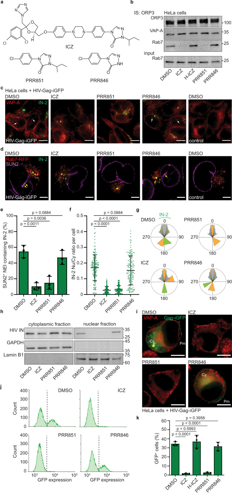
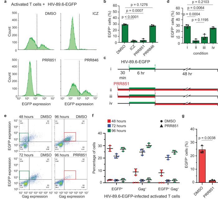
The mechanism of human immunodeficiency virus 1 (HIV-1) nuclear entry, required for productive infection, is not fully understood. Here, we report that in HeLa cells and activated CD4+ T cells infected with HIV-1 pseudotyped with VSV-G and native Env protein, respectively, Rab7+ late endosomes containing endocytosed HIV-1 promote the formation of nuclear envelope invaginations (NEIs) by a molecular mechanism involving the VOR complex, composed of the outer nuclear membrane protein VAP-A, hyperphosphorylated ORP3 and Rab7. Silencing VAP-A or ORP3 and drug-mediated impairment of Rab7 binding to ORP3-VAP-A inhibited the nuclear transfer of the HIV-1 components and productive infection. In HIV-1-resistant quiescent CD4+ T cells, ORP3 was not hyperphosphorylated and neither VOR complex nor NEIs were formed. This new cellular pathway and its molecular players are potential therapeutic targets, perhaps shared by other viruses that require nuclear entry to complete their life cycle.


Conflict of interest statement

The United Kingdom patent application GB2598624A (applicants: M.F.S., G.R., P.D., G.C., A.L., and Technische Universität Dresden; inventors: M.F.S., G.R., P.D., G.C., D.C., and A.L.), European patent application EP3864409A1 (applicants: M.F.S., G.R., A.L. and Technische Universität Dresden; inventors; M.F.S., G.R., D.C. and A.L.) and United States provisional patent number US20210353616A1 (applicants: M.F.S., G.R., A.L. and Technische Universität Dresden; inventors: M.F.S., G.R., D.C., and A.L.) are pending. The patent EP3864409A1/US20210353616A1 is entitled: Inhibition of a tripartite VOR protein complex in multicellular organisms. The patent GB2598624A is entitled: Use of triazole analogues for inhibition of a tripartite VOR protein complex in multicellular organism. These patents are related to the use of itraconazole and triazole analogues to inhibit the VOR protein complex, which prevents the nuclear transfer of materials transported by extracellular particles (e.g., extracellular vesicles and viruses). The authors declare no other competing interests.

Figures

Fig. 1
Fig. 1. VSV-G-pseudotyped HIV-1 is endocytosed and induces type II NEIs.
a HeLa cells were 1-h HIV-Gag-iGFP infected prior to VAP-A and HIV-1 IN (IN-2) immunolabeling. As controls, noninfected cells or no HIV-1 IN antibody were used (Supplementary Fig. 2a). Samples were observed by CLSM and single sections are displayed. Longitudinal (top and bottom panels) or transverse (middle) sections of NEI (arrow) are shown. bd Cells were 1-, 3-, or 5-h infected prior to VAP-A and IN-2 immunolabeling. Nucleoplasmic (Nu) and cytoplasmic (Cy) IN-2 were quantified as defined by regions of interest (ROI, b) and their ratio per cell from one representative experiment (c) or total IN-2 intensity per cell are indicated (d 50 cells per experiment, n = 3). e, f Cells were 3-h infected (or not) prior to VAP-A and SUN2 immunolabeling. Type I (VAP-ASUN2+) and II (VAP-A+SUN2+) NEIs (e green and yellow/red arrows, respectively) were quantified (f 100 cells per experiment, n = 3). g, h Cells were 30-min pretreated with DMSO or DNS (80 µM) prior to 3-h HIV-Gag-iGFP infection followed by VAP-A and HIV-1 IN immunolabeling (g). Note that IN-2 accumulated in NEI (yellow arrow) and/or dispersed into cytoplasm and nucleoplasm in the control, whereas it remained outside the DNS-treated cells (asterisk). Total IN-2 intensity is presented (h, >50 cells per experiment, n = 3). im Colocalization of IN-2 with Rab proteins. Experimental methodology (i). Cells were baculovirus infected to express Rab5/Rab7-RFP and then HIV-Gag-iGFP infected. At different times, cells were processed for ICC for HIV-1 IN and counterstained with DAPI. Noninfected cells expressing Rab5/7-RFP were analyzed (control). Composite images revealed colocalizations of IN-2 and Rab5-RFP (j white arrow) or Rab7-RFP (k green arrow) at early and late time points, which are rendered in a 3D image (l) and quantified using Pearson’s R correlation coefficient (m n = 25 cells per time point). We used a MOI of 2. In all cases, the means ± S.D. and, where appropriate, the individual values for each experiment are shown. P values are indicated. Scale bars, 2 (l), 5 (a, b), 10 (e, g, j, k) µm.
Fig. 2
Fig. 2. Rab7+ late endosomes containing HIV-1 IN enter NEI.
HeLa cells stably transfected with VAP-A-GFP were 1-h HIV-Gag-iGFP infected prior to immunolabeling for HIV-1 IN and Rab7 (a), CD63 (b) or Lamp1 (c). Single sections show transverse (a, c) and/or longitudinal (ac) sections of a NEI. Merge and single channels (insets a1, a2, b1, c1, c2) are presented. IN-2 colocalized with Rab7 or CD63, but not Lamp1 in NEIs (yellow and magenta arrow, respectively). d Noninfected VAP-A-GFP-expressing HeLa cells were Rab7 and Lamp1 immunolabeled. Single transverse section of a NEI is shown with the merge and single channels as indicated. Insets show the absence of Lamp1 in NEI (d1, white arrow) and the colocalization of Rab7 and Lamp1 in the cytoplasmic compartment (d2, pink arrowhead). e, f HeLa cells were 1-h HIV-Gag-iGFP infected prior to immunolabeling for Rab7 and Lamp1. Nuclei were counterstained with DAPI. Single longitudinal section of a NEI is shown with the merge and single channels as indicated (e). Insets show the absence of Lamp1 in NEI (e1) and the colocalization of Rab7 and Lamp1 in cytoplasmic compartment (e2, pink arrowhead). Quantification of NEIs containing specific antigen(s) as indicated in infected cells (f, 50 cells per experiment, n = 3, means ± S.D. are shown). g VAP-A-GFP-expressing HeLa cells were 1-h infected with “bald” virus, lacking VSV-G, prior to Rab7 and HIV-1 IN immunolabeling. A single section shows transverse section of a NEI. Note that most of IN-2 remains outside the cells (asterisk) and the absence of Rab7 in NEI (double arrow). For a lower power view as well as all sections see below Supplementary Fig. 15c. We used a MOI of 2 or equivalent in the case of the “bald” virus. Scale bars, 5 µm.
Fig. 3
Fig. 3. Silencing the VOR complex inhibits HIV-1 integrase transport into NR and productive infection.
ad HeLa cells stably transfected with plasmids carrying scrambled shRNA, shVAP-A, shVAP-B or shORP3 and expressing either ER-GFP (a, upper panels) or Rab7-RFP (a, lower panels, bd) were 1-h HIV-Gag-iGFP infected prior to immunolabeling for either VAP-A, VAP-B, ORP3 or SUN2 and HIV-1 IN (IN-2) as indicated. Samples were observed by CLSM. Single sections are displayed. IN-2 was detected in ER-GFP+ or SUN2+ NEI of scrambled and shVAP-B cells and quantified (a arrow; b 50 NEIs per experiment, n = 3). The ratio of IN-2 in the nucleoplasm versus cytoplasm per cell was determined (c n = 150 cells from three independent experiments). The distribution of IN-2 in perinuclear region of scrambled shRNA, VAP-A-, or ORP3-silenced HeLa cells is shown in a circular histogram (d 10 cells per cell line; color code indicates an individual cell). [For Methods, see Supplementary Fig. 6d]. Cells with dual IN-2 localization are in color. eg HeLa cells were 6-h HIV-Gag-iGFP infected, washed and incubated for 24 h prior to ICC (e) and FC (f, g). Representative fluorescent images for the indicated proteins (e) and FC histograms (f) are displayed. Scrambled shRNA-transfected, noninfected cells were used for gating. Note the reduction in Gag-iGFP expression in cells lacking VAP-A and ORP3. The percentage of GFP+ cells is plotted (g n = 3). Asterisk and arrowhead indicate Gag-iGFP in the cytoplasm (Cy) and plasma membrane (Pm), respectively. Nu nucleus. We used a MOI of 2. Means ± S.D. and individual values for each experiment (b, g) or cell (c) are presented. P values are indicated. Scale bar, 5 (a, top) or 10 (a, bottom, e) µm.
Fig. 4
Fig. 4. The VOR complex inhibition impedes the HIV-1 integrase transport into NR and productive infection.
a Drug structures. b HeLa cells were 90-min incubated with DMSO or various drugs (10 µM) as indicated, detergent solubilized and ORP3 IS. Bound fractions and Rab7 input (1/50) were probed for ORP3, VAP-A, and Rab7 by IB. c HeLa cells were 30-min pretreated with DMSO or drugs (10 µM) before 1-h HIV-Gag-iGFP infection in the presence of drugs. As a control, cells were noninfected. Samples were VAP-A and HIV-1 IN (IN-2) immunolabeled. Single sections are displayed. IN-2 was detected in NEIs of DMSO/PRR846-treated cells (arrow). dg Rab7-RFP-expressing HeLa cells were treated as in c, and IN-2 and SUN2 immunolabeled. The presence of IN-2 in SUN2+ NEI (d arrow) was quantified (e 50 NEIs per experiment, n = 3). The IN-2 nucleoplasm/cytoplasm ratio was determined (f n = 150 cells from three independent experiments). Localization of IN-2 in the perinuclear region is shown using a circular histogram (g 10 cells/condition; color code indicates an individual cell). [For Methods, Supplementary Fig. 6d]. h Drug-treated HeLa cells were 3-h HIV-Gag-iGFP infected, fractionated into cytoplasmic and nuclear fractions and probed for HIV-1 IN by IB. A quarter and the total material of the cytoplasmic and nuclear fractions were loaded. Cytoplasmic GAPDH and nuclear Lamin B1 were used as controls and run in parallel. They are derived from the same experiment (b, h). Molecular mass markers (kDa) are indicated. ik HeLa cells were 30-min pretreated with DMSO or drugs before 6-h HIV-Gag-iGFP infection. They were 24-h incubated in the presence of drugs and then processed for ICC (i) or FC (j, k). For ICC, fixed-cells were VAP-A immunolabeled, while they were trypsinized for FC. Representative images (i) and FC histograms (j) are displayed. DMSO-treated, noninfected cells were used for gating. The GFP+ cells were quantified (k n = 3). We used a MOI of 2. Means ± S.D. and individual values for each experiment (e, k) or cell (f) are presented. P values are indicated. Scale bars, 10 (c, i) or 5 (d) µm.
Fig. 5
Fig. 5. PRR851 impedes the productive infection of native Env-pseudotyped HIV-1 in activated CD4+ T cells.
a, b PHA/IL-2-activated primary CD4+ T cells were 30-min pretreated with 10 µM ICZ, PRR851, PRR846, or DMSO before RetroNectin-based, 6-h HIV-89.6-EGFP infection. Cells were washed and incubated for 48 h in the presence of drugs. Samples were processed for FC and histograms are displayed (a). DMSO-treated, noninfected cells were used for the gating. The percentage of EGFP+ cells is plotted (b n = 3). c, d Representation of alternative protocols (c) and quantification of the EGFP expression in infected PHA/IL-2-activated CD4+ T cells by FC (d n = 3 per condition). In addition to negative and positive controls, i.e., without (#i) or in constant presence of PRR851 (10 µM) (#ii), respectively, the drug was removed after 6-h infection (#iii) or added after (#iv). e, f PHA/IL-2-activated CD4+ T cells were 30-min pretreated with DMSO or 10 µM PRR851 before RetroNectin-based, 6-h HIV-89.6-EGFP infection, washed and further incubated for 48, 72 or 96 h in the presence of drugs. Samples were processed for FC for the expression of EGFP and HIV-1 Gag. Representative FC histograms are displayed (e). DMSO-treated, noninfected cells were used for the gating. The percentage of EGFP+, Gag+, or double-positive cells is plotted (f n = 3). Note that single EGFP+ cells (green boxes) decrease during incubation time, while double-positive cells increase in a drug-dependent manner (red boxes). g PHA/IL-2-activated CD4+ T cells were 30-min pretreated with DMSO or 10 µM PRR851 before 6-h HIV-89.6-EGFP infection by spinoculation instead of the RetroNectin method. After 48 h, samples were processed for FC and the percentage of EGFP+ cells is plotted (n = 3). We used a MOI of 2. In all cases, means ± S.D. and individual value are presented. P values are indicated.
Fig. 6
Fig. 6. Native Env-pseudotyped HIV-1 infection of CD4+ T cells triggers type II NEIs biogenesis.
af PHA/IL-2-activated CD4+ T cells were HIV-89.6-EGFP infected for 3 (a, b) or 6 (ce left y axis, f) hours prior to immunolabeling for SUN2 and VAP-A (ae) or Lamin B1 (f). Nuclei were counterstained with DAPI. In some experiments, cells were 30-min pretreated with PRR851 (10 µM) (c, d). Drugs were present during infection. As a control, cells were noninfected. They were observed by CLSM and serial sections of a representative cell (a) or single section of 10 cells are presented (c). Alternatively, after 6-h infection, cells were 48-h incubated prior to FC (e right y axis, n = 3). The percentage of cells with type I or II NEIs was quantified (b, d, e, 50 cells per experiment, n = 3). Note the presence of NEIs in infected cells without PRR851 (a right, c middle, f arrow). g, h The solidity was evaluated in noninfected and 6-h infected cells. After SUN2 and VAP-A immunolabeling, the surface areas were evaluated (g), and the resulting solidity presented (h, n = 10 cells). i, j Activated T cells were 3-h infected with HIV-89.6-EGFP or corresponding “bald” virus prior to VAP-A and HIV-1 IN (IN-2) immunolabeling. Single section of representative cells is shown (i). IN-2 in induced NEIs (arrows) or in the cytoplasm (asterisks) are indicated. Green dashed lines indicate IN-2 distribution around the cell surface. The percentage of cells with type II NEIs were quantified (j ≥50 cells per condition, n = 3). k, l The presence of IN-2 in NEIs (arrow) and late endosomes of 6 or 3 h-infected cells was observed after immunolabeling for IN-2 and SUN2 (k upper panel) or Rab7 (k lower panel), respectively. Alternatively, cells were immunolabeled for Rab7 or Lamp1 and SUN2 (l). Note that Rab7 (arrows), but not Lamp1, is present in NEIs. We used a MOI of 2 or equivalent for the “bald” virus, unless otherwise stated. In all cases, means ± S.D. and individual values are shown. P values are indicated. Scale bars, 5 µm.
Fig. 7
Fig. 7. HIV-1 infection promotes the VOR complex formation in a microtubule-dependent manner.
a PHA/IL-2-activated CD4+ T cells were noninfected (control) or 6-h HIV-89.6-EGFP infected after 30-min pretreatment with DMSO or 10 µM PRR851. The drug was present during infection. Cells were then solubilized and subjected to ORP3 IS. The bound fractions and the input (1/50) were probed for ORP3, VAP-A, and Rab7 by IB. b Cells were 5-min pretreated with DMSO or nocodazole (1 µM) and then noninfected (control) or 1-h HIV-89.6-EGFP infected, solubilized and processed for ORP3 IS and ORP3, VAP-A and Rab7 IB. All samples came from the same experiment and were run in parallel (a, b). Molecular mass markers (kDa) are indicated. Representative experiments are shown. We used a MOI of 2. Note that VOR complex formation only occurs after HIV-1 infection and is hindered by PRR851 and nocodazole. c Representation of the induction of type II NEIs by virus-laden late endosomes, a process mediated by the interaction of VOR complex proteins, namely ONM-associated VAP-A, cytoplasmic ORP3, and late endosome-associated Rab7 (left panel). Release of viral components from late endosomes into the cytoplasmic core of induced NEIs at the vicinity of the nuclear pore would facilitate their transfer to the nucleoplasm (right panel). PRR851 inhibits the interaction of the VOR complex proteins, and hence the NEI formation. ILV, intralumenal vesicles associated with late endosome/multivesicular body; INM/ONM, inner/outer nuclear membrane; MTOC, microtubule-organizing center; MTs, microtubules.
Fig. 8
Fig. 8. ORP3 hyperphosphorylation is important for VOR complex interactions and NEI formation.
ac Quiescent and PHA/IL-2-activated CD4+ T cells were noninfected (control) or 6-h HIV-89.6-EGFP infected, and then 48-h incubated prior to either ICC (a) or FC (b, c). For ICC, fixed-cells were immunolabeled for VAP-A. Representative images (a) and FC histograms (b) are displayed. Noninfected activated cells were used for the gating. b The percentage of EGFP+ cells is plotted (c n = 3). dg Cells as indicated were noninfected (control) or HIV-89.6-EGFP infected for 1 (df) or 3 (g) hours prior to immunolabeling for HIV-1 IN (IN-2) and Rab7 (df) or SUN2 and VAP-A (g). Nuclei were counterstained with DAPI. A single section of a representative cell (d) or composite images of 25 sections of 10 cells (e) are presented. The localization of Rab7 in the perinuclear region is shown in a circular histogram (f, 10 cells per condition; color code indicates an individual cell). Note the colocalization of IN-2 and Rab7 at MTOC of infected cells (arrow). The percentage of cells with NEIs of type I or II was quantified (g 50 cells per experiment, n = 3). hj Cells as indicated were noninfected (control) or 6-h HIV-89.6-EGFP infected, solubilized and subjected to ORP3 IS. The bound fractions and input (1/50) were probed for ORP3, VAP-A, and Rab7 (h). Note the absence of slower-migrating ORP3 species in quiescent cells (red arrow). The protein ratio of the indicated pair was quantified (i n = 4). Detergent cell lysates were treated with λ-phosphatase (λ) or without (–) prior to ORP3 IB (j). Molecular mass markers (kDa) are indicated. All samples were from the same experiment and were run in parallel (h, j). We used a MOI of 2. Means ± S.D. and individual values of each experiment are shown. P values are indicated. k Representation of type II NEI induction that requires both T cell activation leading to ORP3 hyperphosphorylation (left) and viral infection leading to the accumulation of Rab7+ late endosomes (LE) at the microtubule-organizing center (MTOC, right). INM/ONM, inner/outer nuclear membrane. Scale bars, 5 µm.
Fig. 9
Fig. 9. Sotrastaurin-mediated PKC inhibition prevents the formation of both HIV-1-induced NEIs and the VOR complex.
ac Quiescent CD4+ T cells were pretreated with DMSO (DMSO, –) or 40 µM Sotrastaurin (Sotrast.) for 30 min before their activation with PHA and IL-2 for 48 h. Afterward, they were 6-h HIV-89.6-EGFP infected in the presence of Sotrastaurin prior to SUN2 and VAP-A immunolabeling. Nuclei were counterstained with DAPI. Samples were observed by CLSM. Single section of 10 representative cells are presented. Note the presence of NEIs in the infected cell in the absence of Sotrastaurin (left, arrows). The percentage of cells with type I or II NEIs was quantified (b 50 cells per experiment, n = 3) and the nuclear solidity was determined (c n = 10 cells). d After 6-h infection, cells were incubated for 48 h prior to FC analysis for EGFP expression. Representative FC histograms are displayed. DMSO-treated, noninfected cells were used for the gating. e Cells were treated as in a, solubilized and subjected to ORP3 IS. As a control, cells were noninfected. The input (1/50) and bound fractions were probed for ORP3, VAP-A, and Rab7 by IB. Note the absence of slower-migrating ORP3 immunoreactive species in Sotrastaurin-treated CD4+ T cells regardless of infection (red arrow). OE overexposed blot. All samples were from the same experiment. Molecular mass markers (kDa) are indicated. We used a MOI of 2. In all cases, means ± S.D. and individual values are shown. P values are indicated. Scale bars, 5 µm.

References

    1. Gallo SA, et al. The HIV Env-mediated fusion reaction. Biochim. Biophys. Acta. 2003;1614:36–50. doi: 10.1016/S0005-2736(03)00161-5. - DOI - PubMed
    1. Janas AM, Dong C, Wang JH, Wu L. Productive infection of human immunodeficiency virus type 1 in dendritic cells requires fusion-mediated viral entry. Virology. 2008;375:442–451. doi: 10.1016/j.virol.2008.01.044. - DOI - PMC - PubMed
    1. Chauveau L, et al. HIV fusion in dendritic cells occurs mainly at the surface and is limited by low CD4 levels. J. Virol. 2017;91:e01248–01217. doi: 10.1128/JVI.01248-17. - DOI - PMC - PubMed
    1. Fackler OT, Peterlin BM, Weis K. Lessons from HIV: movement of macromolecules inside the cell. Curr. Mol. Med. 2001;1:1–7. doi: 10.2174/1566524013364167. - DOI - PubMed
    1. Schaeffer E, Soros VB, Greene WC. Compensatory link between fusion and endocytosis of human immunodeficiency virus type 1 in human CD4 T lymphocytes. J. Virol. 2004;78:1375–1383. doi: 10.1128/JVI.78.3.1375-1383.2004. - DOI - PMC - PubMed

Publication types