. 2024:283:249-284.
doi: 10.1007/164_2023_692.

Pharmacology of Compounds Targeting Cation-Chloride Cotransporter Physiology

Affiliations

Pharmacology of Compounds Targeting Cation-Chloride Cotransporter Physiology

Eric Delpire et al. Handb Exp Pharmacol. 2024.

Abstract

Transporters of the solute carrier family 12 (SLC12) carry inorganic cations such as Na+ and/or K+ alongside Cl across the plasma membrane of cells. These tightly coupled, electroneutral, transporters are expressed in almost all tissues/organs in the body where they fulfil many critical functions. The family includes two key transporters participating in salt reabsorption in the kidney: the Na-K-2Cl cotransporter-2 (NKCC2), expressed in the loop of Henle, and the Na-Cl cotransporter (NCC), expressed in the distal convoluted tubule. NCC and NKCC2 are the targets of thiazides and "loop" diuretics, respectively, drugs that are widely used in clinical medicine to treat hypertension and edema. Bumetanide, in addition to its effect as a loop diuretic, has recently received increasing attention as a possible therapeutic agent for neurodevelopmental disorders. This chapter also describes how over the past two decades, the pharmacology of Na+ independent transporters has expanded significantly to provide novel tools for research. This work has indeed led to the identification of compounds that are 100-fold to 1000-fold more potent than furosemide, the first described inhibitor of K-Cl cotransport, and identified compounds that possibly directly stimulate the function of the K-Cl cotransporter. Finally, the recent cryo-electron microscopy revolution has begun providing answers as to where and how pharmacological agents bind to and affect the function of the transporters.

Keywords: Bumetanide; Cation–chloride cotransporters; Chloride dye; Cryo-EM structure; High-throughput screening; Ion binding; Kidney; N-ethylmaleimide; Neurodevelopmental disorders; Protein–ligand complex; Thallium dyes; Thiazide and loop diuretics.


Figures

Figure 1.
Figure 1.
Phylogeny of electroneutral cation-chloride SLC12 transporters. Two functionally uncharacterized orphan members (CCC8-SLC12A9 and CCC9-SLC12A8) separate the Na+-dependent branch (left) from the Na+-independent branch (right). On the Na+-dependent side, there are one Na-Cl cotransporter: NCC (SLC12A3), and two Na-K-2Cl cotransporters: NKCC1 (SLC12A2) and NKCC2 (SLC12A1). Due to high Na+, Cl concentrations in the extracellular versus intracellular spaces, these transporters are typically inward transport mechanisms. Na+-independent transporters are the four K-Cl cotransporters: KCC1 (SLC12A4), KCC2 (SLC12A5), KCC3 (SLC12A6) and KCC4 (SLC12A7). Due to high K+ concentration in the cytosol compared to the extracellular space, the K-Cl cotransporters typically transport ions out of the cell.
Figure 2.
Figure 2.
Properties and structure of hydrochlorothiazide (Compound CID: 3639).
Figure 3.
Figure 3.
Properties and structure of metolazone (Compound CID: 4170).
Figure 4.
Figure 4.
Properties and structure of indapamide (Compound CID: 3702).
Figure 5.
Figure 5.
Properties and structure of furosemide (Compound CID: 3440).
Figure 6.
Figure 6.
Properties and structure of bumetanide (Compound CID: 2471).
Figure 7.
Figure 7.
Properties and structure of torsemide (Compound CID: 41781).
Figure 8.
Figure 8.
Properties and structure of ethacrynic acid (Compound CID: 3278).
Figure 9.
Figure 9.
Properties and structure of DIOA (Compound CID: 5017).
Figure 10.
Figure 10.
Properties and structure of VU0255011 (Compound CID: 25067404) and analogues. Groups were modified on the thiazole ring. As shown by a red arrow, the original hit from the screen had a secondary amide instead of a tertiary N-Me amide. As shown by the second red arrow, the structure of an inactive compound with the 4-methyl group moved to a different position. Finally substituting the secondary amide into a N-cyclopropyl amide resulted in compound VU046271 with higher potency.
Figure 11.
Figure 11.
Properties and structure of N-ethylmaleimide (Compound CID: 4362).
Figure 12.
Figure 12.
Properties and structure of VU-500469 (Compound CID: 18585704).
Figure 13.
Figure 13.
The secondary structure of cation-chloride cotransporters follows the general pattern of the amino acid-polyamine-organocation (APC) superfamily. A. The transmembrane core is composed of an inverted repeat of five transmembrane domains (I – V and VI – X) followed by TMD11 and TMD12. TMD1 and TMD6 helices are disrupted in the middle by a linker, and TMD3 and TMD8 are tilted and longer helices, as they traverse a longer distance in the lipid bilayer. B. Alignment of TMD2-TMD7 and TMB3-TMD8 showing some degree of conservation.
Figure 14.
Figure 14.
Structure of NKCC1 as a cation-chloride cotransporter representative. A. Cartoon (left) and surface(right) representation of human NKCC1 (PDB: 7mxo) showing the dimer. Note the ‘exchange’ of carboxyl-termini with the C-terminus of monomer 1 (cyan) sitting under the core of monomer 2 (yellow), whereas the C-terminus of monomer 2 (orange) sits under the core of monomer 1 (green). The dimer is roughly 90–100 Å x 140 Å in size.
Figure 15.
Figure 15.
A. Surface filled model of a monomer of human KCC2 transporter (PDB: 7d8z). An inhibitory amino-terminal peptide enters the cytosolic cavity of the human KCC2 transporter (PDB: 7d8z). The peptide is composed of alpha helix N1 (yellow), a L12 linker (red), and alpha helix N2 (cyan). Note the presence of cavities (black) at the base of the transmembrane domain. Sitting below the core transporter is the carboxyl-terminus of the adjacent monomer. Note the globular structure with, possibly, a hollow core (black). B. Structure of the carboxyl-terminal domain of a bacterial (Methanosarcina acetivorans) cation-chloride transporter (PDB: 3g40) showing the presence of two domains (domain I in orange and domain II in green). Each domain is composed of 5 b-sheets surrounded by 4 a-helices in so doing forming globular structures. C. Similar structure of the carboxyl-terminus of KCC2 showing that domain II is itself composed of two subdomains separated by a large unresolved (missing) peptide.
Figure 16.
Figure 16.
Cryo-EM structure representation of human KCC2 (A) and NKCC1 (B) transporters. Note the large extracellular domains created by extracellular loops: ECL2 (yellow) and ECL3 (pink) for hKCC2, and ECL2 (blue), ECL3 (yellow), and ECL4 (pink) for NKCC1. C. N-glycosylation sites of ECL3 (for K-Cl cotransporters) and ECL4 (for Na-[K]-Cl cotransporters). The number of residues separating the consensus sites is indicated in red. The last and first residues of TMD5, TMD6 and TMD7, TMD8 are boxed and given in red color over a yellow background.
Figure 17.
Figure 17.
Location of 3 of the 4 ions in NKCC1. A. Residues in NKCC1 that form the K+ and Cl (Cl1) binding sites. Note that the ions are located close to the discontinuity in the helices of TMD1 and TMD6. Multiple residues in TM1a, TM6a, and a highly conserved tyrosine residue in TMD3 coordinate the K+ ion. Three residues in TM1 also coordinate Cl binding. In addition, an ionic interaction between K+ and Cl (at Cl1) further stabilize the ions. Residues in TMD1a, TMD3, and TMD8 coordinate the Na+ binding. B. Alignment of the second transmembrane domain of the 3 NKCC2 variants: NKCC2A, NKCC2B, and NKCC2F. Position of the second transmembrane domain of NKCC1 (yellow) in relationship with the position of Na+ (indicated by an “x”). The distance between residues in TMD2 and Na+ ranges between 13Å and 17Å.
Figure 18.
Figure 18.
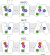
Ion binding to the three main cation-chloride cotransporters: NKCC, NCC, and KCC. While two Cl densities are observed in NKCC (green), only one Cl density is observed for NCC. In the absence of K+, Cl cannot be coordinated at SCl1 (white empty circle). Two Cl densities were however observed in the KCC structure and based on the cryo-EM structure of NCC, we hypothesize that Cl at SCl1 (red) is not released, whereas Cl at SCl2 (green) is transported. Based on the position of the ions and the narrow channel on the outside, for influx (left model), Cl must bind first, followed by K+. On the other hand, since the cavity on the inside is larger, either ion can access its binding site, not restricted by the other. This model is consistent with previous kinetic studies showing strictly ordered binding of ions on the outside, while random binding on the inside.
Figure 19.
Figure 19.
Salt bridge between the NH2+ group of Arg307 in TMD1b and the O group of the glutamic acid of Glu389 in TMD3. The distance between the groups is short: 2.5 Å in human NKCC1 (PDB: 7mxo).
Figure 20.
Figure 20.
Evidence of a pore (tunnel) going through the extracellular domain, the transmembrane domain, and the carboxyl-terminus of NKCC1. The 7mxo structure stripped of its ions was uploaded to MOLEonline to run MOLE 2.5, a tool that allows rapid and fully automated identification of channels, tunnels, and pores in protein structures. This model provides evidence for possible movement of ions or water molecules through the globular carboxyl-terminus of the cotransporter.
Figure 21.
Figure 21.
Two cryo-EM structures of NKCC1 in with bumetanide indicate that the inhibitor can take slightly different positions within the NKCC1 structure: as in PDB: 7smp (A) and in PDB: 7s1x (B). Note that some coordinating residues (underlined) are common in the two structures: Val302, Met303, Ala379, Val385, Pro496.
Figure 22.
Figure 22.
Molecular docking of ML077 (VU0255011) with human KCC1. Note that residue Tyr216 which coordinates the binding of K+ in hKCC1 is the equivalent of Tyr383 in NKCC1, residue that also coordinates K+ binding (and possibly bumetanide binding) in the Na+-dependent transporter. Note that the binding might be biased by a structure that is an inward facing configuration and a different binding pocket could be formed when the transporter is in an outward facing configuration, i.e., similar to what is seen in the cryo-EM structure of NKCC1 in complex with bumetanide.
Figure 23.
Figure 23.
Positions of VU0463271 (A) and DIOA (B) in the cryo-EM structures of KCC1 and KCC3 demonstrate different modes of inhibition. VU0463271 is located in the external pore of the permeation channel, similar to bumetanide. While the thiazole group interacts with the glutamic acid residue responsible for locking the transporter in the inward conformation, the two aromatic rings at the opposite end enter deeper in the channel, interacting with residues in TM1, TM6, and TM10. DIOA, on the other hand, binds between the two monomers interacting with residues of both monomers. The binding of DIOA at that position likely prevents the proper movement of carboxyl-termini and transmembrane domains associated with transport.

Similar articles

Cited by

References

    1. Barber HS(1938) The Mercurial Diuretics. Postgrad Med J 14: 288–294 - PMC - PubMed
    1. Belair EJ, Cohen AI, and Yelnosky J(1972) Renal excretion of metolazone, a new diuretic. Br J Pharmacol 45: 476–479 - PMC - PubMed
    1. Chew TA, Orlando BJ, Zhang J, Latorraca NR, Wang A, Hollingsworth SA, Chen DH, Dror RO, Liao M, and Feng L(2019) Structure and mechanism of the cation-chloride cotransporter NKCC1. Nature 572: 488–492 - PMC - PubMed
    1. Chi X, Li X, Chen Y, Zhang Y, Su Q, and Zhou Q(2020) Cryo-EM structures of the full-length human KCC2 and KCC3 cation-chloride cotransporters. Cell Res 31: 482–484 - PMC - PubMed
    1. Chobanian AV, Bakris GL, Black HR, Cushman WC, Green LA, Izzo JL Jr., Jones DW, Materson BJ, Oparil S, Wright JT Jr., and Roccella EJ(2003) Seventh report of the Joint National Committee on Prevention, Detection, Evaluation, and Treatment of High Blood Pressure. Hypertension 42: 1206–1252 - PubMed

MeSH terms

LinkOut - more resources