Review
. 2023 Sep;19(9):525-541.
doi: 10.1038/s41582-023-00846-7. Epub 2023 Aug 10.

Limbic-predominant age-related TDP43 encephalopathy (LATE) neuropathological change in neurodegenerative diseases

Affiliations
Review

Limbic-predominant age-related TDP43 encephalopathy (LATE) neuropathological change in neurodegenerative diseases

Sukriti Nag et al. Nat Rev Neurol. 2023 Sep.

Abstract

TAR DNA-binding protein 43 (TDP43) is a focus of research in late-onset dementias. TDP43 pathology in the brain was initially identified in amyotrophic lateral sclerosis and frontotemporal lobar degeneration, and later in Alzheimer disease (AD), other neurodegenerative diseases and ageing. Limbic-predominant age-related TDP43 encephalopathy (LATE), recognized as a clinical entity in 2019, is characterized by amnestic dementia resembling AD dementia and occurring most commonly in adults over 80 years of age. Neuropathological findings in LATE, referred to as LATE neuropathological change (LATE-NC), consist of neuronal and glial cytoplasmic TDP43 localized predominantly in limbic areas with or without coexisting hippocampal sclerosis and/or AD neuropathological change and without frontotemporal lobar degeneration or amyotrophic lateral sclerosis pathology. LATE-NC is frequently associated with one or more coexisting pathologies, mainly AD neuropathological change. The focus of this Review is the pathology, genetic risk factors and nature of the cognitive impairments and dementia in pure LATE-NC and in LATE-NC associated with coexisting pathologies. As the clinical and cognitive profile of LATE is currently not easily distinguishable from AD dementia, it is important to develop biomarkers to aid in the diagnosis of this condition in the clinic. The pathogenesis of LATE-NC should be a focus of future research to form the basis for the development of preventive and therapeutic strategies.


Conflict of interest statement

Competing interests

The authors declare no competing interests.

Figures
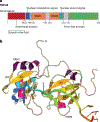
Fig. 1|
Fig. 1|
TDP43 structure, a. Domain architecture of TAR DNA-binding protein 43 (TDP43). Domain boundaries are numbered according to the full-length protein sequence, b. Ribbon representation of the structure of the TDP43 RNA-recognition motif (RRM) construct (amino acids 102–269) in complex with AUG12 RNA (Protein Data Bank ID code 4BS2). Parts a and b reprinted with permission from ref. 19, Cold Spring Harbour Laboratory Press.
Fig. 2|
Fig. 2|. TDP43 pathology.
Sparse (part a), moderate (part b) and frequent (part c) intracytoplasmic neuronal TAR DNA-binding protein 43 (TDP43) inclusions and neurite immunostaining in the anterior temporal pole cortex. The areas depicted in parts a-c are smaller than the 0.25 mm counting frame used to quantify the inclusions. Cytoplasmic TDP43 in neurons and prominent neurite immunostaining shown at high magnification (part d). Scale bars: 25 μm in parts a-c and 50 μm in part d. Reprinted with permission from ref. 34, BioMed Central Ltd.
Fig. 3|
Fig. 3|. Numbers of TDP43 inclusions by TDP43 stage.
Box-and-whisker plots show the total number of TAR DNA-binding protein 43 (TDP43) inclusions per 0.25 mm in eight brain regions (as shown in Fig. 4) by TDP43 stage in 1,160 individuals. Data were obtained at a total magnification of 200× from an area showing the highest density of inclusions in the stated regions. TDP43 inclusions increase progressively in the amygdala by stage. Inclusions in all regions including the anterior temporal pole cortex are maximal in stage 5. Outlier values are indicated by circles. The numbers on the x-axis denote the brain regions: 1, amygdala; 2, entorhinal cortex; 3, CA1 sector of the hippocampus; 4, dentate neurons of the hippocampus; 5, anterior temporal pole cortex; 6, midtemporal cortex; 7, orbitofrontal cortex; 8, midfrontal cortex. Centre line, median; box limits, lower and upper quartiles; whiskers, data range; circles, outliers. Reprinted with permission from ref. 34, BioMed Central Ltd.
Fig. 4|
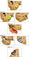
Fig. 4|. The ROSMAP five-stage LATE-NC scheme.
Coronal brain sections showing the regional distribution of phosphorylated TAR DNA-binding protein 43 (TDP43) cytoplasmic inclusions and the percentage of cases showing inclusions at stages 1–5 of the Religious Orders Study and Memory and Aging Project (ROSMAP). This is a cumulative staging system such that any stage from 2 to 5 is rated as positive only if the preceding stage or stages are positive. AMG, amygdala; ATPC, anterior temporal pole cortex; CA1, CA1 sector of the hippocampus; DEN, dentate gyrus; EC, entorhinal cortex; MFC, midfirontal cortex; MTC, midtemporal cortex; OFC, orbitofrontal cortex. Adapted from ref. 34, BioMed Central Ltd.

References

    1. Ou SH, Wu F, Harrich D, Garcia-Martinez LF & Gaynor RB Cloning and characterization of a novel cellular protein, TDP-43, that binds to human immunodeficiency virus type 1 TAR DNA sequence motifs. J. Virol 69, 3584–3596 (1995). - PMC - PubMed
    1. Arai T et al. TDP-43 is a component of ubiquitin-positive tau-negative inclusions in frontotemporal lobar degeneration and amyotrophic lateral sclerosis. Biochem. Biophys. Res. Commun 351, 602–611 (2006). - PubMed
    1. Neumann M et al. Ubiquitinated TDP-43 in frontotemporal lobar degeneration and amyotrophic lateral sclerosis. Science 314, 130–133 (2006). - PubMed
    1. Yokota O et al. Phosphorylated TDP-43 pathology and hippocampal sclerosis in progressive supranuclear palsy. Acta Neuropathol 120, 55–66 (2010). - PMC - PubMed
    1. Brenowitz WD, Monsell SE, Schmitt FA, Kukull WA & Nelson PT Hippocampal sclerosis of aging is a key Alzheimer’s disease mimic: clinical-pathologic correlations and comparisons with both Alzheimer’s disease and non-tauopathic frontotemporal lobar degeneration. J. Alzheimers Dis 39, 691–702 (2014). - PMC - PubMed

Publication types

Substances

Supplementary concepts